Abstract
Background Weakness of the abductor muscles—or even their avulsion—is a potential risk when total hip arthroplasty (THA) is performed using the anterolateral approach. Avulsion of the re-attached gluteus medius leads to a limp, but can also provide an open canal for joint fluid to escape to the trochanteric region. Thus, our hypothesis was that arthrography can be used to diagnose such detachment.
Methods We compared THA arthrographies with peroperative findings retrospectively in 33 patients who had undergone surgical exploration for a muscle reattachment because of a strong suspicion of abductor avulsion at physical examination.
Results After a mean follow-up time of 22 (2–57) months after THA, 14/33 patients had a positive (pathological) arthrogram whereas 19 had a negative (normal) result. All 14 patients with a positive arthrogram were verified to have an avulsion of the abductor muscle at the operation. 10 of the 19 patients with a negative arthrogram had an intact abductor insertion, but 9 had an avulsion. All of these 9 patients with the peroperatively disclosed avulsion had a fibrous capsule, which obstructed the fistula leading from the joint cavity to the trochanteric bursa region.
Interpretation Arthrography is a valuable diagnostic aid in the evaluation of patients with abductor weakness and with Trendelenburg’s gait appearing after a THA performed using the anterolateral approach. A positive finding helps in the operative decision making, but a negative arthrogram is not a reliable predictor.
The anterolateral approach for hip arthroplasty— especially the Hardinge approach—leads to an increased risk of postoperative avulsion of the re-attached abductors, resulting in a limp (Baker and Bitounis Citation1989, Moskal and Mann Citation1996, Pai and D’Ortho Citation1996, Weber and Berry Citation1997). The diagnosis of abductor avulsion is usually based on physical examination (Weber and Berry Citation1997) and the true prevalence of this condition is unknown.
Radiological methods are used for evaluation of suspected failures of totally replaced hips. However, there are no published reports concerning the use of arthrography in the evaluation of possible avulsion of the re-attached abductors. Since arthrography has been in routine use in our hospital in the radiological evaluation of patients with symptoms and signs suggesting the possibility of an abductor avulsion, we performed this retrospective study to examine its accuracy in this setting.
Patients and methods
For this study, data were collected from the medical records of 48 consecutive patients who had undergone arthrography of their replaced hip because of a clinical suspicion of avulsion of the reinserted gluteus medius muscle after primary or revision hip arthroplasty, which had been performed using an anterolateral Hardinge approach. Of these 48 patients, 33 patients with particularly troublesome symptoms underwent a surgical evalution to assess the need for operative repair. The study group consisted of 27 women and 6 men with a mean age of 65 (40–86) years. All patients suffered from osteoarthritis. The index operation, after which the symptoms had developed, was a primary arthroplasty in 25 patients and a revision arthroplasty in 8 patients. The main clinical findings in all of our patients were trochanteric pain in the operated hip, associated with weakness of the abductor muscles and/or a positive Trendelenburg sign. Weakness of the internal rotators was another typical finding. Plain radiographs were normal in all patients and did not disclose any signs of loosening, malposition of the implant components, or heterotopic bone formation. Arthrography of the operated hip was part of the routine preoperative work-up. The reoperation for the suspected abductor avulsion was performed on average 23 (2–63) months after the index operation.
Arthrography
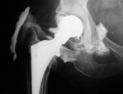
Arthrography was performed successfully in all patients by the technique introduced by Salvati et al. (Citation1971). Using fluoroscopy, the location of the neck of the stem of the prosthesis was localized and marked on the skin. After cleaning of the skin, the area was covered with an operating sheet with its vertical slit placed over the skin mark. The femoral artery was palpated and the skin and the subcutaneous tissue were infiltrated with local anesthetics. A 20–22-gauge (0.9–0.7 mm) disposable needle was directed towards the neck of the stem. Metal-to-metal contact was felt upon contact of the point of the needle with the prosthesis. Synovial fluid, if any, was aspirated and sent for bacteriological tests. Under fluoroscopic guidance, Omnipaque 240 mg I/mL contrast medium was injected to fill the joint space, usually 20–25 mL. The needle was then removed and the hip was passively or actively exercised to spread the contrast. Arthrograms were interpreted to be pathological (positive) if the contrast medium leaked from the joint cavity (limited by a thick pseudocapsule around the prosthesis) to the trochanteric bursa (Figure).
An arthrogram showing leakage of the contrast medium from the pseudojoint cavity to a bursa developed in the trochanter area after total hip arthroplasty, which had been performed using the anterolateral approach. This finding is diagnostic of an avulsion of the abductor muscles from the medial aspect of the greater trochanter.
Clinical examination
The main indication for the reoperations was either a pathological arthrography or, in 19 patients with a negative arthrogram, a strong clinical suspicion of abductor avulsion based on their symptoms and positive clinical signs. Abductor strength was measured against gravity and against a force applied to the patient’s leg by the examiner when the patient was lying on his/her side. Abductor strength on the operated side was compared to that on the healthy side and graded as 0 = negligible, 1 = able to resist gravity, 2 = able to resist moderate force but less than on the healthy side, and 3 = normal strength. Besides the reduced abductor strength (grades 0–2), the weakness of internal rotation of the hip, Trendelenburg sign (Pai and D’Ortho Citation1996), and use of a cane or crutches were registered.
Surgery
After clinical and arthrographic evaluations, a decision to operate was made in 33 cases. Surgical findings were registered and used as the truth, and they were compared to the results of the preoperative arthrographies. If an avulsion was found, the fistula and the bursal lining were excised, the medial aspect of the trochanteric region was roughened, and the abductor muscle was reinserted. The muscle was reinserted to the medial aspect of the greater trochanter, along with the conjoined vastus lateralis tendon, using both unresorbable sutures (Ti-Cron) guided through bone channels and resorbable polyglycolide sutures (Dexon). The offset, the anteversion of the stem, and the position of the acetabular cup were checked to avoid overstrain of abductors. If excessive offset was noticed, the metallic head was changed to a shorter one.
Table 1. Diagnostic accuracy of arthrography in 33 patients with clinically suspected avulsion of abductors
Results (Table)
Of the 33 patients who had persistent symptoms and who underwent surgery, arthrography findings were positive in 14 patients and negative in 19. In all 14 patients with a positive arthrography, the peroperative findings confirmed the avulsion of the anterior part of the gluteus medius muscle. In all such cases the fistuli, which allowed contrast leakage from the joint cavity to the trochanteric bursa, were already clearly visible during the injection of the contrast material and on the arthrograms (Figure). Although leakage was seen without any special exercise, we did not try to aspirate the contrast medium from the joint or bursa, but removed the needle and took the radiograph.
In the 19 patients with negative arthrography findings, the insertion of the gluteus medius was intact in 10 patients, whereas 9 patients had an avulsion. In all of these patients the joint space was constricted by a fibrous capsule, which obstructed the entrance of the joint fistula leading from the joint cavity to the trochanteric bursa. Of 10 patients with an intact abductor at revision, 4 had a loosened stem, 2 had a loosened cup, 1 had a retroversion of the stem, 1 had heterotopic calcifications and cicatrix formation, 1 had herniation of the fascia and adherences, and 1 showed stretching of the gluteus medius muscle after a previous suturation. 1 of the 2 patients with a loosened cup also had denervation of the superior gluteal nerve.
None of the patients had signs of infection in the aspirated joint fluid.
Discussion
Today, the anterolateral and posterior approaches are commonest for THA. The anterolateral approach, described by Hardinge (Citation1982), allows excellent acetabular and proximal femoral exposure and carries a relatively low risk of dislocation (Vicar and Coleman Citation1984, Barber et al.Citation1996, Ritter et al. Citation2001). The main disadvantages of this approach are the relatively high incidence of abductor weakness and heterotopic ossification. Abductor injury is caused by large-size reamers or prosthetic components, especially in revision operations. The mode of approach is perhaps one of the most important risk factors for avulsion. Many surgeons use a modified Hardinge approach, i.e. with detachment of the anterior portion of the gluteus medius muscle only, leaving most of the vastus lateralis tendon intact. In our opinion, it is essential that the incision is placed, according to Hardinge, through the thickest portion of the conjoined tendon area of the gluteus medius and vastus lateralis. This guarantees the most reliable re-attachment. Thus, the correct positioning of the incision may be the prerequisite for both proper re-attachment and the transmission of abductor function. Accordingly, osteosutures are probably less important. Furthermore, an increased offset of the hip and an increased anteversion of the stem increase the risk of abductor avulsion. The Hardinge approach may also be complicated by a partial denervation of the gluteus medius muscle (Ramesh et al. Citation1996, Weale et al. Citation1996).
The Hardinge approach may delay recovery of the abductor muscle strength and can lead to permanent abductor weakness (Barber et al. Citation1996, Pai and D’Ortho Citation1996). Avulsion of the reinserted gluteus muscle, leading to late-onset Trendelenburg gait and trochanteric pain, has also been reported (Weber and Berry Citation1997, Giurea et al. Citation1998).
Abductor avulsion may give long-lasting pain and/or limp in the operated extremity and may present a diagnostic dilemma. Conventional physical examination may not allow reliable diagnosis. Trochanteric pain, reduced abductor strength, and limp developing after THA that has been performed with the anterolateral approach indicate abductor dysfunction, or even avulsion. Abductor dysfunction may also develop in patients operated with the anterolateral approach, particularly if the superior gluteal nerve has been damaged. Muscle avulsion and nerve injury both lead to a similar Trendelenburg gait and muscle atrophy (Twair et al. Citation2003).
Weakness, which is restricted to the abductor muscles, should raise the suspicion of loss of attachment of the gluteus medius muscle. In our series, all patients with a confirmed abductor avulsion had normal plain radiographs. An avulsion opens up a connection between the joint space and the trochanteric bursa. We found that filling of the trochanteric bursa with contrast medium through this pathological channel in arthrography is diagnostic of an avulsion.
We did not observe any false positive arthrography findings. However, the connection between the joint cavity and the trochanteric bursa was sometimes blocked by pseudocapsular tissue, which apparently obstructed the leakage of contrast medium. This led to false negative findings in 9 of the 19 cases. In patients with negative arthrography, the decision to treat surgically must be based on clinical symptoms and signs. In spite of this shortcoming, arthrography is a valuable diagnostic tool.
The total number of patients was 48, but only 33 were operated. The age of the patient, the number of former operations, and the severity of pain and muscle weakness were the main criteria in clinical decision making. In most of the patients who were not operated, with time the symptoms and disability became reduced so that surgical intervention was not warranted. Some patients even refused reoperation when the plain radiographs, joint fluid aspiration, and arthrography showed no signs of loosening or infection.
Contributions of authors
PY: was involved in the design of the study, performed surgery, and drafted the manuscript. KT: performed arthrographies, performed data analyses, and finalized the manuscript. ML: performed arthrographies. YTK: participated in analyzing the material and helped in preparation of the manuscript. TP: was in charge of the surgery team and was involved in the coordination of the patient material.
No competing interests declared.
- Baker A S, Bitounis V C. Abductor function after total hip replacement. An electromydographic and clinical review. J Bone Joint Surg (Br) 1989; 71: 47–50
- Barber T C, Roger D J, Goodman S B, Schurman D J. Early outcome of total hip arthroplasty using the direct lateral v. s. the posterior surgical approach. Orthopaedics 1996; 19: 873–5
- Giurea A, Paternostro T, Heinz–Peer G, Kaider A, Gott–sauner–Wolf F. Function of reinserted abductor muscles after femoral replacement. J Bone Joint Surg (Br) 1998; 80: 284–7
- Hardinge K. The direct lateral approach to the hip. J Bone Joint Surg (Br) 1982; 64: 17–9
- Moskal J T, Mann J W. A modified direct lateral approach for primary and revision total hip arthroplasty. A prospective analysis of 453 cases. J Arthroplasty 1996; 11: 255–66
- Pai V S, D'Ortho M S. Significance of the Trendelenburg test in total hip arthroplasty. Influence of lateral approaches. J Arthroplasty 1996; 11: 174–9
- Ramesh M, Byrne J M, McCarthy N, Jarvis A, Mahalingham K, Cashman W F. Damage to the superior gluteal nerve after the Hardinge approach to the hip. J Bone Joint Surg (Br) 1996; 7: 903–6
- Ritter M A, Harty L D, Keating M E, Faris P M, Meding J B. A clinical comparison of the anterolateral and posterolateral approaches of the hip. Clin Orthop 2001, 385: 95–9
- Salvati E A, Freiberger R H, Wilson P D, Jr. Arthrography for complications of total hip replacement. A review of thirty–one arthrograms. J Bone Joint Surg (Am) 1971; 53: 701–9
- Twair A, Ryan M, O'Connell M, Powell T, O'Byrne J, Eustace S. MRI of failed total hip replacement caused by abductor muscle avulsion. AJR Am J Roentgenol 2003; 181: 1547–50
- Vicar A J, Coleman C R. A comparison of the anterolateral, transtrochanteric, and posterior surgical approaches in primary total hip arthroplasty. Clin Orthop 1984, 188: 152–9
- Weale A E, Newman P, Ferguson I T, Bannister G C. Nerve injury after posterior and direct lateral approaches for hip replacement. A clinical and electrophysiological study. J Bone Joint Surg (Br) 1996; 78: 899–902
- Weber M, Berry D J. Abductor avulsion after primary total hip arthroplasty. Results of repair. J Arthroplasty 1997; 12: 202–6