Abstract
Background and purpose With Ganz periacetabular osteotomy, the osteotomized acetabular fragment is reoriented in an adducted, extended, and rotated position. The acetabular fragment is fixated with 2 screws and the patients are allowed 30 kg of weight bearing immediately after surgery. We were interested in examining the stability of the reoriented acetabulum after Ganz osteotomy; thus, the migration of the acetabular fragment was assessed by radiostereometry.
Patients and methods 32 dysplastic patients (27 females; 32 hips) were included in the study. Median age was 39 (20–57) years. Radiostereometric examinations were done at 1 week, 4 weeks, 8 weeks and 6 months. Data are presented as mean (SD).
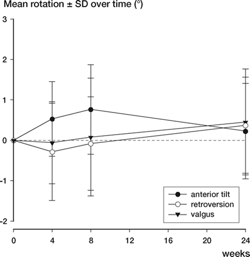
Results 6 months postoperatively, the acetabular fragment had migrated 0.7 (0.8) mm medially, and 0.7 (0.5) mm proximally. Mean rotation in adduction was 0.5° (1.3). In other directions, mean migration was below 0.5 mm/°. There were no statistically significant differences in migration at 8 weeks and 24 weeks postoperatively regarding translation and rotation.
Interpretation Due to the limited amount of migration, we find our postoperative partial weight-bearing regime safe.
If not treated, hip dysplasia in adults leads to secondary osteoathritis in half of the cases by the time the patient reaches the age of 50 (Wiberg Citation1939, Cooperman et al. Citation1983). The Ganz periacetabular osteotomy (Ganz et al. Citation1988) has become a widely used treatment for hip dysplasia due to the inherent stability associated with a partially intact posterior column (Shiramizu et al. Citation2004, Aminian et al. Citation2005). The osteotomy results in an increased acetabular load-bearing area (Mechlenburg et al. Citation2004) and may improve the pressure distribution over the available cartilage surface (Armand et al. Citation2005, Tsumura et al. Citation2005). At the operation, the osteotomized acetabular fragment is reoriented in an adducted, extended, and rotated position (Søballe 2003). The acetabular fragment is fixated with 2 screws and the patient is allowed 30 kg of weight bearing with two crutches until 8 weeks postoperatively, after which full weight bearing is allowed. At our institution, the amount of 3D reorientation of the acetabular fragment is based on preoperative radiographically measured angles. We were interested in examining the stability of the reoriented acetabulum after Ganz osteotomy, and the migration of the acetabular fragment was assessed by radiostereometric analysis (RSA). Also, we hypothesized that the acetabuli that underwent the highest amount of reorienta-tion—evaluated by center-edge angle of Wiberg (Wiberg Citation1939) and by the acetabular index of the weight-bearing zone (Tönnis Citation1987) measured pre- and postoperatively—would also be the ones that migrated the most postoperatively.
Patients and methods
The study was accepted by the local ethical committee. After obtaining signed consent, 27 women and 5 men presenting 32 dysplastic hips were included in the study. Median age was 39 (20–57) years. The patients were scheduled for Ganz periacetabular osteotomy and they had the following radiological and clinical characteristics: centeredge angle < 25° (Wiberg Citation1939), osteoarthritis of degree 0 or 1 according to the classification of Tönnis (Citation1987), closed growth zones in the pelvis, symptomatic and painful hip, and a minimum of 110° flexion in the hip joint. Patients in whom the dysplasia might have been caused by neurological illnesses, Legg-Calvé-Perthes’ disease, or sequelae after earlier hip surgery were excluded from the study. Also, patients for whom an intertrochanteric femoral osteotomy was necessary were excluded from the study.
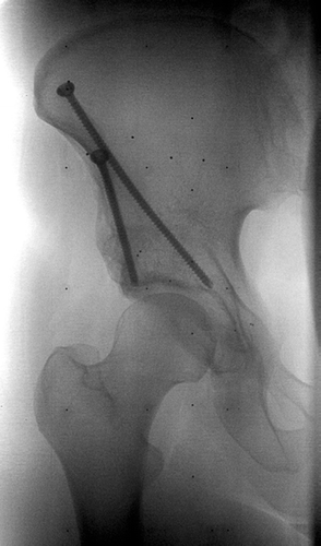
At the time of the operation, tantalum markers were inserted into the acetabular fragment (5 markers (1 mm) were spread well apart) and 5 markers (0.8 mm) were inserted into the iliac bone above the fragment (). At the beginning of the study, the markers in the iliac bone were inserted fairly close to each other but as we realized that this was not optimal, the markers were inserted further apart. When using RSA, a precise and accurate estimation of the migration is obtained (Vrooman et al Citation1998, Valstar et al. Citation2000, Valstar Citation2001). Radiostereometric examinations were done at 1 week, 4 weeks, 8 weeks and 6 months. The patients were mobilized on the second day after surgery and the first stereometric examination was performed after mobilization. Comparison of the images provided an estimation of the micromotion in three dimensions as a function of time.
Figure 1. Radiostereometric image with bone markers.
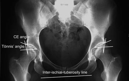
Two angles were measured on pre- and postoperative AP radiographs: CE angle (center-edge) which assesses the superior coverage of the acetabulum (Wiberg Citation1939) and Tönnis’ angle (the acetabular index of the weight-bearing zone) (Tönnis Citation1987) which evaluates the orientation of the acetabular roof (). The CE angle is obtained by drawing a vertical line through the femoral head perpendicular to the horizontal line extending through the center of both femoral heads. A line is then drawn from the center of the femoral head to the most superolateral point of the acetabulum (Delaunay et al. Citation1997). The CE angle is normally above 25° (Wiberg Citation1939). The acetabular index of the weightbearing zone is the angle formed by a line parallel to the inter-teardrop line and a line from the lateral point to the medial point of the weight-bearing portion of the acetabulum. We chose however, to replace the inter-teardrop line with an interischial tuberosity line, as the position of the teardrop is changed after Ganz osteotomy. The Tönnis angle is normally below 10° (Tönnis Citation1987).
Figure 2. Preoperative radiograph with CE angle and Tönnis'angle.
Radiostereometric examinations
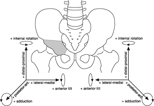
Radiostereometric examinations were done with the patient in a supine position. A calibration box (Carbon Box Aarhus; Medis, Leiden, the Netherlands) placed beneath the patient was used to create a 3D coordinate system of the tantalum markers. Two fixed roentgen tubes with a 40-degree angle between them were positioned above the patient. The patient was exposed to the two simultaneously firing roentgen tubes (150 microSv, 96 kV and 13 mAs). Digital radiographs (Fuji FCR AC – 3CS/ ID) were obtained and the DICOM files were evaluated by RSA-CMS (Medis). This software package performs the RSA procedure automatically. The first radiostereometric examination served as a reference baseline. All subsequent evaluations of migration were related to the position of the acetabular fragment relative to the iliac bone at the time of the evaluation. Migration of the acetabular fragment was expressed as translation and rotation of the center of gravity of the markers inserted into the acetabular fragment ().
Figure 3. Axes of rotation and translation of the acetabular fragment.
Double examinations
The first 7 patients were examined twice, with complete repositioning of the patient and set-up in order to obtain an estimate of precision of the method used.
Results
No postoperative complications were encountered (i.e. no infections, deep vene thrombosis, neurovascular complications, or delayed union). After radiostereometric examinations had started, no patients who had been included were subsequently excluded from the study but 1 patient did not attend the final examination.
The migration of the acetabular fragment after Ganz periacetabular osteotomy is shown in . The translation and rotation over time is shown in and . Tested by paired t-test, there were no statistically significant differences in migration 8 weeks and 24 weeks postoperatively regarding both translation and rotation (with p-values ranging between 0.08 and 0.44). The precision was estimated from double examination of 7 patients (). Our measurement precision, expressed as the maximum standard deviation, was 0.58 mm in translation and 0.56° in rotation. The mean error of rigid body which accounts for marker stability was 0.07. The 3D distribution of markers in the bone is indicated by the condition number, which was 30.23 for the acetabular fragment and 31.94 for the iliac bone.
Figure 4. Mean translation and SD over time.
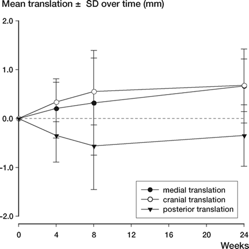
Figure 5. Mean rotation and SD over time,
. Migration of acetabular fragment 6 months after Ganz periacetabular osteotomy (n=32)
. The precision as estimated from 7 double examinations
Tested by Pearson’s coefficient of correlation, there was no correlation between the amount of reorientation (measured by change in radiographic angles) and the migration of the acetabular fragment in all directions (p = 0.16–0.85) ().
. Radiographic evaluation of preoperative and postoperative radiographs from 32 hips
Discussion
We have not found any reports dealing with RSA in Ganz periacetabular osteotomy. We consider RSA to be an objective measure of osteotomy migration and find this important to evaluate, as there has been some concern about loss of correction and failure of fixation in relation to performing periacetabular osteotomies. We found that 6 months postoperatively the mean translation of the acetabular fragment was less than 0.7 mm and the mean rotation was less than 0.5°. As there was no statistically significant difference between migration at 2 and 6 months postoperatively, we conclude that the osteotomies were stable 2 months postoperatively.
In a biomechanical study by Babis et al. (Citation2002), periacetabular osteotomies were created on 6 pelvises from cadavers and fixed randomly with 3 screws from the iliac crest, or with 2 screws supplemented with a transverse screw to the ilium. The pelvises were loaded up to 130 kg in a simulated push-off phase of the gait cycle. An average displacement of the pubic osteotomy of 13 mm was found in both cases and the authors concluded that neither type of fixation provides enough stability to allow immediate weight bearing after periacetabular osteotomy. However, at our institution patients are only allowed to bear 30 kg until 2 months postoperatively and our clinical study clearly demonstrates that acetabular migration is very limited, although the osteotomies in our study were fixated with 2 screws only.
Yassir et al. (Citation2005) tested the stability of Ganz periacetabular osteotomy on 8 cadaver hips under simulated weight-bearing conditions using 3D analysis of fragment displacement and angular rotation. The mechanical loading approximated partial weight bearing in an adult. The acetabular fragment fixated with 2 screws from the iliac wing, as in our study, became displaced by less than 1.5 mm and rotated less than 2.5 degrees. The displacement and angulation found in that study is also higher than what we found. According to our data, acetabular migration postoperatively does not constitute a clinical problem in terms of loss of correction or failure of fixation, and consequently we find our postoperative partial weight-bearing regime safe.
Radiostereometric measurements of one pelvic osteotomy were performed as early as 1978 (Hansson et al. Citation1978) with the purpose of obtaining information about the position of the acetabulum pre- and postoperatively, but that study did not evaluate acetabular migration in the time period after surgery. RSA studies estimating migration after fractures have found slightly higher (Ragnarsson et al. Citation1992, Ahl et al.Citation1994) or much higher (Mattsson and Larsson Citation2003, Citation2004) migration compared to the amount of migration we found.
Precision is defined as the closeness of agreement between repeated independent test results obtained under stipulated conditions. The precision of RSA in our study as calculated from double examinations was not as high as that reported by Vrooman et al. (Citation1998) who, in an RSA study of knee prosthesises, found the maximum standard deviation in translation to be 0.11 mm and in rotation 0.24°. Madanat et al. (Citation2005) estimated the precision of RSA in a fracture model of the distal radius. They reported that under optimal laboratory conditions, translations of 200 µm can be measured with a precision of 2–6 µm and rotations of 0.5° can be measured with a precision of 0.025–0.096°. The precision of this method applied to patients will be lower than under laboratory conditions because it is impossible to position the patient and the roentgen tubes in exactly the same way under consecutive radiostereometric examinations. Furthermore, the optimal distribution of markers is more difficult to obtain in a patient during surgery than in a phantom. Also, in the patient the markers may be loose or hidden behind a screw, which reduces the number of markers available for estimation of migration.
The pre- and postoperatively measured angles in our study correspond well with what other groups have found (Matta et al Citation1999, Siebenrock et al. Citation1999, Valenzuela et al. Citation2004, Pogliacomi et al. Citation2005). Our hypothesis about a possible correlation between the amount of reorientation and the degree of migration of the acetabular fragment proved to be incorrect. In another study, we have tested whether a correlation might exist between the amount of migration and bone density in the acetabulum (Mechlenburg 2006, unpublished data). This was not the case, and probably a whole range of factors affects migration of the acetabular fragment postoperatively.
In conclusion, due to the very limited degree of migration, we find our postoperative partial weightbearing regimen after Ganz osteotomy to be safe.
This study was financially supported by the Danish Rheumatism Association, the Aase and Ejnar Danielsen Foundation, the Marie and M.B. Richter Foundation, and Dr Søren Segel and Johanne Wiibroe Segel’s Research Foundation.
Contributions of authors
IM: collected and analyzed data and wrote the article. SK: supervised throughout study period. LR: responsible for RSA examinations and supervised. KS: operated the patients and supervised.
References
- Ahl T, Dalen N, Lundberg A, Wykman A. Biodegradable fixation of ankle fractures. A roentgen stereophotogrammetric study of 32 cases. Acta Orthop Scand 1994; 65: 166–70
- Aminian A, Mahar A, Yassir W, Newton P, Wenger D. Freedom of acetabular fragment rotation following three surgical techniques for correction of congenital deformities of the hip. J Pediatr Orthop 2005; 25: 10–3
- Armand M, Lepisto J, Tallroth K, Elias J, Chao E. Outcome of periacetabular osteotomy: joint contact pressure calculation using standing AP radiographs, 12 patients followed for average 2 years. Acta Orthop 2005; 76: 303–13
- Babis G C, Trousdale R T, Jenkyn T R, Kaufman K. Comparison of two methods of screw fixation in periacetabular osteotomy. Clin Orthop 2002, 403: 221–7
- Cooperman D R, Wallensten R, Stulberg S D. Acetabular dysplasia in the adult. Clin Orthop 1983, 175: 79–85
- Delaunay S, Dussault R G, Kaplan P A, Alford B A. Radiographic measurements of dysplastic adult hips. Skeletal Radiol 1997; 26: 75–81
- Ganz R, Klaue K, Vinh T S, Mast J W. A new periacetabular osteotomy for the treatment of hip dysplasias Technique and preliminary results. Clin Orthop 1988, 232: 26–36
- Hansson L I, Olsson T H, Selvik G, Sunde´n G. A Roentgen stereophotogrammetric investigation of innominate osteotomy (Salter). Acta Orthop Scand 1978; 49: 68–72
- Madanat R, Makinen T J, Moritz N, Mattila K T, Aro H T. Accuracy and precision of radiostereometric analysis in the measurement of three-dimensional micromotion in a fracture model of the distal radius. J Orthop Res 2005; 23: 481–8
- Matta J M, Stover M D, Siebenrock K. Periacetabular osteotomy through the Smith-Petersen approach. Clin Orthop 1999, 363: 21–32
- Mattsson P, Larsson S. Stability of internally fixed femoral neck fractures augmented with resorbable cement. A prospective randomized study using radiostereometry. Scand J Surg 2003; 92: 215–9
- Mattsson P, Larsson S. Unstable trochanteric fractures augmented with calcium phosphate cement A prospective randomized study using radiostereometry to measure fracture stability. Scand J Surg 2004; 93: 223–8
- Mechlenburg I, Nyengaard J R, Romer L, Soballe K. Changes in load-bearing area after Ganz periacetabular osteotomy evaluated by multislice CT scanning and stereology. Acta Orthop Scand 2004; 75: 147–53
- Pogliacomi F, Stark A, Wallensten R. Periacetabular osteotomy. Good pain relief in symptomatic hip dysplasia, 32 patients followed for 4 years. Acta Orthop 2005; 76: 67–74
- Ragnarsson J I, Eliasson P, Karrholm J, Lundstrom B. The accuracy of measurements of femoral neck fractures. Conventional radiography versus roentgen stereophotogrammetric analysis. Acta Orthop Scand 1992; 63: 152–6
- Shiramizu K, Naito M, Asayama I, Yatsunami M. A quantitative anatomic characterization of the quadrilateral surface for periacetabular osteotomy. Clin Orthop 2004, 418: 157–61
- Siebenrock K A, Scholl E, Lottenbach M, Ganz R. Bernese periacetabular osteotomy. Clin Orthop 1999, 363: 9–20
- Soballe K. Pelvic osteotomy for acetabular dysplasia. Acta Orthop Scand 2003; 74: 117–8
- To¨nnis D. Congenital dysplasia and dislocation of the hip in children and adults. Springer, Berlin, Heidelberg, New York 1987
- Tsumura H, Kaku N, Ikeda S, Torisu T. A computer simulation of rotational acetabular osteotomy for dysplastic hip joint: does the optimal transposition of the acetabular fragment exist. J Orthop Sci 2005; 10: 145–51
- Valenzuela R G, Cabanela M E, Trousdale R T. Sexual activity, pregnancy, and childbirth after periacetabular osteotomy. Clin Orthop 2004, 418: 146–52
- Valstar E R. Digital roentgen stereophotogrammetry: Development, validation, and clinical application. Leiden University. 2001, Thesis/Dis-sertation
- Valstar E R, Vrooman H A, Toksvig-Larsen S, Ryd L, Nelissen R G. Digital automated RSA compared to manually operated RSA. J Biomech 2000; 33: 1593–9
- Vrooman H A, Valstar E R, Brand G J, Admiraal D R, Rozing P M, Reiber J H. Fast and accurate automated measurements in digitized stereophotogrammetric radiographs. J Biomech 1998; 31: 491–8
- Wiberg G. A measuring method for distinguishing between a normal and a maldeveloped acetabulum. Acta Chir Scand 1939; 83: 28–38
- Yassir W, Mahar A, Aminian A, Newton P, Wenger D. A comparison of the fixation stability of multiple screw constructs for two types of pelvic osteotomies. J Pediatr Orthop 2005; 25: 14–7