Abstract
Background Arthroscopically-assisted reconstruction of the anterior cruciate ligament with hamstring tendons has achieved widespread acceptance; however, the anatomy of these tendons may cause technical problems at harvesting.
Methods We studied the anatomy of the fascial band between semitendinosus and gastrocnemius and the distance between the semitendinosus insertion and the origin of this band in 23 knees from cadavers (17 male). The length of the semitendinosus tendon and the width of the fascial band were also recorded.
Results Fascial attachment was detected in all cadavers except 1. The mean width of the band was 2.6 (1–4) cm. The mean distance from the insertion of the semitendinosus to the fascial band was 7 (6–8) cm. The mean length of the semitendinosus tendon was 22 (18–26) cm.
Interpretation A better understanding of the anatomy of the hamstring tendons will reduce the risk of a disappointing complication right at the start of the operation.
Bone-patellar tendon-bone (BTB) and hamstring tendon reconstruction techniques are the most commonly used methods in ACL reconstruction. The lower harvest morbidity and preservation of the extensor mechanism have made hamstring autografts an attractive alternative to BTB (Chen et al. Citation2003).
Although a great deal of information is available about BTB harvesting, information about donor site morbidity from hamstring autografts is scarce. One controversial issue is the graft site morbidity while harvesting hamstring tendons or BTB graft. The incidence of postoperative anterior knee pain, patellofemoral pain, crepitus, or lack of knee extensor strength has been reported to be less with hamstring reconstruction than with patellar tendon graft (Ibrahim et al. Citation2005, Papastergiou et al. Citation2006).
The thigh fascia is divided into superficial and deep layers that enclose the sartorius muscle and tendon. The first layer is the sartorius fascia and the second one is the superficial collateral ligament. The semitendinosus (ST) and the gastrocnemius (G) tendons lie between these two layers (Warren and Marshall Citation1979). After opening the sartorial fascia, the ST level is reached. The deep portion of this fascia adheres to the gracilis and ST tendons, and commonly forms a dense 3–4-cm band around the tendons, approximately 8–10 cm proximal to the tendon insertion. The largest and most constant band originates from ST to G fascia (Warren and Marshall Citation1979). The risk of premature tenotomy while harvesting the semitendinosus graft has been reported in some series, and this fascial band is one of the reasons for this complication since it can potentially divert the course of the tendon stripper (Chen et al. Citation2003, Prodromos et al. Citation2005).
We measured the mean length of the fibrous band of the ST to the G, the mean total ST tendon length, and the mean distance from the insertion of the ST to the fascial attachment
Material and methods
23 right knees from fresh cadavers (17 male) were used for the study. All cadavers were adult, with a mean age of 39 (25–49) years. As the study was performed in a forensic center, all cadavers were fresh without any kind of embalming solution.
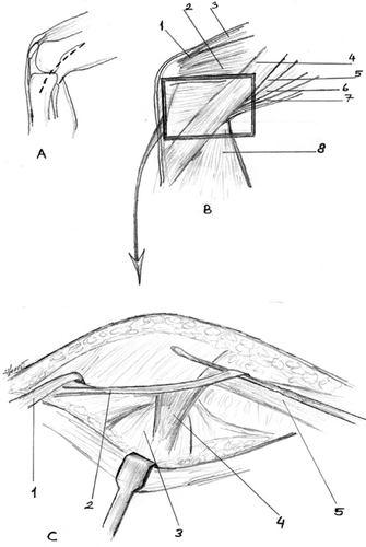
After a routine forensic autopsy, we marked the tibial tuberosity. An oblique incision starting next to the tuberosity was made on the medial hamstrings. The incision was carried down to the subcutaneous tissue to the crural fascia and was lengthened proximally in order to see the fascial band. The sartorius was incised along the course of the ST and gracilis tendons as the first fascial layer. After this layer, the ST tendon was exposed. The superficial medial collateral ligament was preserved during dissection. At this step, the most distal periostal insertion part of the tendon was elevated and this point was accepted as the starting point (). The whole ST tendon was dissected, including the musculotendinous area.
Figure 1. Drawing of anatomical dissection. A. Skin incision. B. Gross anatomical structures of the medial part of the knee. 1: quadriceps tendon; 2: vastus medialis muscle; 3: quadriceps muscle; 4: sartorius muscle; 5: semimembranosus muscle; 6: gracilis muscle; 7: semitendinosus muscle; 8: medial head of the gastrocnemius muscle. C. Schematic illustration of the cadaveric dissection specimen. 1: gracilis muscle is cut and reflected; 2: tendon of semitendinosus muscle; 3: medial head of the gastrocnemius muscle; 4: vincula; 5: semimembranosus muscle.
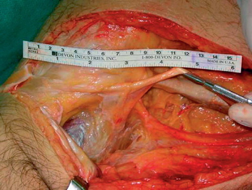
The distance from the insertion of the ST to the fascial band () and the width of the band was measured. The ST was totally resected with a tendon stripper, and their lengths, including the musculotendinous parts, were also recorded. At the most proximal area, where tendon and muscle converge, even the thinnest part was included in the measurement.
Figure 2. The view of the fascial band from ST to gastrocnemius. Compare with markings and legends in .
The study was approved by the ethics committee of the Forensic Medicine Council, Ministry of Justice, Istanbul.
Results
We detected fascial attachments in all cadavers except one (cadaver no. 9). The mean width of the fibrous band between the ST and the G was 2.56 (1–4; SD 0.94) cm. The mean total ST tendon length was 22 (18–26) cm. The mean distance from the insertion of the ST to the fascial band was 7 (6–8) cm.
Discussion
Arthroscopically-assisted reconstruction with ham string tendons has achieved widespread acceptance; however, some technical problems can be observed while tendon harvesting. Initially, open dissection was the method of choice for graft harvesting, but with the advent of arthroscopically-assisted ACL reconstruction, tendon strippers have provided an effective means of harvesting the tendon graft using a small incision (Ferrari and Ferrari Citation1991). Premature division of the tendon graft is the potential disadvantage of this technique, resulting in a shorter graft and possibly necessitating an alternative graft choice (Pagnani et al. Citation1993, Solman and Pagnani Citation2003). The fascial band from ST to G is one of the reasons for this complication. Studies concerning this fascial band show that this anatomical structure is variable both in size and placement (Warren and Marshall Citation1979, Ferrari and Ferrari Citation1991, Pagnani et al. Citation1993, Solman and Pagnani Citation2003, Tillett et al. Citation2004). The common belief is that 10 cm proximal to the insertion of pes anserinus is a safe distance to avoid encountering this band, but Candal-Couto and Deehan (Citation2003) found that the band originated more than 10 cm proximally in 8 of 10 cadavers. In our study, we could see that the band started within 6–8 cm and ended within 8–12 cm of the insertion point of pes anserinus, so the surgeon must be careful between these ranges when using the tendon stripper.
Usually there are two fascial bands involved with the ST tendon. One is close to the insertion of the tendon. We did not measure this distal band in the present study, as it is visible through the routine incision for graft harvesting and would not cause any complications during graft harvesting.
Measurement of the tendon, including the musculotendinous, part is not an accurate assessment of tendon length as the tendon is thicker proximal to the junction. The distance between the start of the musculotendinous junction and the tendinous termination also varies. There is good palpable tendon at the beginning, while at the end the tissue is tenuous. Zarins and Rowe (Citation1986) cited an average length of 21 cm for the attached semitendinosus, but they did not report from where this measurement was taken (Zarins and Rowe Citation1986). We measured the tendon just at the most distal point of the insertion and found the mean length to be 22 cm, which is in accordance with the results of Zarins and Rowe.
We conclude that it is important to recognize and know the anatomical details of fascial attachment to the G while harvesting ST, in order to prevent a disappointing complication right at the start of the operation.
Special thanks to Mustafa Ozdemir MD for his drawings.
Contributions of authors
HK and IU: obtained the cadavers. IT, HK and IU: measured the anatomical structures. IT and NK: wrote the manuscript.
- Candal-Couto J J, Deehan D J. The accessory bands of Gracilis and Semitendinosus: an anatomical study. Knee 2003; 10(4)325–8
- Chen L, Cooley V, Rosenberg T. ACL reconstruction with hamstring tendon. Orthop Clin North Am 2003; 34: 9–18
- Ferrari J D, Ferrari D A. The semitendinosus: anatomic considerations in tendon harvesting. Orthop Rev 1991; 20: 1085–8
- Ibrahim S A, Al-Kussary I M, Al-Misfer A R, Al-Mutairi H Q, Ghafar S A, El Noor T A. Clinical evaluation of arthroscopically assisted anterior cruciate ligament reconstruction: patellar tendon versus gracilis and semitendinosus autograft. Arthroscopy 2005; 21: 412–7
- Pagnani M J, Warner J J, O'Brien S J, Warren R F. Anatomic considerations in harvesting the semitendinosus and gracilis tendons and a technique of harvest. Am J Sports Med 1993; 21: 565–71
- Papastergiou S G, Voulgaropoulos H, Mikalef P, Ziogas E, Pappis G, Giannakopoulos I. Injuries to the infrapatellar branch(es) of the saphenous nerve in anterior cruciate ligament reconstruction with four-strand hamstring tendon autograft: vertical versus horizontal incision for harvest. Knee Surg Sports Traumatol Arthrosc 2006; 14(8)789–93
- Prodromos C C, Han Y S, Keller B L, Bolyard R J. Posterior mini-incison technique for hamstring anterior cruciate ligament reconstruction graft harvest. Arthroscopy 2005; 21: 130–7
- Solman C G, Jr, Pagnani M J. Hamstring tendon harvesting Reviewing anatomic relationships and avoiding pitfalls. Orthop Clin North Am 2003; 34: 1–8
- Tillett E, Madsen R, Rogers R, Nyland J. Localization of the semitendinosus - gracilis tendon bifurcation point relative to the tibial tuberosity: an aid to hamstring tendon harvest. Arthroscopy 2004; 20: 51–4
- Warren R F, Marshall J L. The supporting structures and layers on the medial side of the knee: an anatomical analysis. J Bone Joint Surg (Am) 1979; 61: 56–62
- Zarins B, Rowe C. Combined anterior cruciate- ligament reconstruction using semitendinosus and iliotibial tract. J Bone Joint Surg (Am) 1986; 68: 160–77