Abstract
Background Treatment of proximal humerus fractures in elderly patients is challenging because of reduced bone quality. We determined the in vitro characteristics of a new implant developed to target the remaining bone stock, and compared it with an implant in clinical use.
Methods Following osteotomy, left and right humeral pairs from cadavers were treated with either the Button-Fix or the Humerusblock fixation system. Implant stiffness was determined for three clinically relevant cases of load: axial compression, torsion, and varus bending. In addition, a cyclic varus-bending test was performed.
Results We found higher stiffness values for the humeri treated with the ButtonFix system—with almost a doubling of the compression, torsion, and bending stiffness values. Under dynamic loading, the ButtonFix system had superior stiffness and less K-wire migration compared to the Humerusblock system.
Interpretation When compared to the Humerusblock design, the ButtonFix system showed superior biomechanical properties, both static and dynamic. It offers a minimally invasive alternative for the treatment of proximal humerus fractures.
The optimal treatment of displaced proximal humerus fractures continues to be a subject of some controversy (Hessmann and Rommens Citation2001, Lill and Josten Citation2001). In elderly patients, implant anchorage can be particularly challenging due to reduced cancellous bone mass and trabecular connectivity, which are a age-related phenomena (Delling Citation1974, Delling and Amling Citation1995). A review of various implant fixation techniques demonstrated that the majority of current implants tend to target the central region of the humeral head, where bone stock is reduced and bone quality has diminished (Hepp et al. Citation2003). As a consequence, loss of implant fixation may occur and this may require more invasive treatments to achieve mechanical stability.
The ButtonFix is a new minimal invasive K-wire osteosynthese device concept based on the idea of the Humerusblock (Resch et al. Citation2001). In the ButtonFix system, 4 K-wires are placed into those regions of the humeral head where bone stock has been determined to be optimal (Hepp et al. Citation2003).
There is concern, however, that such minimally invasive implants may be too fiexible and not sufficiently stable to allow healing, particularly in complex fracture situations. In addition, migration of the wires has been associated with K-wire osteosynthesis (Lyons and Rockwood Citation1990, Schindele et al. Citation1999). We used a previously developed experimental method (Lill et al. Citation2003) to determine the stiffness and stability of the new Button-Fix implant concept for the treatment of proximal humeral fractures in elderly patients, and compared it to an implant that is currently in clinical use (Humerusblock).
Material and methods
Implants
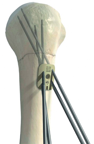
The following 2 implants were examined: 1) ButtonFix (Synthes, Switzerland) consisting of a PEEK (Poly-ether-ether-ketone) plate (26 × 20 × 3 mm) with 4 holes and 4 K-wires. The stainless steel K-wires have a diameter of 2.5 mm and a length of 280 mm. The K-wire is threaded over a distance of 100 mm behind the trochar tip. The K-wires are inserted into the humeral head through the plate using an aiming device. The plate serves to anchor the K-wires (). 2) Humerusblock (Synthes, Switzerland) consisting of a stainless steel block (diameter 16 mm and height 12 mm), a 3.5-mm cortical screw, and 2 K-wires. The block is affixed to the humeral shaft with the cortical screw. The Kwires are inserted through the pre-drilled holes in the Humerusblock. Fixation is achieved by threads on the K-wire that interface with the block (n.b. there are no threads at the tip-end of the K-wires inserted into the bone) (Resch et al. Citation2001).
Sample preparation and protocol
The experimental design has been reported previously and is summarized only briefiy here (Lill et al. Citation2003). 6 fresh pairs of humeri, with soft tissues removed, were obtained from human cadavers (Table). The specimens were stored at –20°C until further processing. To rule out any bone defects, the specimens were examined graphically in the anterior-posterior and lateral views. No focal bone disease or osteoarthritis was apparent. For 2 of the humeral pairs, the age of the cadavers was unknown. 3 left and 3 right humeri were treated with the ButtonFix implants. The contralateral humeri were treated with Humerusblock implants.
For the purpose of uniformity, the humeral shaft of each specimen was cut 170 mm below the base of the cranial calotte. The humeral head (proximal 30 mm) and shaft (distal 20 mm) were potted in methyl acrylate; 2 K-wires (1.8 mm) were inserted into the humeral head and at the distal end of the shaft to provide additional stability during potting. Prior to the osteotomy, the fixation devices were implanted by an experienced surgeon according to the manufacturer’s guidelines (Synthes, Switzerland). Transverse osteotomies (5-mm gap) at the surgical neck were then created 40 mm below the cranial calotte of each specimen using a thin oscillating saw in combination with a chisel, to carefully remove the bone around the K-wires. During testing at room temperature, all specimens were wrapped in saline-soaked gauze to avoid desiccation.
Mechanical test set-up
Mechanical testing was performed using an electromechanical material-testing machine (type 1455; Zwick, Germany). Mechanical testing was carried out under displacement control. Preliminary analysis ensured that all experiments were carried out within the linear range of the load-dis-placement curve. A custom-made jig was used to mount the specimens in the testing machine (Duda et al. Citation2000).
Implant stiffness
Fixation stiffness was calculated from the loads and resulting relative movement for the following three cases of load: axial compression, torsion under an axial preload, and medial-lateral (varus) bending induced by mounting the construct eccentrically (100 mm) and loading with an axial compressive displacement of 4 mm. In addition to the quasistatic load cases, a fourth cyclic test consisting of 1,000 cycles was performed with medial-lateral bending under displacement control. For each load case and specimen, the measurement was repeated 5 times.
Relative movements of the proximal and distal fracture fragments were determined using a threecamera optical measuring system (Workstation; Vicon Motion Systems Ltd., UK). Rectangular frames, each with four refiective markers (one out of plane), were attached either side of the fracture gap. Relative movements at the fracture gap were calculated from the spatial coordinates of the markers using software developed in-house. Relative movement was described in terms of three translations (x, y, z) and three rotations (x, y, z). The accuracy of measurement was estimated to be within ±0.08 mm and ± 0.1° for all three movements and rotations, respectively. The load and moments produced were recorded by a load cell mounted in the material-testing machine. For the cyclic test, measurements were made of the maximum load (Mx) during the first cycle, load level at 300 cycles, load reduction due to migration or loosening throughout the 1,000 cycles, and the slope of the load-cycles curve between cycles 700 and 1,000.
Statistics
Statistical comparison between the groups was performed using the Wilcoxon test for paired nonparametric data (SPSS version 12.0). A p-value of ≤ 0.05 was considered significant.
Results
Bone quality and clinical results
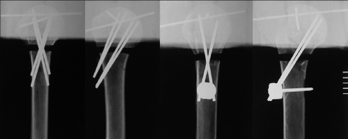
The preoperative radiographs showed no pathological changes to the bone structure. In all humeri, an even distribution of the trabecular structure and a homogenous cortex could be seen. The implantation of both fixation systems took place in a manner similar to the clinical situation and without complication. Radiographs taken after implantation confirmed good positioning of the implants ().
Implant stiffness
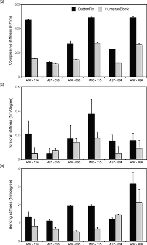
The stiffness of the ButtonFix system (349, SD 145 N/mm) under axial compression was higher that that of the Humerusblock system (179, SD 70 N/mm; p = 0.03). This trend could also be seen for the individual humeral pairs (. Whilst the intra-specimen variability was low, as indicated by the relatively small deviation bars, the differences in the stiffness values between the individual specimens were relatively high.
Figure 3. Compression (a), torsion (b), and bending stiffness (c) of the humeral pairs treated with ButtonFix and Humerusblock implants.

In torsion, the stiffness of the ButtonFix (0.19, SD 0.10 Nm/°) was nearly double that of the Humerusblock (0.10, SD 0.05 Nm/°; p = 0.05) (. Again, this pattern could also be seen for the individual specimens. Even though the intra-specimen variability was slightly higher in torsion, it was still smaller than the observed interspecimen variability.
In bending, the intra-individual variation in stiffness was again relatively low in comparison to the inter-individual variability (. With a stiffness of 1.78 (SD 0.69) Nm/°, the ButtonFix system was superior to the Humerusblock which had an average stiffness of 1.03 (SD 0.57) Nm/° in bending (p = 0.05).
Cyclic testing
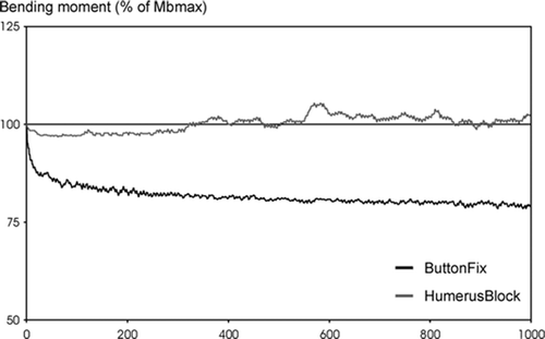
The 6 humeri treated with the ButtonFix system showed a characteristic hysteresis curve and progression of the maximum load during cyclic testing (). After an initial decrease in the bending moment, the maximal load remained almost constant. The following values were determined from the cyclic testing: Mbmax 2.31 (SD 0.49) Nm, Mb300 1.78 (SD 0.32) Nm, slope –0.00015 (SD 0.00012) Nm/cycle, ΔMb 24% (SD 6.8). In contrast to the ButtonFix system, the humeri treated with the Humerusblock showed uncharacteristic behavior during the cyclic testing (). Over the course of the cyclic test, little variation was seen in the bending moment. For this reason, it was not meaningful to determine the parameters of the cyclical testing (Mb300, Slope, ΔMb) with the exception of the maximum bending moment, Mbmax 2.55 (SD 0.90) Nm, which was not significantly different to that determined for the ButtonFix (p = 0.8).
Discussion
The ButtonFix system is a new minimally invasive osteosynthesis device for percutaneous fixation of proximal humerus fractures. The device has been designed to optimize implant anchorage by ensuring placement of the K-wires in regions of the humeral head where bone stocks are optimal. However, there are concerns as to whether such minimally invasive implants are sufficiently stable to allow healing. We determined the stiffness and stability of this new implant concept and found higher stiffness values for the humeri treated with the ButtonFix system than with the Humerblock system.
To determine the stability behavior over time, the constructs were subjected to cyclic bending loads. After a distinct, but not extreme, decrease in the fixation stability over the first cycles, humeri treated with the ButtonFix system appeared to reach a stable level. In contrast, cyclic loading of the Humerusblock group led initially to a very small loss of stability, but after some time an unexpected increase in stability was observed. The reason for this was found to be a distinct and continuous migration of the K-wires in the humeral head, ending with perforation of the calotte and impingement on the surrounding hard PMMA material used for fixing the ends of the humeri for testing. This was found to be the cause of the atypical loading behavior of the Humerusblock under dynamic loading conditions.
Comparison with conventional implants previously tested by our group (Humerus T Plate (HTP; Synthes, Bochum, Germany), Unreamed Humerus Nail (UHN; Synthes USA, Paoli, USA) and the angle-stable Locking Compression Plate Proximal Humerus (LCP-PH; Mathys, Bettlach, Switzerland)) revealed slightly lower compression and torsional stiffness values for the ButtonFix (Lill et al. Citation2003). However, in bending the ButtonFix system reached similar values to those exhibited by the HTP and LCP-PH. Under cyclic loading, the ButtonFix carried loads similar to the loads carried by all previously tested implants. Over time, the ButtonFix even tended to have the lowest load reduction and also a more stable mechanical behavior compared to the other implants (Lill et al. Citation2003).
The ButtonFix represents a new concept of fracture stabilization: a guided placement of the Kwires allows the regions of the humeral head with the best bone stock to be targeted, making it particularly suitable for the treatment of osteoporotic bone. In addition, the tensioning in the plate created by the K-wires pulling in different directions leads to further stabilization. The crossed-screw technique with the tension band represents a modification of the well-known screw osteosynthesis. The grip of the screws in the humeral head is improved by the crossing technique. The tensionband wiring also antagonizes the traction force of the rotator cuff. While the Humerusblock has shown some clinical success, under the critical conditions simulated in this study, the static stability of the implant was inferior to that of the Button-Fix. Also, the performance of the Humerusblock in cyclic testing was marred by the observed failure and migration of the K-wires. The problem of migration of the K-wires is solved in the ButtonFix implant by a novel anchoring concept, which does not require additional fixation elements (Resch et al. Citation2001). This concept has a number of advantages compared to other percutaneous techniques. The guide instrument allows a simple positioning of the plate and minimizes the risk of incorrectly inserting the K-wires compared to manual positioning. In addition, due to the minimally invasive insertion of the K-wires, the soft tissues are subjected to only minor trauma.
The pairs of humeri used in this study did not show any signs of bone defects. The inter-speci-men differences in stiffness observed in each group can be explained by age- and sex-related differences in the mechanical properties of bone (Lill et al. Citation2002). As expected, constructs with older or female humeri in both groups tended to have lower stiffness values.
Previous test sequences have consisted of stiffness determination of the implants, predominantly in bending or torsion, as with tests on humeral shaft fractures (Sehr and Szabo Citation1988, Henley et al. Citation1991, Dalton et al. Citation1993, Weinstein et al. Citation1994, Rajesh et al. Citation2000, Ruch et al. Citation2000). Axial stiffness examinations have rarely been performed (Blum et al. Citation1998, 2000). Our investigation was carried out under standardized laboratory conditions (Duda et al. Citation1998). The boundary conditions applied represent critical fracture displacement: subsequent sintering, malrotation of the humeral head, and varus displacement. The number of test cycles was specified as 1,000 since it has been shown in preliminary tests and by others (Wheeler and Colville Citation1997) that the highest reduction of load and loss of fracture stabilization occurs in the first 100 cycles. In this study, an osteotomy was used to investigate fixation of surgical neck fractures. Thus, the results of this study may not be directly applicable to more complex fractures.
This study was supported by a grant from the AO/ASIF Foundation, Switzerland (AO Collaborative Research Center).
Contributions of authors
This study was conceived by NPS, SML, RB and GND. RM: was involved in the study design and realization of the device prototypes. GND and DRE: performed the research and collected and analysed the data. DRE: wrote the paper, incorporating input from all authors.
References
- Blum J, Rommens P M, Janzing H, Langendorff H S. Retrograde nailing of humerus shaft fractures with the unreamed humerus nail. An international multicenter study. Unfallchirurg 1998; 101: 342–52
- Blum J, Machemer H, Hogner M, Baumgart F, Schlegel U, Wahl D, Rommens P M. Biomechanics of interlocked nailing in humeral shaft fractures. Comparison of 2 nail systems and the effect of interfragmentary compression with the unreamed humeral nail. Unfallchirurg 2000; 103: 183–90
- Dalton J E, Salkeld S L, Satterwhite Y E, Cook S D. A biomechanical comparison of intramedullary nailing systems for the humerus. J Orthop Trauma 1993; 7: 367–74
- Delling G. Age-dependent bone changes (author’s transl). Klin Wochenschr 1974; 52: 318–25
- Delling G, Amling M. Biomechanical stability of the skeleton--it is not only bone mass, but also bone structure that counts. Nephrol Dial Transplant. 1995; 10: 601–6
- Duda G N, Kirchner H, Wilke H.-J, Claes L. A method to determine the 3-D stiffness of fracture fixation devices and its application to predict inter-fragmentary movement. J Biomech 1998; 31: 247–52
- Duda G N, Kassi J P, Hoffmann J E, Riedt R, Khodadadyan C, Raschke M. Mechanical behavior of Ilizarov ring fixators. Effect of frame parameters on stiffness and consequences for clinical use. Unfallchirurg 2000; 103: 839–45
- Henley M B, Monroe M, Tencer A F. Biomechanical comparison of methods of fixation of a midshaft osteotomy of the humerus. J Orthop Trauma 1991; 5: 14–20
- Hepp P, Lill H, Bail H, Korner J, Niederhagen M, Haas N P, Josten C, Duda G N. Where should implants be anchored in the humeral headfi Clin Orthop. 2003, 415: 139–47
- Hessmann M H, Rommens P M. Osteosynthesis techniques in proximal humeral fractures. Chirurg 2001; 72: 1235–45
- Lill H, Josten C. Conservative or operative treatment of humeral head fractures in the elderlyfi. Chirurg 2001; 72: 1224–34
- Lill H, Hepp P, Gowin W, Oestmann J W, Korner J, Haas N P, Josten C, Duda G N. Age-and gender-related distribution of bone mineral density and mechanical properties of the proximal humerus. Fortschr Röntgenstr 2002; 174: 1544–50
- Lill H, Hepp P, Korner J, Kassi J P, Verheyden A P, Josten C, Duda G N. Proximal humeral fractures: how stiff should an implant befi A comparative mechanical study with new implants in human specimens. Arch Orthop Trauma Surg 2003; 123: 74–81
- Lyons F A, Rockwood C A, Jr. Migration of pins used in operations on the shoulder. J Bone Joint Surg (Am) 1990; 72: 1262–7
- Rajesh M, Manning P, Neumann L, Parry M, Wallace W. The effect of bone quality on intra-medullary fixation of the proximal humerus using a retrograde nail. Osteoporos Int 2000; 11: 45
- Resch H, Hubner C, Schwaiger R. Minimally invasive reduction and osteosynthesis of articular fractures of the humeral head. Injury 2001; 32: SA25–32
- Ruch D, Glisson R, Marr A, Russell G, Nunley J. Fixation of three-part proximal humeral fractures: a bio-mechanical evaluation. J Orthop Trauma 2000; 14: 36–40
- Schindele S, Hackenbruch W, Sutter F, Schaerer M, Leutenegger A. Migration von Kirschner-Drähten nach operativer Stabilisierung von Verletzungen im Bereich der Schulter - Vier Fallberichte. Swiss Surg 1999; 5: 281–5
- Sehr J R, Szabo R M. Semitubular blade plate for fixation in the proximal humerus. J Orthop Trauma 1988; 2: 327–32
- Weinstein D, Gomez M, Hawkins R. Biomechanical comparison of tension-band wiring versus plating in the fixation of three-part fractures of the proximal humerus. Orthop Trans 1994; 18: 3
- Wheeler D L, Colville M R. Biomechanical comparison of intramedullary and percutaneous pin fixation for proxi mal humeral fracture fixation. J Orthop Trauma 1997; 11: 363–7


