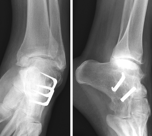
A 56-year-old patient with a history of cerebral palsy, pes equinovarus adductus, and coronary bypass presented at our outpatient clinic with pain in his left foot and ankle. A left subtalar arthrodesis had been performed 6 years previously, because of osteoarthritis. Plain radiographs showed extensive osteoarthritis of the ankle joint and pes equinovarus (). A pantalar arthrodesis was performed under general anesthesia and a tourniquet was used. Through a medial and lateral approach, a corrective ankle arthrodesis was performed to correct the pes equinovarus adductus. A condylar plate and lag screw osteosynthesis were used.
Figure 1. Extensive osteoarthritis of the ankle joint after a subtalar fusion had been performed. Note the pes equinovarus adductus deformation.
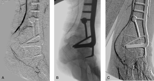
After removing the tourniquet, there was no capillary refill. Doppler signals were absent for the posterior tibial artery and the dorsal pedal artery. 2,500 units of heparin were administered i.v. Digital subtraction angiography (DSA) was performed on the operating table. There was a preexisting proximal occlusion of the anterior tibial artery. The peroneal artery was patent up to the distal tibial metaphysis. There was partial filling of the dorsal pedal artery through collaterals of the peroneal artery. DSA showed an occlusion of the posterior tibial artery. At first, a release of the tarsal tunnel was contemplated. However, a closer look at the native angiogram suggested that the occlusion was due to external compression by the posterior talar process. To restore the patency of the posterior tibial artery, the posterior talar process was removed with a chisel using the existing medial incision. Immediately afterwards, capillary refill and Doppler signals of the the foot returned to normal. Control angiography showed a patent posterior tibial and plantar artery (). Thus, bypass surgery was avoided.
Figure 2. A. Digital subtraction angiography (DSA), showing occlusion of the posterior tibial artery. B. The native angiogram showing that the occlusion was due to external compression by the posterior talar process. C. Control angiography showing a patent posterior tibial artery, after removal of the posterior talar process.
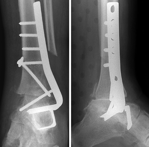
The postoperative course was uneventful ().
Figure 3. Postoperatively, the pantalar arthrodesis and removal of the posterior talar process.
Discussion
Arterial occlusion in elective orthopedic surgery is rare, and appears most often after total knee arthroplasty (Wilson et al. 2003). Occlusion or injury of vessels around the ankle is also rare, and to date has only been described after severe trauma of the ankle (Pai 1999, Chaugle et al. 2004) and after ankle arthroscopy (Salgado et al. 1998, Marianni et al. 2001, Darwish et al. 2004). In our case, the occlusion was caused by stretching and impingement of the posterior tibial artery over the posterior talar process. Because of the pre-exist-ing occlusion of the anterior tibial artery, arterial supply of the foot depended on the posterior tibial and peroneal arteries. In order to correct the pes equinovarus the ankle had to be corrected by fusing the subtalar joint in a neutral position, thereby causing traction to the posterior tibial artery over the prominent posterior talar process. Fortunately, the occlusion was detected while the patient was still on the operating table and under general anesthesia. Rapid diagnostics by Doppler ultrasound and angiogram and intervention by removal of the prominent posterior talar process prevented persistent critical ischemia and limb loss. When performing diagnostic angiography for vascular injury in orthopedic or trauma surgery, the native images may add important information to the digital subtraction images.
Contributions of authors
JT: Case study, research, and writing of manuscript. HV: supervisor and review of manuscript. NK: supervisor and review of manuscript. TPR: assessment of radiographs and writing of text on radiology.
- Chaugle A P D Batty, Simms M H, Hodgkinson J P. Ankle sprain: an unexpected complication. Injury 2004; 35(8)823–4
- Darwish A, Ehsan O, Marynissen H, Al-Khaffaf H. Pseudoaneurysm of the anterior tibial artery after ankle arthroscopy. Arthroscopy 2004; 20(6)63–4
- Marianni P P, Mancini L, Giorgini T. Pseudoaneurysm as a complication of ankle arthroscopy. Arthroscopy 2001; 17(4)400–2
- Pai V S. Traumatic aneurysm of the perforating peroneal artery following ankle fracture. J Foot Ankle Surg 1999; 38(6)417–9
- Salgado C J, Mukherjee D, Quist M A, Cero S. Anterior tibial artery pseudoaneurysm after ankle arthroscopy. Cardiovasc Surg 1998; 6(6)604–6
- Wilson J S, Wilson J, Miranda A, Johnson B L, Shames M L, Back M R, Bandyk D F. Vascular injuries associated with elective orthopaedic procedures. Ann Vasc Surg 2003; 17(6)641–4