Abstract
Background and purpose There have been few reports on the long-term function after shoulder arthrodesis. We report the outcome after shoulder arthrodesis with plate fixation in 18 patients who were followed for 3–15 years.
Methods 25 patients with a median age of 64 (19–75) years were operated with a shoulder arthrodesis between 1982 and 2003. Standard AO surgical technique with plating was used in all patients. 18 of the patients were examined retrospectively after a mean of 8 (3–15) years. 6 of the other patients had died and 1 refused examination.
Results Radiologically, all but two arthrodeses fused completely. The remaining two were partially fused, within the glenohumeral joint or between humerus and acromion. The mean Oxford shoulder score was 32 and the mean ASES shoulder index was 59. Nine patients had intermittent or continuous pain; their mean pain score on a visual analog scale was 3. One patient had been re-operated after 4 months because of severe pain and 1 was operated due to a humeral shaft fracture after 8 months. 1 patient suffered from a complex regional pain syndrome. No infections occurred.
Interpretation In this patient series there were few complications after shoulder arthrodesis, and the longterm functional results were acceptable.
For decades, arthrodesis of the shoulder joint has been a solution to problems that have been difficult to solve by other means. Current indications include failed arthroplasty, bone deficiency of the proximal humerus after tumor resection, chronic infection, severe instability, posttraumatic brachial plexus injury, and paralysis of the deltoid muscle and rotator cuff (Clare et al. Citation2001). Other indications are inflammatory arthritis with severe rotator cuff involvement, irreparable rotator cuff tears, and painful osteoarthritis in patients in need of strength more than movement (González-Diaz et al. Citation1997). Several techniques for fixation of the arthrodesis, both external and internal, have been described. Stable internal plate fixation with one or two plates has become the method of choice (Clare et al. Citation2001).
All methods for shoulder arthrodesis have, however, been associated with relatively high complication rates. Nonunion has been frequent and postoperative soft tissue problems with infection rates up to 14% have been reported (Richards et al. Citation1993, Groh et al. Citation1997, Rühmann et al. Citation1999, Citation2005, Rühmann Citation2002), as well as frequent postoperative fractures of the humeral shaft (Cofield and Briggs Citation1979, Groh et al. Citation1997, Richards et al. Citation1993, Rühmann et al. Citation2005).
Several authors have reported on function after shoulder joint arthrodesis (Rowe Citation1974, Citation1983, Cofield and Briggs Citation1979, Richards et al. Citation1985, Citation1988, Rühmann et al. Citation1999, Diaz et al. Citation2003, Nagy et al. Citation2004), but measures in terms of standardized function scores are rarely given. We report the objective and subjective long-term results of shoulder arthrodesis for 18 patients.
Patients and methods
25 patients (mean age 64 (19–75) years, 14 women) were operated with a shoulder arthrodesis between 1982 and 2003 in Martina Hansens Hospital or Ullevaal University Hospital, Norway. Surgery was performed using an extended deltopectoral approach with the patient in the lateral position. After removal of cartilage and subchondral bone, the aim was to position the humerus so that the hand could reach the mouth and the anterior perineum without looking specifically at exact angles. Standard AO surgical technique according to Richards et al. (Citation1985) with one stable plate and compression screws over the joint was used for internal fixation of the glenohumeral and acromiohumeral joints. Perioperatively, it is difficult to adjust and secure the preoperatively planned angles due to the high mobility of the scapula, and several suggestions have been put forward on how to ensure these angles. We prefer to secure the arthrodesis temporarily using drill pins, ensuring that the patient’s hand can reach the mouth, the contralateral axilla and the anterior perineum, and that there is a minimum of external rotation to 0°. The drill pins are then interchanged with compression screws, and the plate adapted (). Intraoperative fluoroscopy to evaluate the position of the arthrodesis was not used.
Figure 1. Shoulder arthrodesis with stable plate fixation.
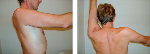
Figure 2. Shoulder arthrodesis. Postoperative flexion and abduction.
The indications for surgery were fracture sequelae with secondary osteoarthritis, including both pseudarthrosis and humeral head avascular necrosis in 13 patients. 2 of the patients with pseudarthrosis had previously suffered from a postoperative infection; the infection had subsided after removal of internal fixation devices, soft tissue debridement, and systemic antibiotic treatment prior to the shoulder arthrodesis. 5 patients had severe primary osteoarthritis with a nearly ankylotic shoulder joint. 3 patients were operated due to deltoid dysfunction and recurrent shoulder instability. Of these 3 patients, 1 had iatrogenic axillary nerve palsy with deltoid paralysis as a result of earlier ORIF of a major tubercle fracture, 1 patient had lost her deltoid function after a total acromionectomy, and 1 had severe sequelae after a cerebral stroke with total paralysis of the deltoid and rotator cuff muscles. 1 patient was operated due to a chronic posterior shoulder joint dislocation resulting from an epileptic attack. 1 patient had the arthrodesis performed after a failed revision arthroplasty, 1 was operated due to shoulder joint tuberculosis, and 1 after a gunshot wound.
Of the 25 patients, 6 patients had died at the time of follow up. The medical records of these patients were examined; all arthrodeses had healed without complications. 1 patient refused re-examination. The remaining 18 patients were examined clinically after an average of 7.5 (3–15) years by an independent examiner (SD). The American Shoulder and Elbow Surgeons (ASES) shoulder index (Richards et al. Citation1994) including VAS pain score (0–10, with 0 representing no pain) and activities of daily living, and the Oxford shoulder score (Dawson et al. Citation1996) were registered. Radiographs of both shoulders were taken.
Results
Demographic data, complications, and functional results of all patients are summarized in the Table.
Radiographically, 16 of the 18 arthrodeses were completely fused. 1 was fused between the acromion and humerus only, and the other in the upperthird part of the glenohumeral joint. Both of these patients were free of pain, and the arthrodesis was stable at physical examination.
Data of the patients
1 patient was reoperated after 1 week due to diastasis in the arthrodesis, which healed thereafter. 2 patients had humeral shaft fractures below the plate. 1 occurred after 8 months, was operated, and was reoperated 2 months later due to fixation failure. After 14 years, the patient still had a nonunion of the humeral shaft, but she refused further surgery. The other fracture occurred after 6 months and healed after nonoperative treatment.
The implants had been removed in 6 patients due to tenting of the skin and related discomfort. In 1 of these patients from whom the plate had to be removed after 12 months, the arthrodesis was secured with 3 new screws due to questionable radiographical healing; the course was then uncomplicated. 1 patient had a lateral clavicle resection performed after 3 years due to osteoarthritis of the AC joint. None of the patients had postoperative infection.
The most serious complication was one case of complex regional pain syndrome (CRPS). This patient was previously operated with a total acromionectomy, and before the arthrodesis she had a total of 7 operations performed on her shoulder, including 2 failed stabilization procedures. 4 months after the index operation, due to severe pain a re-arthrodesis was performed with removal of the screws and the plate, autologous bone grafting, and fixation with 2 new compression screws. Perioperatively, however, neither instability of the arthrodesis nor any other findings could explain the pain. 3 months later, yet another operation was performed in the same patient; an exostosis was resected. 1 month later, the 2 compression screws were removed. There was no instability, and radiographically the arthrodesis was healed. CRPS was later diagnosed.
At follow-up, 9 patients reported intermittent or continuous pain during activity or periodic pain from their shoulder during the night. The pain was localized anterolaterally and related to the muscle insertions on the acromion, the major and lesser tubercle, and the anterior proximal part of the humerus in 6 of the patients; 4 of them also had periscapular muscular pain provoked by movement of their arm. The CRPS patient had generalized pain around the shoulder at rest, and movement aggravated the pain. The other 2 patients reporting pain only had distinct pain related to palpation of or pressure against the implant. The mean pain score (VAS) of the 9 patients was 3 (1–8). The patient with the CRPS used high doses of opioids daily, 2 patients used paracetamol daily, and 1 patient used ibuprofen sporadically. The other patients had no need for analgesics.
14 of the patients had intact or only minimally reduced strength compared to the contralateral shoulder as judged clinically, 4 patients had considerable reduction of strength in their shoulder flexion, and the CRPS patient also had a considerable reduction of internal rotation and abduction strength.
Mean postoperative abduction was 59° (20–80), flexion was 72° (20–90), external rotation was 5° (-30 to 50), and internal rotation from the major trochanter to the level of the Th12 posterior spinous process. The clinical positioning of the humerus postoperatively was in 18° (10–40) of abduction, 11° (0–20) of flexion and 23° (-20 to 40) of internal rotation, as evaluated clinically. All patients could reach to their mouth. 8 patients managed to reach their perineum, and 7 of them could also comb their hair.
The mean Oxford score of the 18 patients was 32 (15–49) points (where the best score is 12). Mean ASES score was 59 (15–95) (where the best score is 100).
One of the patients would not have chosen to have the arthrodesis performed if she could have chosen again; 2 others stated that they would probably not. The other 15 patients were satisfied with their shoulders relative to their preoperative condition.
Discussion
Nowadays, shoulder arthrodesis is used in relatively few cases according to the indications outlined above. Matsen et al. (Citation1998) stated that arthrodesis is seldom, if ever, performed due to arthritis of the shoulder. Today, some of the patients presented in this retrospective study would therefore obviously have been treated with a shoulder arthroplasty.
Several surgical approaches and fixation techniques for shoulder arthrodesis have been reported. The intraarticular glenohumeral arthrodesis involves fixation of the glenohumeral joint, while the extraarticular acromiohumeral arthrodesis is performed between the acromion and the humeral head only—and was previously used mainly for infections, especially tuberculosis (Matsen et al. Citation1998). The most commonly used technique is a combined extra- and intraarticular technique with acromiohumeral and glenohumeral fixation (Clare et al. Citation2001).
Several methods for internal fixation are available (Richards et al. Citation1985, Morgan and Casscells Citation1992, Mohammad 1998). During the last decade, stable internal plate fixation with one plate (the Richards technique) or two plates (the Müller technique) has been the method of choice (Clare et al. Citation2001) as long as the periarticular soft tissues allow the extensive dissection involved. This fixation is stable, with no need for a postoperative cast or brace. Dynamic compression plates were described in the original method (Richards et al. Citation1985), but other plates may be used—such as pelvic reconstruction plates, which may be easier to contour (Clare et al. Citation2001, Diaz et al. Citation2003, Richards et al. Citation1988, Rühmann et al. Citation1999). Screws or pins alone or in combination with other implants have also been an option (Cofield and Briggs Citation1979, Morgan and Casscells Citation1992, Richards et al. Citation1988, Rybca et al. Citation1979), and tension band osteosynthesis has been suggested when severe osteoporosis is present (Blauth et. al Citation1975). These methods require postoperative immobilization in a spica cast or brace, but may give satisfactory results.
Complications related to the shoulder arthrodesis are frequent. Nonunion is common; of 651 arthrodeses performed with different fixation techniques, 61 (9%) resulted in a nonunion (Rühmann Citation2002). Clare et al. (Citation2002) stated, however, that nonunion may be less of a problem and may be successfully solved by secondary bone grafting, and that less nonunions were reported using the current AO fixation technique. Rühmann et al. (Citation2005) also demonstrated that nonunions occur less frequently in plate arthrodesis than in screw arthrodesis. In our series of patients, operated with internal fixation using one plate and compression screws, all healed with no wound complications. The 2 arthrodeses that were only partly fused radiographically had no subjective symptoms of a nonunion.
Postoperative infections have been described in 0–14% of shoulder arthrodeses (Cofield and Briggs Citation1979, Schrøder and Frandsen Citation1983, Richards et al. Citation1988, Citation1993, Rühmann et al. Citation1999, Citation2005). We routinely used perioperative antibiotic prophylaxis and did not experience any infections.
Fractures around the arthrodesed shoulder are also frequently described; humeral shaft fractures are reported in up to 9% of the cases (Cofield and Briggs Citation1979, Rühmann et al. Citation2005). 2 of our patients suffered a fracture of the humeral shaft distal to the plate. Cofield and Briggs (Citation1979) reported 8 humeral shaft fractures distal to a shoulder arthrodesis, which all healed after immobilization with brace.
Removal of the implants due to prominence of the internal fixation irritating the overlying soft tissue is common (Cofield and Briggs Citation1979, Diaz et al. Citation2003, Richards et al. Citation1993). To counteract this problem, which is especially related to the acromion area, we have recently performed a partial osteotomy of the superomedial part of the acromion and bent it downwards to meet with the humeral head. This enables a smoother curvature of the plate and also ensures maximum contact area between the concave glenoid and the convex humeral head, since it is not necessary to translate the humeral head upwards to increase the contact with the acromion.
Functionally, the most critical complication after shoulder arthrodesis is malpositioning of the extremity (Clare et al. Citation2001), which may cause shoulder pain (Groh et al. Citation1997). Traction neuritis of the suprascapular nerve may also occur with excessive abduction (González-Diaz et al. Citation1997). Correction of a malpositioned arthrodesis may result in substantial relief of pain and improved ability to perform activities of daily living (Groh et al. Citation1997). The most serviceable position was previously believed to be 50° of true abduction (humerus making an angle of 50° with the vertebral border of the scapula), 20° of flexion, and 25° of internal rotation (Barr et al. Citation1942). During the last decades, however, several authors have preferred less abduction and more internal rotation. A clear consensus of exact angles has not been reached.
When evaluating the functional results in the arthrodesed patients, the Constant shoulder score (Constant and Murley Citation1987), being probably the most widely used score when evaluating shoulder arthroplasties, is unsuitable. The arthrodesed patients lack the ability to elevate their arm to 90° as required for the scoring (Moseley Citation1953). We therefore chose to use the ASES shoulder index (Richards et al. Citation1994) and the Oxford shoulder score (Dawson et al. Citation1996). Gartsman et al. (Citation2000) reported an ASES shoulder index of 65 (15–94) after hemiarthroplasty in patients with degenerative osteoarthritis, while Levine et al. (Citation1997) reported an ASES shoulder index of 68 in a similar patient population. We found an ASES shoulder index of 59 (15–95) after arthrodesis. Even though our patient groups are not directly comparable, our results indicate that the functional outcome after shoulder arthrodesis is acceptable—and furthermore, few serious complications occurred. Thus, shoulder arthrodesis performed with stable plate fixation may be a good solution for solving complex and difficult shoulder problems that are unsuitable for shoulder arthroplasty.
No competing interests declared.
Contributions of authors
Both authors contributed equally to the manuscript.
- Barr J S, Freiberg J A, Colonna P C, Pemberton P A. Research Committee of American Orthopeadic Association. A survey of end results on stabilization of the paralytic shoulder. J Bone Joint Surg (Am) 1942; 24(3)699–707
- Blauth W, Hepp W R, . Arthrodesis of the shoulder joint by traction absorbing wiring. The arthrodesis in the restoration of working ability, G Chapchal, et al. Georg Thieme, Stuttgart 1975; 30
- Clare D J, Wirth M A, Groh G I, Rockwood C A. Shoulder arthrodesis. J Bone Joint Surg (Am) 2001; 83(4)593–600
- Clare D J, Wirth M A, Groh G I, Rockwood C A. Shoulder arthrodesis. J Bone Joint Surg (Am) 2002; 84(5)874–6
- Cofield R H, Briggs B T. Glenohumeral arthrodesis. Operative and long-term functional results. J Bone Joint Surg (Am) 1979; 61(5)668–77
- Constant C R, Murley A H. A clinical method of functional assessment of the shoulder. Clin Ortop Relat Res 1987, 214: 160–4
- Dawson J, Fitzpatrick R, Carr A. Questionnaire on the perceptions of patients about shoulder surgery. J Bone Joint Surg (Br) 1996; 78(4)593–600
- Diaz J A, Cohen S B, Warren R F, Craig E V, Allen A A. Arthrodesis as a salvage procedure for recurrent instability of the shoulder. J Shoulder Elbow Surg 2003; 12(3)237–41
- Gartsman G M, Roddy T S, Hammerman S M. Shoulder arthroplasty with or without resurfacing of the glenoid in patients who have osteoarthritis. J Bone Joint Surg (Am) 2000; 82(1)26–33
- González-Diaz R, Rodríguez-Merchán E C, Gilbert M S. The role of shoulder fusion in the era of arthroplasty. Int Orthop 1997; 21(3)204–9
- Groh G I, Williams G R, Jarman R N, Rockwood C A. Treatment of complications of shoulder arthrodesis. J Bone Joint Surg (Am) 1997; 79(6)881–7
- Levine W N, Djurasovic M, Glasson J M, Pollock R G, Flatow E L, Bigliani L U. Hemiarthroplasty for glenohumeral osteoarthritis: results correlated to degree of glenoid wear. J Shoulder Elbow Surg 1997; 6(5)449–54
- Matsen F A, Rockwood C A, Jr, Wirth M A, Lippitt S B. Glenohumeral artritis and its management. The shoulder, CA Rockwood, Jr, F A Matsen, 3rd. WB Saunders, Philadelphia 1998; 2: 879–88
- Mohammed N S. A simple method of shoulder arthrodesis. J Bone Joint Surg (Br) 1998; 80(4)630–3
- Morgan C D, Casscells C D. Arthroscopic-assisted glenohumeral arthrodesis. Arthroscopy 1992; 8(2)262–6
- Moseley H F. Examination of the shoulder. Measurement of power. Shoulder Lesions, H F Moseley. Churchill Livingston, Edinburgh 1953; 3: 29
- Nagy L, Koch P, Gerber C. Functional analysis of shoulder arthrodesis. J Shoulder Elbow Surg 2004; 13(4)386–95
- Richards R R, Waddell J P, Hudson A R. Shoulder arthrodesis for the treatment of brachial plexus palsy. Clin Orthop 1985, 198: 250–8
- Richards R R, Sherman R M P, Hudson A R, Waddell J P. Shoulder arthrodesis using a pelvic reconstruction plate – a report of eleven cases. J Bone Joint Surg (Am) 1988; 70(3)416–21
- Richards R R, Beaton D, Hudson A R. Shoulder arthrodesis with a plate fixation: a functional outcome analysis. J Shoulder Elbow Surg 1993; 2(5)225–39
- Richards R R, Bigliani L U, Gartsman G M, Iannotti J P, Zuckerman J D. A standardized method for the assessment of shoulder function. J Shoulder Elbow Surg 1994; 3(6)347–52
- Rowe C R. Re-evaluation of the position of the arm in arthrodesis of the shoulder in the adult. J Bone Joint Surg (Am) 1974; 56(5)913–22
- Rowe C R. Arthrodesis of the shoulder used in treating painful conditions. Clin Orthop 1983, 173: 92–6
- Rühmann O. Pseudartrosis after shoulder arthrodesis. J Bone Joint Surg (Am) 2002; 84(5)874–6
- Rühmann O, Gossé F, Wirth S, Schmolke S. Reconstructive operations for the paralyzed shoulder in brachial plexus palsy: concept of treatment. Injury 1999; 30(9)609–18
- Rühmann O, Scmolke S, Bohnsack M, Flamme C, Wirth C J. Shoulder arthrodesis: Indications, technique, results and complications. J Shoulder Elbow Surg 2005; 14(1)38–50
- Rybca V, Raunio P, Vainio K. Arthrodesis in the shoulder in rheumatoid arthritis. J Bone Joint Surg (Br) 1979; 61(2)155–8
- Schrøder H A, Frandsen P A. External compression arthrodesis of the shoulder joint. Acta Orthop Scand 1983; 54(4)592–5