Abstract
Background and purpose The aim with high tibial valgus osteotomy (HTO) is to correct the mechanical axis in medial compartmental osteoarthritis of the knee. Loss of operative correction may threaten the long‐term outcome. In both a lateral closing‐wedge procedure and a medial opening‐wedge procedure, the opposite cortex of the tibia is usually not osteotomized, leaving 1 cm of bone intact as fulcrum. A fracture of this cortex may, however, lead to loss of correction; this was examined in the present study.
Patients and methods We used a prospective cohort of 92 consecutive patients previously reported by Brouwer et al. (Citation). The goal in that randomized controlled trial, was to achieve a correction of 4 degrees in excess of physiological valgus. In retrospect, we evaluated the 1‐year radiographic effect of opposite cortical fracture. Opposite cortical fracture was identified on the postero‐anterior radiographs in supine position on the first day after surgery.
Results 44 patients with a closing‐wedge HTO (staples and cast fixation) and 43 patients with an opening‐wedge HTO (non‐angular‐stable plate fixation) were used for analysis. 36 patients (four‐fifths) in the closing‐wedge group and 15 patients (one‐third) in the opening‐wedge group had an opposite cortical fracture (p < 0.001). At 1 year, the closing‐wedge group with opposite cortical fracture had a valgus position with a mean HKA angle of 3.2 (SD 3.5) degrees of valgus. However, the opening‐wedge group with disruption of the opposite cortex achieved varus malalignment with a mean HKA angle of 0.9 (SD 6.6) degrees of varus.
Interpretation Fracture of the opposite cortex is more common for the lateral closing wedge technique. Medial cortex disruption has no major consequences, however, and does not generally lead to malalignment. Lateral cortex fracture in the medial opening‐wedge technique, with the use of a non‐angular stable plate, leads more often to varus malalignment.
High tibial valgus osteotomy (HTO) is a generally accepted treatment for medial unicompartmental osteoarthritis of the knee with varus alignment, especially in younger, active patients (Virolainen et al. Citation2004). A successful outcome for the osteotomy relies on proper patient selection, and achievement and maintenance of adequate operative correction (Hernigou et al. Citation1987, Berman et al. Citation1991, Spahn et al. Citation2006a). The two most commonly used surgical techniques are the closing‐wedge HTO with fibular osteotomy and the opening‐wedge HTO with plate fixation, the second of which has become popular more recently. Avoidance of fracture of the opposite cortex in HTO can be difficult, and is generally limited by the angular size of the wedge. A fractured medial cortex in a lateral closing‐wedge osteotomy may lead to progressive movement of the distal tibia into a varus position (Kessler et al. Citation2002). Instability at the opening‐wedge osteotomy site due to disruption of the lateral cortical hinge may possibly result in displacement of the osteotomy, and in this way it may contribute to recurrent varus deformity (Miller et al. Citation2005).
Opposite cortical fracture in HTO is an adverse event for which one cannot randomize the patient. The highest practicable level of evidence for evaluation of the consequences of intraoperative opposite‐cortex fracture in HTO would thus be a retrospective analysis of a well‐designed randomized controlled trial. We therefore used a cohort of 92 patients who had been included in a prospective level‐I study on two different techniques of HTO, which has been described extensively by Brouwer et al. (Citation2006). The objective of the present analysis was to determine the effects of opposite cortical fracture on whole‐leg alignment, for both closing‐wedge and opening‐wedge osteotomy.
Patients and methods
The patients included in this study were part of a randomized, controlled, and consecutive trial published by Brouwer et al. (Citation2006), comparing lateral closing‐wedge and medial opening‐wedge osteotomy in 92 patients. The goal in that trial was to achieve a correction of 4 degrees in excess of physiological valgus. For the closing‐wedge group, a slotted wedge resection guide of Allopro (Zimmer, Winterthur, Switzerland) was used under fluoroscopic guidance. The anterior part of the proximal fibular head was resected and the osteotomy was fixated with 2 staples. After surgery, a standard cylinder plaster cast was applied for 6 weeks. The opening‐wedge HTO was created with the Puddu HTO instrumentation (Arthrex, Naples, FL), and performed under fluoroscopic guidance to control the correction during the surgical procedure; the osteotomy was fixated with the non‐angular stable Puddu plate. If the wedge was more than 7.5 mm, the open wedge was filled with bone from the ipsilateral iliac crest. All patients were mobilized on the first postoperative day, and partial weight bearing was allowed for 6 weeks.
This analysis focuses on the radiographic effect of opposite cortical fracture on the whole‐leg alignment in both techniques at the 1‐year follow‐up. Standardized radiography was performed preoperatively, on the first day postoperatively, and 12 months after surgery.
1 patient was lost to follow‐up (closing‐wedge), and in 3 cases (2 closing‐wedge and 1 opening‐wedge) we were unable to retrieve the radiographs taken 1 day after surgery to determine opposite cortical fracture. In another patient (opening‐wedge), the 1‐year postoperative whole‐leg radiograph had not been done because of emergency treatment of an unrelated condition. Thus, the study population consisted of 87 of the 92 patients originally included ().
Table 1. Baseline characteristics of the total study population and separately for the two intervention groups
Measurements
The grade of radiographic osteoarthritis was scored according to Ahlbäck (Citation1968) and measured on standard short posteroanterior radiographs in standing position. The mechanical axis (hip‐knee‐ankle (HKA) angle) was measured on a whole‐leg radiograph in standing position before surgery and 1 year after surgery (Brouwer et al. Citation2003). Opposite cortical fracture of the osteotomy site was scored from the posteroanterior radiographs in supine position on the first day after surgery by one assessor (TMR), who was blinded as to the 1‐year postoperative radiographic outcome ( and ). When cortical disruption was noted in the lateral closing‐wedge group, we also looked for a gap. A gap was defined as more than 2 mm width between the disrupted opposite cortex fragments ().
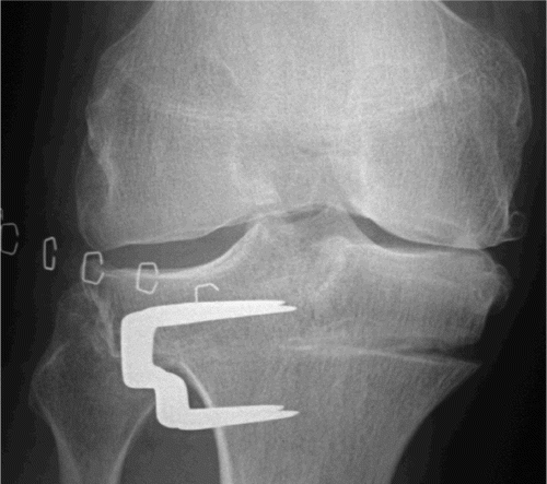
Figure 1. Closing‐wedge HTO. Opposite cortical fracture on the first day after surgery (radiograph in supine position).

Figure 2. Opening‐wedge HTO. Opposite cortical fracture on the first day after surgery (radiograph in supine position).
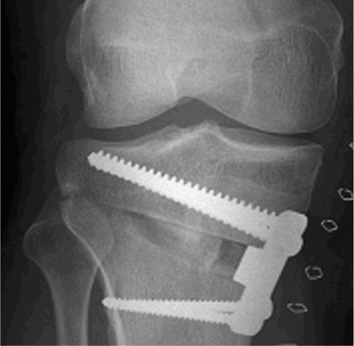
Figure 3. Opposite medial cortical fracture with a gap in closing‐wedge HTO on the first day after surgery (radiograph in supine position).
Statistics
Distribution of HKA angle was examined by the Shapiro‐Wilk test. The HKA data were not normally distributed; thus, the Mann‐Whitney‐Wil‐coxon U‐test was used to analyze between‐group differences. The chi‐squared test was used to compare the percentages of opposite cortical fractures between the groups. We used SPSS version 10.1 and p‐values of < 0.05 were considered to be statistically significant.
Results
44 patients had a closing‐wedge HTO and 43 patients had an opening‐wedge HTO. 36 patients (0.8) in the closing wedge group, and 15 patients (0.4) in the opening wedge group were found to have an opposite cortical fracture; this difference between the two osteotomy techniques was highly significant (p < 0.001). The relative risk of an opposite cortical fracture in closing‐wedge HTO compared to opening wedge HTO was 8 (95% CI: 3–23).
Closing‐wedge HTO
For the closing‐wedge group, the mean preopera‐tive HKA angle was 6.7 (SD 2.9) degrees of varus in patients with a cortex fracture and 6.0 (SD 4.1) degrees of varus in those without opposite cortical disruption. At the 1‐year follow‐up, the mean postoperative HKA angle was 3.2 (SD 3.5) degrees of valgus in the closing‐wedge group with cortex fracture and 2.5 (SD 3.7) degrees of valgus without cortex fracture; these differences were not statistically significant (). Of the 36 patients with a medial cortex fracture, 18 showed no gap. In 18 patients, we observed a gap of more than 2 mm between the opposite cortex fragments. When a gap was seen on the radiograph from the first postoperative day, the mean 1‐year postoperative HKA angle was more in valgus compared to the group with opposite cortex fracture without a gap: 4.3 (SD 3.4) and 2.1 (SD 3.4) degrees of valgus, respectively (p = 0.05) (). In the group with a gap, 7 patients had a valgus of more than 4 degrees as compared to 6 patients without a gap; this difference was not significant. No significant deviation from the planned 4 degrees in excess of physiological valgus was seen in any of the subgroups at the 1‐year follow‐up.
Table 2. Whole‐leg alignment (HKA angle) for closing‐ and opening‐wedge high tibial osteotomy groups with or without opposite cortical fracture. Values are mean (SD)
Table 3. Whole‐leg alignment (HKA angle) for closing‐wedge osteotomy, stratified according to medial cortex fracture pattern in 36 cases with opposite cortical fracture. Values are mean (SD)
Opening‐wedge HTO
Patients in the opening‐wedge group with and without opposite cortical fracture showed no significant difference in the mean preoperative HKA angle of varus (4.9 degrees (SD 3.2) and 5.5 degrees (SD 2.5), respectively). 1 year postoperatively, the mean HKA angle was 0.9 (SD 6.7) degrees of varus in patients with cortex fracture, and 2.3 (SD 4.0) degrees of valgus in patients without cortex disruption (). This difference in postoperative alignment between the two groups almost reached statistical significance (p = 0.057) 1 year after surgery. The group of patients with opposite cortical fracture had less accurate correction, with significant deviation from the planned 4 degrees in excess of physiological valgus at the 1‐year follow‐up (p = 0.04). In patients without cortex fracture, there was no statistically significant deviation from the planned correction 1 year after surgery.
Discussion
Opposite cortical fracture in closing‐ and opening‐wedge HTO appears to be an operative complication that is not entirely preventable when correcting a large varus deformity. Pape et al. (Citation2004) reported that the capacity for plastic deformation of the medial cortex of the proximal tibia might have been exceeded in closing osteotomies with a larger wedge size (> 8 degrees), leading to a non‐displaced fracture during the operation. Böhler et al. (Citation1999) argued that valgus correction of the tibia plateau by removal of a wedge of as much as 10 degrees is possible without fracturing the medial cortex. However, another cadaver study by Kessler et al. (Citation2002) permitted far less maximal angular correction (7 degrees for both the closing‐ and the opening‐wedge technique) that can be applied to human tibias without fracturing the cortex at the apex of the wedge. This was confirmed by Spahn et al. (Citation2003) who reported results from a series of 55 patients treated with opening‐wedge HTO using the Puddu spacer plate (mean wedge correction angle of 9.7 (SD 1.9) degrees), with 8 lateral cortical fractures. In our patients, the main aim was to achieve a correction of 4 degrees in excess of physiological valgus at the 1‐year follow‐up, which led to a mean correction angle of 11 (SD 3.1) degrees in the closing wedge and 9 (SD 2.8) degrees in the opening wedge group. The magnitude of the wedge sizes may be the reason for the high rate of unintentional opposite cortical fracture seen in both the closing‐wedge (four‐fifths) and opening‐wedge technique (one‐third). Also, with the closing‐wedge technique it can be difficult to remove the wedge completely, especially at its apex at the medial side. Consequently, closing the wedge may cause fracturing at the medial osteotomy site. This probably explains the significantly higher rate of opposite cortical fracture compared to the opening‐wedge technique.
Limitations of the study
The study patients were part of a randomized, controlled and consecutive trial on two osteotomy techniques (Brouwer et al. Citation2006). In clinical studies, there have been few reports on the effect of opposite cortical fracture in HTO that may lead to loss of correction. We realize that subgroup analysis in such a trial has its statistical limitations. However, it is impossible to randomize for adverse events such as opposite cortical fracture in HTO. With this analysis, we focused on the 1‐year radiological effect of intraoperative disruption of the opposite cortex in both techniques. One limitation may be that we did not take a whole‐leg radiograph on the first day postoperatively because the patient could not stand on the operated leg; thus, we cannot discriminate between insufficient correction and loss of correction during the 1‐year follow‐up.
Closing‐wedge HTO
It has been recommended that one should maintain the medial cortex of the proximal tibia in lateral closing‐wedge HTO, to provide sufficient stability and avoid a reduction in cortical contact area of tibial segments (Miniaci et al. Citation1989, Coventry et al. Citation1993). Progressive movement into a varus position may otherwise occur (Myrnerts Citation1980, Insall et al. Citation1984). However, our data do not support this statement. The desired correction was achieved even more often in the closing‐wedge group with medial cortex fracture and a gap between the medial cortex fragments (). With the closing‐wedge technique, posteromedial bony remnants may act like a more lateral hinge when closing the wedge, and probably cause fracture and gaping at the medial osteotomy site with pronounced valgisation. Pape et al. (Citation2004) detected no loss of valgus correction on full weight‐bearing standing radiographs in patients with a fractured medial cortex of the proximal tibia after a closing‐wedge procedure either; they used an L‐shaped rigid plate, which is supposed to offer high primary stability. Radiostereo‐metric findings indicate a less stable situation for closing‐wedge osteotomy when bone staples with a plaster cast are used (Magyar et al. Citation1999). However, Harrison and Waddell (Citation2005) did not note any change in femoral‐tibial alignment with the use of staples and a long leg cast.
Opening‐wedge HTO
Whether or not the lateral cortex has been harmed largely dictates the stability after opening‐wedge HTO, regardless of implant design (Stoffel et al. Citation2004). Agneskircher et al. (Citation2006) also stated that in composite tibias, independently of the implant, axial loading is well tolerated with an intact lateral cortical bridge. Our results seem to confirm these findings in a clinical setting. With the use of the non‐angular stable Puddu plate, 1‐year after surgery the mean HKA angle was 2.3 degrees of valgus in the opening‐wedge group without cortex fracture (). In our study, however, the opposite tibial cortex was fractured in 15 of 43 patients, and they had less accurate correction with a mean HKA angle of 0.9 degrees of varus at the 1‐year follow‐up. Hernigou et al. (Citation1987) reported lateral cortex fracture as a complication of medial opening‐wedge HTO, resulting in displacement of the osteotomy and recurrent varus malalignment before osteotomy union in 12% of all their patients. In our series, 4 patients with lateral cortex disruption were reoperated within 1 year—2 patients because of nonunion and 2 patients requiring revalgisation osteotomy due to the recurrent varus deformity (Brouwer et al. Citation2006). Disruption of the lateral cortex causes increased micromotion at the osteotomy site, and this instability most likely contributes to the high incidence of delayed union and nonunion after medial HTO (Miller et al. Citation2005). The Puddu plate proved unable to oppose the instability, which was also reported by others (Spahn et al. Citation2003). Mechanical studies have shown that when the lateral cortex is injured, angle‐stable implants provide superior primary stability compared to the Puddu plate (Stoffel et al. Citation2004, Agneskircher et al. Citation2006). The angle‐stable design protects the lateral cortex and prevents lateral displacement. Furthermore, the use of a spacer with an angle‐stable plate seems to increase primary stiffness even more (Spahn et al. Citation2006b). When unintentional opposite cortical fracture occurs, we suggest the use of an angle‐stable implant, which has the best biomechanical properties in internal fixation after medial opening‐wedge HTO.
Conclusion
We conclude that fracture of the opposite cortex in HTO is much more common in the lateral closing‐wedge technique than in the medial opening‐wedge technique. Nevertheless, medial cortex fracture in closing‐wedge osteotomy with the use of staples and plaster has no major consequences, and can be managed successfully in the vast majority of patients. It does not generally lead to recurrence of varus malalignment 1 year after surgery. However, lateral cortex fracture in the medial opening‐wedge technique is an unstable situation. The use of a non‐angular stable Puddu plate seems to provide insufficient primary stability and leads more often to varus malalignment.
- Agneskircher J D, Freiling D, Hurschler C, Lobenhoffer P. Primary stability of four different implants for opening wedge high tibial osteotomy. Knee Surg Sports Traumatol Arthrosc 2006; 14: 291–300
- Ahlbäck S. Osteoarthritis of the knee. A radiographic investigation. Acta Radiol Diagn (Suppl 277) 1968; 7–72
- Berman A T, Bosacco S J, Kirshner S, Avolio A, Jr. Factors influencing long-term results in high tibial osteotomy. Clin Orthop 1991, 272: 192–8
- Böhler M, Fuss F K, Schachinger W, Wölfl G, Knahr K. Loss of correction after lateral closing wedge high tibial osteotomy: A human cadaver study. Arch Orthop Trauma Surg 1999; 119: 232–5
- Brouwer R W, Jakma T S C, Bierma‐Zeinstra S M A, Ginai A Z, Verhaar JAN. The whole leg radiograph: standing versus supine for determining axial alignment. Acta Orthop Scand 2003; 74: 565–8
- Brouwer R W, Bierma‐Zeinstra S M A, van Raaij T M, Verhaar JAN. Osteotomy for medial compartment arthritis of the knee using a closing wedge or an opening wedge controlled by a Puddu plate: a one-year randomised, controlled study. J Bone Joint Surg (Br) 2006; 88: 1454–9
- Coventry M B, Ilstrup D M, Wallrichs S L. Proximal tibial osteotomy: a critical long-term study of eighty-seven cases. J Bone Joint Surg (Am) 1993; 75: 196–201
- Harrison M M, Waddell J P. A comparison of plate versus staple-and-cast fixation in maintaining femoral tibial alignment after valgus tibial osteotomy. Can J Surg 2005; 48(1)33–8
- Hernigou P, Medevielle D, Debeyre J, Goutallier D. Proximal tibial osteotomy for osteoarthritis with varus deformity. A ten to thirteen-year follow-up study. J Bone Joint Surg (Am) 1987; 69: 332–54
- Insall J N, Joseph D M, Misika C. High tibial osteotomy for varus gonarthrosis: a long-term follow-up study. J Bone Joint Surg (Am) 1984; 66: 1040–8
- Kessler O C, Jacob H A C, Romero R. Avoidance of medical cortical fracture in high tibial osteotomy: improved technique. Clin Orthop 2002, 395: 180–5
- Magyar G, Toksvig-Larsen S, Lindstrand A. Changes in osseous correction after proximal tibial osteotomy. Radio-stereometry of closed- and open-wedge osteotomy in 33 patients. Acta Orthop Scand 1999; 70: 473–7
- Miller B S, Dorsey WOP, Bryant C R, Austin J C. The effect of lateral cortex disruption and repair in the stability of the medial opening wedge high tibial osteotomy. Am J Sports Med 2005; 33: 1552–7
- Miniaci A, Ballmer F T, Ballmer P M, Jakob R P. Proximal tibial osteotomy. A new fixation device. Clin Orthop 1989, 246: 250–9
- Myrnerts R. Failure of the correction of varus deformity obtained by high tibial osteotomy. Acta Orthop Scand 1980; 51: 569–73
- Pape D, Adam F, Rupp S, Kohn D. Stability, bone healing, and alignment loss after closing-wedge high tibial osteotomy. A roentgen stereometric analysis. Orthopaede 2004; 33: 208–17
- Spahn G. Complications in high tibial (medial opening wedge) osteotomy. Arch Orthop Trauma Surg 2003; 124: 649–53
- Spahn G, Kirschbaum S, Kahl E. Factors that influence high tibial osteotomy results in patients with medial gonarthri-tis: a score to predict results. Osteoarthritis and Cartilage 2006a; 14: 190–5
- Spahn G, Mückley T, Kahl E, Hofmann G O. Biomechani-cal investigation of different internal fixations in medial opening-wedge high tibial osteotomy. Clin Biochem 2006b; 21: 272–8
- Stoffel K, Stachowiak G, Kuster M. Open wedge high tibial osteotomy: biomechanical investigation of the modified Arthrex Osteotomy Plate (Puddu Plate) and the TomoFix Plate. Clin Biochem 2004; 19: 944–50
- Virolainen P, Aro H T. High tibial osteotomy for the treatment of osteoarthritis of the knee: a review of the literature and a meta-analysis of follow-up studies. Arch Orthop Trauma Surg 2004; 124: 258–61