Abstract
Background and purpose Polymer technology has provided solutions for filling of bone defects in situations where there may be technical or biological complications with autografts, allografts, and metal prostheses. We present an experimental study on segmental bone defect reconstruction using a polymethylmethacrylate‐(PMMA‐) based bulk polymer implant prosthesis. We concentrated on osteoconductivity and surface characteristics.
Material and methods A critical size segment defect of the rabbit tibia in 19 animals aged 18–24 weeks was reconstructed with a surface porous glass fiber‐reinforced (SPF) prosthesis made of polymethylmethacrylate (PMMA). The biomechanical properties of SPF implant material were previously adjusted technically to mimic the properties of normal cortical bone. A plain PMMA implant with no porosity or fiber reinforcement was used as a control. Radiology, histomorphometry, and scanning electron microscopy (SEM) were used for analysis of bone growth into the prosthesis during incorporation.
Results The radiographic and histological incorporation model showed good host bone contact, and strong formation of new bone as double cortex. Histomorphometric evaluation showed that the bone contact index (BCI) at the posterior surface interface was higher with the SPF implant than for the control. The total appositional bone growth over the posterior surface (area %) was also stronger for the SPF implant than for controls.
Both bone growth into the porous surface and the BCI results were related to the quality, coverage, and regularity of the microstructure of the porous surface.
Interpretation Porous surface structure enhanced appositional bone growth onto the SPF implant. Under load‐bearing conditions the implant appears to function like an osteoconductive prosthesis, which enables direct mobilization and rapid return to full weight bearing.
Current methods for repair of segmental defects in long bones rely mostly on metal prostheses or allograft bone. In clinical trials with large allografts, the nonunion rate has been reported to be as high as 11–32%, and long‐term survival of metal prostheses has been reported to be less than 60% (Aho et al. Citation1994, Aldlyami et al. Citation2005). Recent research has led to a multitude of synthetic biomaterials as candidates for this purpose (Fujibayashi et al. Citation2003, Wang Citation2003, Yoneda et al. Citation2005). The new materials have been mainly composed of structural block ceramics, for example calcium phosphate/ hydroxyapatite, coral, bioactive glass, and glassceramics (Ohgushi et al. Citation1989, Walsh et al. Citation2003, Gil‐Albarova et al. Citation2005). Metals or block ceramics alone are not biologically or mechanically ideal for reconstruction bridging of diaphyseal bone defects because of the mismatch in mechanical properties relative to those of bone (Wang Citation2003, Aldlyami et al. Citation2005).
The inability of metals and polymers alone to allow direct bone union has led to research into new and more complex composite materials, which combine resorbable or non‐resorbable polymers and bioactive fillers in vehicle models (Kitsugi et al. Citation1989, Shinzato et al. Citation2000, Lu et al. Citation2001, Flautre et al. Citation2001, Miyazaki et al. Citation2003). Improved osteoconduction and osteoinduction has been achieved through addition of various growth factors (most often bone morphogenetic proteins (BMPs)) to polymer carrier materials (Gao et al. Citation1996, Lane et al. Citation1999, Seeherman and Wozney Citation2005, Yoneda et al. Citation2005). Advancements in the creation of a suitable biomaterial for segmental bone defect repair are promising, but there are still problems concerning bone growth and mechanics of the interface area.
Biocompatibility, osteoconductivity, and bio‐mechanical properties are important for all bone substitutes, but good biomechanical properties are essential for segmental defect reconstruction in load‐bearing applications. Our group has concentrated on improving the already well‐known, current orthopedic use of polymers by combining them with modern fiber‐reinforced composite technology to develop a new implant material. The SPF implant is a non‐degradeable material with a porous surface to enable bone ingrowth. It has a fiber‐reinforced stem, with mechanical properties adjusted to match those of cortical bone (Aho et al. Citation2004). The present study was a 20‐week follow‐up of critical size segment defects in the rabbit tibia that had been repaired with the SPF implant, with histomorphometric analysis of host bone ingrowth/on‐growth and detailed SEM analysis of the surface structure of the implants.
Material and methods
Implant preparation
The SPF implant stems with artificial marrow canals were pre‐cured in molds as previously described (10 ± 2 mm length, 6 ± 1 mm diameter), simulating the cross‐sectional triangular form of rabbit tibia (Aho et al. Citation2004). Autopolymerizing polymethylmethacrylate (PMMA) resin (PMMA powder/MMA monomer liquid: Palapress; Heraeus Kulzer, Wehrnheim, Germany) was used as core material. Continous unidirectional E‐glass fibers (53–55% SiO2) (Stick; Stick Tech Oy, Turku, Finland) (21%‐weight, pre‐impregnated with PMMA) running along the long axis of the implant were added for reinforcement of the implant, by placing the fibers in the molds before the core was polymerized as previously described (Vallittu Citation1999, Aho et al. Citation2004) ().
Figure 1. Schematic drawing representing the SPF implant, with surface porosity and E-glass fiber-reinforced core. Macroscopic picture of the implant.
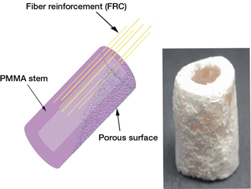
The porous surface was created by a tetrahydrofuran solvent‐foaming method as previously reported (Aho et al. Citation2004). Control implants were made of plain PMMA, with surfaces ground with a metal file to remove the shiny/polished surface created by the mold‐casting process (). All implants were stored for at least 24 hours at 37°C in distilled water to wash out the MMA monomers and residual solvent (Vallittu et al. Citation1995).
Figure 2. SEM photomicrographs showing the surface of the SPF implant and the control implant, (a) Ideal surface porosity (between arrows) on SPF implant with unidirectional fiber-reinforced stem. (b) Variable surface quality of porosity (arrows) on SPF implant. (c) Porosity in the intramedullary canal of SPF implant with higher magnification (x250) detail. (d) Ideal porosity layer at the end of SPF implant (between arrows). (e) Control (PMMA) implants: filed end and higher magnification detail. (f) Side of control implant with slightly uneven filed surface (arrowheads).
Mechanical properties
Fiber‐reinforced composite used in SPF implants had 3‐point bending flexural test and Young's modulus of 320 (SD 91) MPa and 11.7 (1.6) GPa, respectively, which are close to values for human cortical bone (255 (28) MPa and 13 (2.1) GPa) (Reilly and Burstein Citation1975, Lotz et al. Citation1991). Plain PMMA used as the control implant had much lower values (64 (16) MPa and 3.2 (0.8) GPa, respectively) (Aho et al. Citation2004). Corresponding values for rabbit cortical bone are approximately 101 MPa and 4.5 GPa (3‐point bending and Young's modulus, respectively) (Gondret et al. Citation2005).
Surface characteristics and microstructure covering the implants
The surface structure of the implant was evaluated in vitro by SEM and in vivo from histological samples. For the SEM study, 5 SPF and control implants were put into liquid nitrogen for approximately 2 min and then split longitudinally, and the other halves were cut in the middle with a diamondcoated water‐cooled band saw. The freshly formed and cleaned surfaces were coated with carbon film (BalTec CED 030; BalTec, Balzers, Liechtenstein) for analysis by SEM (JEOL 5000; Jeol, Tokyo, Japan) ().
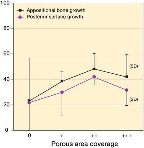
Porosity of the surface covering the implant was qualitatively classified under a light microscope by visual inspection of the anterior and posterior edges of the histological sections. Assessment was made from all animals according to the following score: 0 for no porosity, + for < 1/3 (less than 33%) coverage, ++ for 1/3–2/3 (between 33% and 66%) coverage, +++ for > 2/3 (more than 66%) coverage. The relationship between bone growth and degree of porosity coverage is presented in .
Figure 8. Graph showing the relationship between porosity observed and measured growth of bone onto the implant.
Animal experiments
Surgery.
32 New Zealand White female rabbits weighing 3.4— 4 kg were used as test animals under the permission of the provincial government of southwestern Finland (no. 51124/7624). The observation times were 4 weeks (n = 5), 8 weeks (n = 5) and 20 weeks (n = 10) for the SPF implant. A total of 12 control PMMA implants were operated, with 4 animals for each time point. The operations were carried out under anesthesia induced intramuscularly by combination of midazolam hydrochloride 5 mg, medetomidine hydrochloride 0.8 mg, and ketamine hydrochloride 50 mg. Postoperative pain was treated with subcutaneus injections of buprenorfin hydrochloride (∼0.15 mg/day for 3 days).
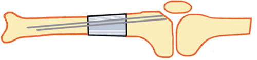
The total segment defect (10 ± 2 mm in length) was created on the proximal portion of the left tibia of each animal with a water‐cooled circulating surgical saw. The defect was replaced with the implant and fixed with intramedullary K‐wires (1.0–1.4 mm, 2–3 per operated animal) in the present series of follow‐up to 20 weeks ().
The animals were allowed free mobilization and had the full use of the operated limb from the first postoperative day. They were killed by intravenous overdose of phenobarbital and the tibias were removed and separated from soft tissues. The specimens were fixed in 75% ethanol for histological processing and radiography.
Figure 3. Schematic illustration of positioning and fixation of the implant into the rabbit tibia.
Radiography.
Plain non‐screen contact radiographs (35 kV and 175 mA) were taken of the whole tibias. Radiological incorporation was determined, evaluating the bone growth qualitatively around the implant and at the bone‐implant junctions ().
Figure 4. Radiographs of the operation site at 20 weeks. (A) Formation of bridging callus (arrows); anteroposterior view. (B) Posteriorly, strong callus formation; double cortex (long arrows). Anteriorly, resorptive rounding and a gap at the distal junction (arrowheads), bridging callus over the proximal junction; lateral view. Both images show the intra-medullary K-wires still in place.
Histology and histometry of the incorporation model.
For light microscopic studies, the harvested bones were embedded in acrylic resin (Technovit; Kulzer GmbH, Wehrheim, Germany) and 20‐|im thickness non‐decalcified sections of the tibias were prepared by cutting‐grinding method, and stained with van Gieson stain (Donath and Breuner Citation1982) ().
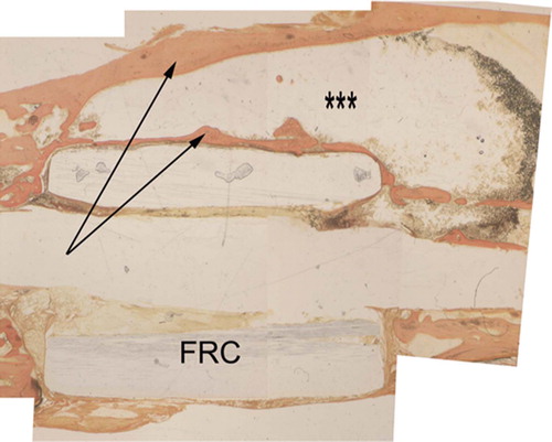
Figure 5. Histological image at 20 weeks, with van Giesson stain (reconstructed from 5 pictures). Longitudinally sectioned sample showing double cortex (arrows), with marrow (***), fiber reinforcement (FRC), and intramedullary canal.
Histomorphometry was done using a computer‐assisted analysis system (Microscale TC; Digithurst Ltd., Royston, UK) to measure bone growth at different anatomical areas: both longitudinal surfaces, junctions (interface) between the implant and the host cortical bone, and the marrow canal.
Total appositional bone growth (ABG) over the posterior surface is the percentage area of bone from the whole area over the posterior cortex of the implant covered by tissue.
The porous surface bone growth (PSG) is the percentage area of bony ingrowth into the porous surface determined from the area rising 0.4 mm over the stem on the posterior cortex, which is the average thickness of the porous surface evaluated from the SEM images. The bone growth into the marrow canal (IMG) is percentage of bone from the total canal area within the implant.
The bone contact index (BCI) is the percentage from the sum of contact lines between bone and implant—at cortical junctions, posterior and anterior surface—of the corresponding total length of the implant ().
Figure 6. Schematic model showing the different areas included in the histometrical measurements.
Statistics
Statistical analysis of the data was done using SPSS software. One‐way analysis of variance (ANOVA), Mann‐Whitney U‐test, and Student's t‐test were performed to determine significant differences in test results. We considered p‐values less than 0.05 to be significant and 95% confidence intervals (CI) are given.
Results
Notes on the surgery
The SPF implant was easy to handle, i.e. easy to shape and size by drilling and sawing during surgery. No animals were lost due to breakage of the implant or infection. One animal from the SPF group (8 weeks) was killed during the follow‐up, at the third postoperative day. This was due to instability of the whole limb and the animal was excluded.
Analysis of surface microstructure
Regarding the surface porosity and thickness of the porous surface layer, the pores ranged from 10— 500 μm in diameter and the pores were found to be interconnected. The majority of the pores were in the size range 50–80 μm (). The average thickness of the surface was 0.4 (0– 0.8) mm ( and ), and it varied between different parts of the implant – being thinnest or even absent at the junction ends of the implant ( and ). Control PMMA implants also showed some uneven surface texture due to the manual grinding ( and ).
At surgery, before implantation some of the SPF implants showed bald surface spots macroscopically, where the porous surface had been shed because of the weak attachment of the surface layer.
Table.
Radiography
Abundant growth of new bone as appositional tube‐like callus formation around the implant was observed, especially on the posterior cortical side. Strong bony bridging callus over the junctions was seen already at 4 weeks, and it developed or remodeled to lamellar bone by 20 weeks. Anterior cortex showed the least bone formation, and even some resorption in the form of rounding of the edges of the cortexes ().
Histology: the incorporation model
No adverse inflammatory cell reactions were seen. New bone formation was evident at 4–8 weeks as bridging trabeculous bone growing from the host periosteum junctions, creating a tube‐like external callus around the implant (). Lamellar bone was evident in all specimens at 20 weeks. 4 of 9 SPF implants showed growth of bone into the medullary canal at 20 weeks. On the posterior side of the specimens (in 6/10) we found a formation of double cortex, i.e. 2 perpendicular longitudinal lamellar bony bridges spanning the length of the implant and an empty space in between them. This is shown both on the radiographs and in the histological samples ( and ).
Slight bone resorption in the form of rounded lacunae could be seen at the junctional cortexes of some specimens; however, osteoclast cells were seldom observed in these areas. Resorption apparently resulted from incomplete operational alignment between the sawed cortex and the end of the implant leading to loading imbalance during weight bearing, illustrated by slight resorptive bone loss. Intervening fibrous tissue was found in some places at junctions between the implant and host cortex bone, particularly in cases of the above‐mentioned technical/operational gap and line mismatch.
Histomorphometry
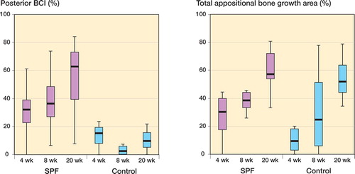
The bone contact index (BCI) at the posterior cortex was higher for SPF implants at 20 weeks than in the control group (p = 0.01) and already markedly higher at 4 weeks (p = 0.06). These results were confirmed in the non‐parametric analysis of the group data by Mann‐Whitney test. ANOVA showed a clear group difference in favor of the SPF implant, independently of implantation time.
The total appositional bone growth over the posterior side was clearly higher with the SPF implant early on (26% (SD 21) vs. 10% (SD 10) at 4 weeks, and 38% (SD 13) vs. 32% (SD 38) at 8 weeks), but diminished towards 20 weeks (59% (SD 15) vs. 55% (SD 19)). There were no statistically significant differences between the groups.
In other areas of new bone growth (anterior surface and medullary canal), the differences were not statistically significant, although the measured averages were higher for the SPF implant than for the controls in most cases. The lowest BCI values at 20 weeks (9.8% (SD 7.8)) were measured for the SPF implant at the junction areas, which illustrates the irregularity and uneven quality of the porous surface (Table,). A relationship between degree of coverage of the porous area, the surface microstructure, and the amount of bone growth onto the implant was found ().
Figure 7. Panels show the bone-implant contact (BCI) and appositional growth at the posterior side of the implant.
Discussion
Uneven surface of an implant—for example, due to porosity—has previously been shown to enhance bone attachment to implant material (Flautre et al. Citation2001, Itälä et al. Citation2001, Hacking et al. Citation2003). Consistent with this, we found that the degree of coverage of the porous surface was related to the amount of bone growth ().
The porous surface structure of the implant is likely to play a key role in bone growth, although it appears to lack regularity when evaluated by
SEM and by histological studies. This variability in porosity could also be detected in early SEM studies (Aho et al. Citation2004), but the effects—variation in the degree of bone growth into the porous surface (PSG) and the low BCI at the junctions—were only seen after the 20‐week follow‐up period. The loss of porosity after implantation is thought by the researchers to cause the low values of BCI at the junction areas during healing. This is thought to be due to: (1) incomplete fitting between the implant interfaces and the sawed cortexes, (2) insufficient compression and stability at the bone‐implant interfaces, (3) the irregularity of the porosity (–) and possible movements at the junction during weight bearing. In recent studies by our co‐workers (Mattila et al. Citation2004), the uniformity of the porous surface has been improved technically by short fiber additives.
A noteworthy observation was also the abundant bone growth into the intramedullary canals of both of the implants, SPF implant's porous and PMMA implant's drilled canals. As well as the relatively high bone growth seen on the control implant, can be explained with the uneven (i.e. not as smooth as polished) surface created by drilling or filing with a rasp.
Notable results in the SPF implants in our incorporation model were the difference in BCI at the posterior side (p = 0.01 at 20 weeks) and good total appositional bone growth (APG) over the porous surface. The APG was highest at the dorsal longitudinal side of the implant where the blood supply and muscle coverage were optimal, in contrast to the anterior side. A formation of two parallel cortexes at the posterior side of the implant was observed and termed a double cortex. This was noticed both in histological and radiographic evaluation of the samples ( and ). It can be explained by the remaining periosteal cells in the muscle insertion after sharp dissection during removal of the bone segment. Other possible stimulatory factors could be the relative motion/movement during weight bearing and good blood supply from the surrounding muscles. The same phenomenon has also been witnessed in large osteoarticular allografts in humans evaluated by CT scanning, and it was thought to be a biomechanical response to loading (Mattila et al. Citation1995, Aho et al. Citation1996). The incorporation model of the SPF‐implant (the bridging bone growth, external callus over the junction areas, and the appositional surface bone growth (as double cortex or neo‐cortex)) resembles the repair of large structural human allografts and experimental segment repair in dogs (Delloye Citation1990, Aho et al. Citation1996, Enneking and Campanacci Citation2001). The host bone resorption lacunae at junction areas described by the latter authors correspond to few cortical resorption signs observed in our studies.
Reconstruction of segmental bone defects with resorbable or non‐resorbable biomaterials has been associated with many complications: nonunion, resorption of the implant that is too fast, connective tissue formation, breakage/fragmentation, stress shielding, etc. (Ohgushi et al. Citation1989, Okada et al. Citation1999, den Boer et al. Citation2003). There are a growing number of reports in the literature of growth factors (BMP, TGF‐b) being used to improve bone growth in different bone defect models (Gerhart et al. Citation1993, Teixeira and Urist Citation1998). Experimental models using biodegradable ceramics (with or without growth factors) rely on regular degradation and new ingrowth of bone for mechanical stability, and this can present problems concerning the biomechanical properties of the newly formed bone and the mechanical stability of the whole model (Lane et al. Citation1999, Seeherman and Wozney Citation2005, Yoneda et al. Citation2005).
Fiber‐reinforced composites have already been widely explored in restorative and prosthetic dentistry, where E‐glass fiber‐reinforced PMMA‐based polymers have gained wide acceptance because of their good mechanical properties (Kim and Watts Citation2004, Dyer et al. Citation2005). Promising clinical results at an early stage using these composite materials as bone substitutes have recently been achieved in the field of head and neck surgery (Tuusa et al. 2005). Residual monomers (MMA) leaching from PMMA‐based fiber‐reinforced polymers have even been associated with cytotoxic effects, but this has been proven to be clinically irrelevant after adequate prosessing and preoperative storage (Vallittu et al Citation1995, Miettinen and Vallittu Citation1997).
The present SPF implant did not have any of the problems associated with the mechanical properties of an implant (breaking, stress shielding, etc.) because of its matched modulus of elasticity and flexural strength with normal cortical bone (Reilly and Burstein Citation1975, Lotz et al. Citation1991). The use of non‐degradable biostable osteoconductive implant material, like a permanent prosthesis, gives immediate benefits concerning loading forces and early incorporation of the implant into host bone.
In conclusion, this study has shown that the SPF implant based on glass fiber‐reinforced PMMA with surface porosity provided a prosthesis for segment defects of long bones in a weight‐bearing model. It contained a porous surface, which enhanced bridging of bone over the interface area and allowed ingrowth of host bone. Osteoconductivity and biomechanically equivalent properties with bone suggests that the material is potentially useful for bone reconstruction purposes.
Contributions of authors
MH: primary surgeon, sample preparation, SEM imaging, analysis of data (histology, histomorphometry, radiography, statistics) and the first author. AJA: leader of the research team, co‐analysis of histology and histomorphometry (first observer), manuscript writing. PL: manufacture and testing of the implants (mechanics), SEM imaging assistant. JR: statistical analysis, surgery. JG: surgery, sample preparation, histological co‐analysis. NS: surgery, histomorphometrical co‐analysis (second observer). PKV: head of the Biomaterials Research group, implant development, and materials consultant.
Financial support for this study was received from Finnish Funding Agency for Technology and Innovation (TEKES), the University of Turku Foundation, and HUCH‐Jorvi Hospital Research Grants. This work belongs in part to the activity of the Bio‐ and Nanopolymers Research Group at the Center of Excellence, Academy of Finland.
No competing interests declared.
- Aho A J, Ekfors T, Dean PB, Aro H T, Ahonen A, Nikkanen V. Incorporation and clinical results of large allografts of the extremities and pelvis. Clin Orthop 1994, 307: 200–13
- Aho A J, Ekfors T, Knuuti J, Mattila K, Heikkilä J. Repair of massive allografts: Histological, nuclear medicine and CT-studies. In: Orthopaedic allograft surgery, A A Czitrom, H Winkler. Springer‐Verlag Wien, New York 1996; 67–73
- Aho A J, Hautamäki M P, Mattila R, Alander P, Strandberg N, Rekola J, Gunn J, Lassila L V. Surface porous fiber-reinforced composite bulk bone substitute - In vitro studies and in vivo evaluation of segment defect. Cell Tissue Banking 2004; 5(4)213–21
- Aldlyami E, Abudu A, Grimer R J, Carter S R, Tillman R M. Endoprosthetic replacement of diaphyseal bone defects. Long-term results. Int Orthop 2005; 29: 25–9
- Delloye C H. The Bridging capasity of a cortical bone defect by different bone and crafting materials and diaphyseal distraction lengthening: An experimental study. Thesis. Catholic University of Louvain, Belgium, Louvain 1990
- den Boer F C, Wippermann B W, Blokhuis T J, Patka P, Bakker F C, Haarman H J. Healing of segmental bone defects with granular porous hydroxyapatite augmented with recombinant human osteogenic protein-1 or autolo-gous bone marrow. J Orthop Res 2003; 21(3)521–8
- Donath K, Breuner G. A method for the study of undecalci-fied bones and teeth with attached soft tissues. The Sage-Schliff (sawing and grinding) technique. J Oral Path 1982; 11(4)318–26
- Enneking W F, Campanacci D A. Retrieved human allografts: a clinicopathological study. J Bone Joint Surg (Am) 2001; 83(7)971–86
- Dyer S R, Lassila L V J, Jokinen M, Vallittu P K. Effect of cross-sectional design on the modulus of elasticity and toughness of fiber-reinforced composite materials. J Pros-thet Dent 2005; 94(3)219–26
- Flautre B, Descamps M, Delecourt C, Blary M C, Hardouin P. Porous HA ceramic for bone replacement: Role of the pores and interconnections - experimental study in rabbit. J Mat Sci: Materials in Medicine 2001; 12: 679–82
- Fujibayashi S, Kimb H, Neoa M, Uchida M, Kokubo T, Nakamura T. Repair of segmental long bone defect in rabbit femur using bioactive titanium cylindrical mesh cage. Biomaterials 2003; 24: 3445–51
- Gao T J, Lindholm T S, Kommonen B, Ragni P, Paronzini A, Lindholm T C. Enhanced healing of segmental tibia defects in sheep by a composite bone substitute composed of tricalcium phosphate cylinder, bone morphogenetic protein, and type IV collagen. J Biomed Mat Res 1996; 32: 505–12
- Gerhart T N, Kirke-Head C A, Kriz MJ, Holtrop M E, Hennig G E, Hipp J. Healing segmental bone defects in sheep using recombinant human bone morphogenetic protein. Clinic Orthop 1993, 293: 317–26
- Gil-Albarova J, Salinas A J, Bueno-Lozano A L, Roman J, Aldini-Nicolo N, Garcıa-Barea A. The in vivo behaviour of a sol-gel glass and a glass-ceramic during critical diaphyseal bone defects healing. Biomaterials 2005; 26(21)4374–82
- Gondret F, Larzul C, Combes S, de Rochambeau H. Carcass composition, bone mechanical properties, and meat quality traits in relation to growth rate in rabbits. J Anim Sci 2005; 83: 1526–35
- Hacking S A, Harvey E J, Tanzer M, Krygier J J, Bobyn J D. Acid-etched microtexture for enhancement of bone growth into porous-coated implants. J Bone Joint Surg (Br) 2003; 85(8)1182–9
- Itälä A, Nordstrom E G, Ylanen H, Aro H T, Hupa M. Creation of microrough surface on sintered bioactive glass microspheres. J Biomed Mat Res 2001; 56(2)282–8
- Kim S H, Watts D C. The effect of reinforcement with woven E-glass fibers on the impact strength of complete dentures fabricated with high-impact acrylic resin. J Prosthet Dent 2004; 91(3)274–80
- Kitsugi T, Yamamuro T, Kokubo T. Bonding behavior of a glass-ceramic containing apatite and wollastonite in seg-mental replacement of the rabbit tibia under load-bearing conditions. J Bone Joint Surg (Am) 1989; 71(2)264–72
- Lane J M, Tomin E, Bostrom M P G. Biosynthetic bone grafting. Clin Orthop 1999, 367: 107–17
- Lotz J C, Gerhart T N, Hayes W C. Mechanical properties of metaphyseal bone in the proximal femur. J Biomechanics 1991; 24(5)317–25
- Lu W W, Cheung K M, Li Y W, Luk KD K, Holmes A D, Zhu Q A. Bioactive bone cement as a principal fixture for apinal burst fracture. An in vitro biomechanical and morphologic study. Spine 2001; 26(24)2684–91
- Mattila K T, Heikkilä J T, Aho A J, Manner I K, Dean P B. Massive osteoarticular knee allografts; structural changes evaluated with CT. Radiology 1995; 196(3)657–60
- Mattila R H, Lassila L V J, Vallittu P K. Production and structural characterisation of porous fibre-reinforced composite. Composites Part A. Appl Scie Manufact 2004; 35: 631–6
- Miettinen V M, Vallittu P K. Release of residual methyl methacrylate into water from glass fibre-poly(methyl methacrylate) composite used in dentures. Biomaterials 1997; 16: 181–5
- Miyazaki T, Ohtsuki C, Kyomoto M, Tanihara M, Mori A, Kuramoto K. Bioactive PMMA bone cement prepared by modification with methacryloxypropyltrimethoxysilane and calcium chloride. J Biomed Mat Res 2003; 67(4-A)1417–23
- Ohgushi H, Goldberg V M, Caplan A I. Repair of bone defects with marrow cells and porous ceramic. Experiments in rats. Acta Orthop Scand 1989; 60: 334–9
- Okada Y, Kawanabe K, Fujita H, Nishio K, Nakamura T. Repair of segmental bone defects using bioactive bone cement: Comparison with PMMA bone cement. J Biomed Mat Res 1999; 47(3)353–9
- Reilly D T, Burstein A H. The elastic and ultimate properties of compact bone tissue. J Biomechanics 1975; 8(6)393–405
- Seeherman H, Wosney J M. Delivery of bone morphoge-netic proteins for orthopedic tissue regeneration. Cytokine Growth Factor Rev 2005; 16: 329–45
- Shinzato S, Kobayashi M, Mousa W F, Kamimura M, Neo M, Kitamura Y. Bioactive polymethyl methacrylate-based bone cement: comparison of glass beads, apatite-and wollastonite-containing glass-ceramic, and hydroxy-apatite fillers on mechanical and biological properties. J Biomed Mat Res 2000; 51(2)258–72
- Teixeira J O C, Urist M R. Bone morphogenetic protein induced repair of compartmentalized segmental diaphy-seal defects. Arch Orhop Trauma Surg 1998; 117: 27–34
- Tuusa S M-R, Lassila L V J, Matinlinna J P, Peltola M J, Vallittu P K. Initial adhesion of glass-fiber reinforced com-postie to surface of porcine calvarial bone. J Biomed Mat Res 2005; 75(A)334–42
- Vallittu P K. Flexural properties of acrylic resin polymers reinforced with unidirectional and woven glass fibers. J Prosthet Dent 1999; 81: 318–26
- Vallittu P K, Miettinen V M, Alakuijala P. Residual monomer content and its release into water from denture base materials. Dent Mat 1995; 11(6)338–42
- Walsh W R, Chapman-Sheath P J, Cain S, Debes J, Bruce W M J, Svehla M J. A resorbable porous ceramic composite bone graft substitute in a rabbit metaphyseal defect model. J Orthop Res 2003; 21: 655–61
- Wang M. Developing bioactive composite materials for tissue replacement. Biomaterials 2003, 24: 2133–2151
- Yoneda M, Terai H, Imai Y, Okada T, Nozakiet K, Inou H. Repair of an intercalated long bone defect with a synthetic biodegradable bone-inducing implant. Biomaterials 2005; 26(25)5145–52