Forearm fractures are one of the most common injuries in children (Bailey et al. Citation1989, Kramhoft and Bodtker Citation1988). Growth disturbance or growth arrest of an injured physis after distal radius fractures occur in 4–7% of cases (Lee et al. Citation1984). The resulting deformity resembles Madelung's deformity, and is also called pseudo‐Madelung's deformity (). This deformity leads to ulnocarpal impaction and dorsal dislocation of the distal radioulnar joint (DRUJ). Several treatment options such as lengthening of the radius and shortening of the ulna or epiphysiodesis of the distal ulna have been described (Villa et al. Citation1990, Abe et al. Citation1996, Hove and Engesaeter Citation1997, Waters et al. Citation1997, Pennigetal. Citation1999).
The Taylor spatial frame (TSF) is a hexapod‐based external ring fixator, which is widely used to perform six‐axis deformity corrections of the lower limb (Eidelman et al. Citation2006, Rozbruch et al. Citation2006). TSF planning is web‐based (www.spatialframe.com), but its use is only available for the lower extremities. This technical note shows how to apply the TSF to the arms to correct pseudo‐Madelung deformities.
Method and patients
Defining the nomenclature
To correct bony deformities with the TSF, one must determine the deformity parameters, the frame parameters, and mounting parameters for the web‐based planning program. The 6 deformity parameters and the 4 mounting parameters use the anatomic nomenclature for the lower extremities. To use the TSF on the forearm, one must transfer the nomenclature of the deformity parameters and the mounting parameters to the nomenclature of the forearm (). With the transferred nomenclature, one can correct forearm deformities with the “long bone” correction mode in the planning program for the lower limb.
Figure 1. A. Sketch of the deformity and explanation of the nomenclature transferred from the lower limb to the forearm. IR: internal rotation; ER: external rotation; Pro: pronation; Sup: supination. B. Frame assembly with a proximal reference ring and the struts turned upside down for better visualization of the patient. The Master Tab is 0° anterior. Transfixation of the wrist is removed after lengthening.
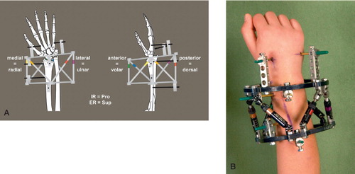
Frame assembly
The TSF was assembled with two 105‐mm full rings, selecting the proximal ring and the proximal fragment as reference (). The reference ring must be placed orthogonal to the proximal reference fragment (proximal radius). In addition, one can use the distal ring and fragment (distal radius) as the reference; however, the orthogonal placement of the distal reference ring to the short distal radius fragment is more difficult. For ring fixation, Rancho Cubes and 4.0‐mm half pins were used for the radius. Two‐angled half pins were used in each segment. The wrist was transfixed during the lengthening to avoid radiocarpal impaction. Thus, 3.0‐mm half pins were used at the base of the second and third metacarpals ().
Osteotomy
A multiple drill‐hole osteotomy was performed in the metaphyseal bone. We used a small dorsal approach to the radius with a subperiosteal preparation.
Distraction
Distraction at the osteotomy site was initiated on day 5, with 0.5–1.2 mm per day, and divided into 2 phases over 24 h. The progress of correction was followed with radiographs. Additional low‐dose CT scans were used for better visualization of the DRUJ and its reduction ( and ). At the end of the correction, we locked the frame with threaded rods.
Patients
2 boys (patient 1, 13 years old, and patient 2, 14 years old) were seen in our clinic with progressive pseudo‐Madelung deformities after an epiphysial fracture of the distal radius at age 12. They complained of radial deviation of the wrist () and restricted pronation and supination (Table). There were no neurovascular deficits, normal pinch and grasp, and full range of motion of all finger joints. In both patients, there was pain but no clicking during rotation in the dorsal dislocated DRUJ and ulnocarpal joint. Skeletal maturity (RUS, TW3 method) corresponded to the patient's age ( and ).
Figure 2. Patient 1. A and B. Pseudo‐Madelung deformity of the right distal radius after physial injury and closed radial growth plate. At the time of correction, the ulnar growth plate was in stage G according to the Tanner and Whitehouse scale. C and D. After deformity correction, with an almost normal functioning wrist and anatomically reduced DRUJ 1 year after correction.
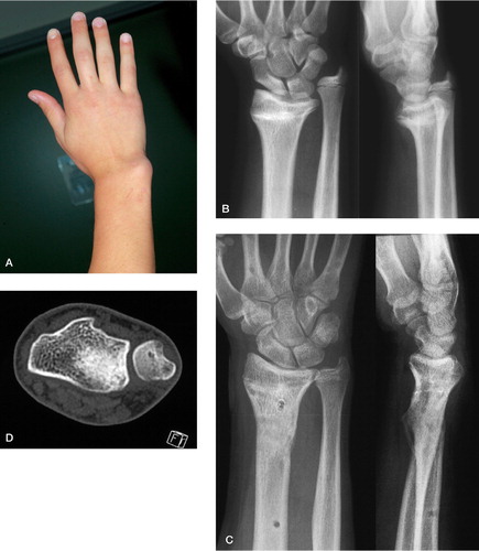
Figure 3. Patient 2. Before (A) and after (B) correction of the distal radius deformity of the right forearm. The radial growth plate was partially closed (> 50%) at the time of correction and the ulnar growth plate was at stage G according to the Tanner and Whitehouse scale. The DRUJ was reduced anatomically in the low‐dose CT at the end of correction (C).
Results
The multiplanar deformitiy of the distal radius could be corrected anatomically with the Taylor spatial frame in both patients ( and , and and ). No frame changes or frame modifications were necessary for deformity correction. The distraction time, the time until frame removal, and the healing index of each patient are listed in . Patient 2 was slightly overcorrected because of some growth in the distal ulnar growth plate (). During the distraction, each patient had 2 low‐dose CT scans for better visualization of the radiocarpal and radioulnar joints. The web‐based planning program was adjusted twice until total deformity correction was achieved. No further immobilization after frame removal was required. The 1‐year follow‐up showed an anatomically aligned forearm/hand relation with increased pronation and supination compared to the preoperative range of motion (). The wrist and especially the DRUJ were stable and reduced at the follow‐up examination ( and ). The patients did not complain about any pain or functional deficits in the hand.
Radial deformities, distraction data and ROM
Discussion
The overall goal for surgical procedures in posttraumatic deformities of the distal radius with shortening and dislocation of the DRUJ is to restore the anatomic relationship between the distal radius and ulna, and between the distal radius and carpus. Several operative procedures have been proposed to realign the relation between forearm and hand. Acute correction of the distal radius with iliac bone graft interposition and plate fixation is a commonly used and accepted procedure (Hove and Engesaeter Citation1997, Waters et al. Citation1997). The main disadvantages of this acute procedure are the need for an iliac bone graft with donor site morbidity, requirement for operative hardware removal, and radiocarpal impaction due to acute lengthening. Furthermore, it is difficult to control the proper amount of correction intraoperatively.
Gradual corrections using callus distraction with external unilateral or Ilizarov ring fixators have been used more frequently in severe forearm shortening, especially in children with Madelung deformities (Villa et al. Citation1990, Bader and Grill Citation2000, Houshian et al. Citation2004, Hosny Citation2005, Matsuno et al. Citation2006). The gradual correction of deformities allows soft tissue to adapt slowly to the lengthened bone. The correction of multiplanar deformities with unilateral or standard Ilizarov ring fixators is time consuming because of frequent frame modifications. By transferring the nomenclature of the lower limbs to the forearm, we applied the TSF for the treatment of radial deformities. Our 2 patients with pseudo‐Madelung's deformities were corrected by one operation with gradual correction of the rotational, angulational, and translational component of the deformity using simultaneous lengthening. The DRUJ was reduced gradually to its anatomic position and confirmed by low‐dose CT scans, and the range of motion for prosupination was increased to near‐normal range. However, reducing the DRUJ and increasing the limited prosupination can increase the risk of secondary degenerative changes when some incongruence is left in the DRUJ after correction. However, the 1‐year follow‐up CT scan in patient 1 did not show any joint narrowing or incongruence. Patient 2 did not accept a CT scan at the 1‐year follow‐up. The mechanical accuracy of the TSF by manual adjustment of the 6 struts during full multiplanar deformity correction has been measured to be within 0.7° and 2 mm (Paley Citation2002). This high accuracy and also low‐dose CT scans are crucial for reduction of DRUJ anatomically and for achievement of a good range of prosupination.
In conclusion, the power of the Taylor spatial frame, with the ability to move 2 fragments precisely, can be transferred to the forearm. This permits correction of multiplanar radial deformities simultaneously without any need for frame modifications of rotational and translational deformities, which is necessary with the standard Ilizarov system.
Contributions of authors
DS: patient care, surgery, and preparation of the manuscript; JG: patient care and follow‐up investigations; GM: initiator of the procedure; MG: surgery.
No competing interests declared.
- Abe M, Shirai H, Okamoto M, Onomura T. Lengthening of the forearm by callus distraction. J Hand Surg (Br) 1996; 21(2)151–63
- Bader B, Grill F. Ulnar lengthening in osteochondroma (multiple cartilagenous exostoses) of the forearm. Hand-chir Mikrochir Plast Chir 2000; 32(5)321–7
- Bailey D A, Wedge J H, McCulloch R G, Martin A D, Bern-hardson S C. Epidemiology of fractures of the distal end of the radius in children as associated with growth. J Bone Joint Surg (Am) 1989; 71(8)1225–31
- Eidelman M, Bialik V, Katzman A. Correction of deformities in children using the Taylor spatial frame. J Pediatr Orthop B 2006; 15(6)387–95
- Hosny G A. Forearm Lengthening. J Orthopaed Traumatol 2005; 6: 132–7
- Houshian S, Schroder H A, Weeth R. Correction of Made-lung's deformity by the Ilizarov technique. J Bone Joint Surg (Br) 2004; 86(4)536–40
- Hove L M, Engesaeter L B. Corrective osteotomies after injuries of the distal radial physis in children. J Hand Surg (Br) 1997; 22(6)699–704
- Kramhoft M, Bodtker S. Epidemiology of distal forearm fractures in Danish children. Acta Orthop Scand 1988; 59(5)557–9
- Lee B S, Esterhai J L, Jr, Das M. Fracture of the distal radial epiphysis. Characteristics and surgical treatment of premature, post-traumatic epiphyseal closure. Clin Orthop 1984, 185: 90–6
- Matsuno T, Ishida O, Sunagawa T, Suzuki O, Ikuta Y, Ochi M. Radius lengthening for the treatment of Bayne and Klug type II and type III radial longitudinal deficiency. J Hand Surg (Am) 2006; 31(5)822–9
- Paley D. Six-axis deformity analysis and correction. In: Principles of deformity correction, D Paley. Springer, Berlin, Heidelberg, New York 2002
- Pennig D, Gausepohl T, Mader K. Radio-radial external fixation for correction of malunited distal radius fracture. Handchir Mikrochir Plast Chir 1999; 31(4)227–33
- Rozbruch S R, Fragomen A T, Ilizarov S. Correction of tibial deformity with use of the Ilizarov-taylor spatial frame. J Bone Joint Surg (Am) (Suppl 4) 2006; 88: 156–74
- Villa A, Paley D, Catagni M A, Bell D, Cattaneo R. Lengthening of the forearm by the Ilizarov technique. Clin Orthop 1990, 250: 125–37
- Waters P M, Van Heest A E, Emans J. Acute forearm lengthenings. J Pediatr Orthop 1997; 17(4)444–9