
ABSTRACT
Percutaneous coronary intervention for bifurcated anatomy, particularly at the proximal left coronary artery site, requires guide catheters (GC) of at least 6 french and preferably larger in diameter. We describe a new trans-radial approach more suitable for small artery size: the simultaneous use of both radial arteries for double cannulation of the LMCA with 5F GC: each GC will target either the LM/LAD or the LM/CX artery (or LM-LAD/LM-LAD-1st diagonal branch) stenoses. The technique successfully was applied to 5 cases. When the technique was used for distal left main coronary artery stenoses (3 cases), a special crogss-like configuration obtained when guide catheters, coronary wires and balloons kissed was observed.
Bigger is better ? Think …twice!
Introduction
Left main coronary artery (LMCA) disease is associated with poor outcomes if not revascularized. Consistently good clinical results for LMCA disease have been obtained with coronary artery bypass grafting (CABG) surgery. Percutaneous LMCA interventions may be a reasonable alternative in cases where open chest surgery risk is prohibitive (Citation1,Citation2).
Unprotected LMCA disease is one of the most challenging situations faced by interventional cardiologists. In many instances patient is critically ill, needs fast relief of ischemia and is unable to tolerate further ischemic burden, even for a short period of time. Lesions in unprotected LMCA disease are technically complex: careful attention has to be paid for seating safely the guide catheter (GC), particularly in case of ostial stenoses, and to avoid additional vessel injury during cannulation as well as materials insertion/withdrawal. As for any bifurcation lesions, side branch angulation added to lesion eccentricity may lead to serious difficulties in wiring one or more branches of the LMCA. Short LMCA associated with severe proximal LAD artery disease when involving a large first diagonal branch affords the same level of complexity and clinical severity as a distal LMCA disease. In these situations, the usual presence of large vessels necessitates the use of large-sized balloons and stents, and frequently simultaneous use of several stents/balloons or additional imaging techniques/materials including intravascular ultrasound (IVUS) or optical coherence tomography (OCT). Large-or very large-bore GCs (≥7F) are used for addressing such lesions. However, the use of large catheters implies the need for a large vascular access and the femoral route. Transradial access (TRA) is considered a safer route for percutaneous coronary intervention (PCI) (Citation3–Citation5) but its use is limited by the radial artery size. Owing to the development of dedicated materials (e.g., Sheathless Eaucath GC from Asahi, Japan) and techniques, it is now possible to push the limit of TRA such that 7F GCs can be advanced and manipulated through TRA (Citation6). Nevertheless, not all radial arteries will accept such interventions: some cannot be cannulated using these large-bore catheters at the time of the procedure and others will occlude thereafter.
We hypothesized that the dilemma of the need for a large-bore GC and the desire to reduce the burden on the vascular access site may be solved through the use of a double wrist artery access (radial or ulnar) and double 5F GC technique: this results in a potential combined lumen size of 10F and each GC will target separately either the LM-LAD artery or the LM-CX artery (or LM-LAD artery/LM-1st Diagonal branch). The “double wrist-double 5F GC technique for PCI” was applied to 5 consecutive cases of distal unprotected LMCA disease (3 cases), LMCA equivalent disease (1 case) and one proximal (+mid) LAD artery diseased involving a large first diagonal branch. We had to report an unexpected finding when addressing the distal LMCA in such a way.
Methods
Patient selection
The DW-DG TRA-PCI was considered for patients
(a) With critical proximal LCA disease thought to require kissing technique and large materials/large GCs for addressing bifurcation anatomy;
(b) With critical medical condition favouring PCI over CABG surgery (and favouring transradial vascular approach for minimizing the bleeding/procedural related risk);
(c) With small body size or difficult vascular access site or medical condition favouring radial approach.
Procedures and material selection
Both radial (or ulnar) arteries were cannulated with 5FS Glidesheaths from Terumo Inc.
The LCA was cannulated with usual 5F GC favouring a dedicated radial curve (Ikari Left 3.5) for the right wrist and a different shape (EBU 3.5) for the left wrist. The EBU 3.5 was typically positioned first with the intention to directly wire the larger vessel. The Ikari Left 3.5 GC was thereafter positioned for wiring the second vessel. Depending of the GC at works, each GC was disengaged/re-engaged;
Dye injection possibility and pressure control was continuously maintained for both GC.
Standard 0.014”coronary wires, buddy wiring or multiple wiring techniques when requested were used;
There was no size restriction for catheter-balloons as 5F GCs accept sizes up to 5.0 mm (one catheter-balloon per GC);
There was little restriction for stent’s sizing: 5F GCs accept 5 mm bare metal stents and 4.0 mm DES (one stent delivery system for each GC). 4.0 mm DES may be delivered with the presence of a buddy wire. 3.0 mm BVS from Abbott may be delivered in a 5F GC (without buddy wiring);
Provisional stenting was the by default strategy;
Heparine was used (8.000 to 10.000 UI) with provisional Tirofiban (high bolus dose + i.v. infusion post PCI [4 h]).
Usual procedural variables were recorded (fluoroscopy time, volume of contrast, dose area product) and usual laboratory tests following PCI were performed (EKG, CPK-Mb, Troponine, creatinine, hemoglobin levels);
Prolonged dual anti-platelet medication was prescribed.
TR-Band compression device was used for hemostasis on both wrists.
Outcome
Success was defined as angiographic success on all planned lesions without conversion to larger GC or another arterial access and clinical success was defined as relief of the critical medical condition, discharge from the hospital and more than 3 months of sustained good clinical results, without evidence of recurrent ischemia, new myocardial infarction, new episode of congestive heart failure requiring hospitalization. Occurrence of bleeding and vascular access problems as need of blood transfusions were verified until hospital discharge.
Results
Since October 2015, 5 patients () met the inclusion criteria: critical medical conditions: severe COPD (Citation3), severe aortic valve stenosis (Citation1), advanced age (Citation3) and frailty (Citation4) were associated with severe proximal LCA disease: 3 distal LM diseases (, , ), one LM equivalent disease () and one proximal LAD artery disease involving a large first diagonal branch (). Angiographic success and hospital discharge without complication were obtained for all patients. There was no need for conversion to larger GC or another arterial access. No bleeding nor vascular problems occurred. Re hospitalization happened for 2: the first patient for acute pneumonia at 1 month, medically managed, and the fifth one patient was hospitalized at week 3 for recurrent heart failure; she was also managed medically and recovered fully (class 1 at 6 month).
Table 1.
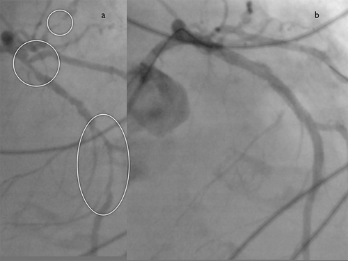
Figure 1. (a) Case 1. Caudal right anterior oblique (RAO) coronary angiograms obtained in 2005 and 2015 revealing the appearance of a severe Medina 1,1,1 LMCA lesion. Result after provisional stenting. (b) Case 1. A triple X-kiss pattern is discovered at the balloons, Wires and Guiding catheters levels. Balloons and wires X-kissing was unexpected.
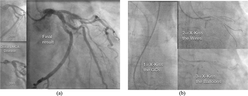
Figure 2. (a) Case 2. Caudal RAO and cranial LAO views before (a, b) and after stenting (c,d) a severe Medina 1,1,1 LMCA lesion, CX artery is a small-sized vessel. (b) Case 2. The triple X-kiss pattern (GCs, Wires and balloons) is again observed.
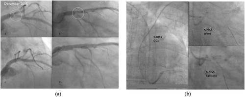
Figure 3. (a) Case 3. Cranial and caudal RAO views of a severe Medina 1,1,1 LMCA lesion before (a, b) and after T stenting (c, d). Early emergence of the first OM branch from the proximal CX artery and the large size of the vessels can be observed. View of the final X-kiss balloon inflation after T stenting (e). (b) Case 3. Triple X-kiss pattern again present. Triple wiring (LAD artery, CX artery, and first OM branch) was used for the trifurcated distal LMCA lesion.
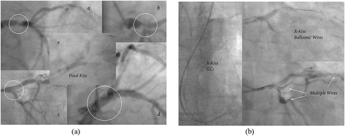
Figure 4. Case 4. Caudal RAO views before and after LAD/CX artery stenting. Final kiss balloons view (“V-Pattern”), only the GCs X-kissed.
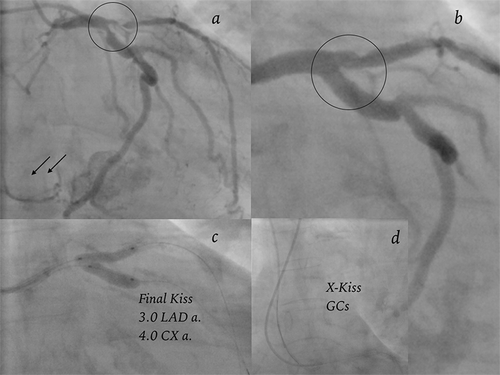
Figure 5. Case 5. Cranial RAO (40–10°) view of LCA, before and after distal, proximal and mid LAD artery stenting (P O T for LAD/Diag 2 and LAD/Diag 1 arteries).
Provisional stenting allowed a one stent strategy for 3 cases. T stenting was used for the third patient and the distal LM equivalent (patient 4) required stenting on both branches. A distal LAD artery stenting was added for patient 4. Fluoroscopic time and use of contrast was well in the range of usual complex PCI, all vascular access were worry free.
Interestingly, we observed a special cross-like configuration (“X-kiss”) obtained when guide catheters, coronary wires and balloons kissed for the 3 patients with distal LM disease (, ), )). For the remaining two patients, the “X-Kiss” pattern was only observed at the guiding catheters level.
All patients are alive more than 6 months after the index procedure, the LV function is notably improved for the last two cases and the severity of the aortic stenosis of patient 3 was re assessed as moderate and surgery was dismissed. Cases description is added in appendix.
Discussion
We report two findings from our experience.
First, all our 5F GC driven procedures were successful. It includes the usual ease of management regarding the vascular access site, thanks to the reduced sized of the 5F Terumo Glidesheath introducers (virtual 4F). As current 5F GCs allow the passage of bare-metal stents as large as 5 mm (Citation7,Citation8), well sized stents were correctly deployed at the planned position by using the recommended techniques for bifurcation management (Citation9,Citation10). Final kissing balloon (KB) involving large balloons was performed (as in case 4 when a 3.0 and a 4.0 mm balloon were used for a final cumulated size of at least 7.0 mm). Of course, our cases would have been successfully performed through one transfemoral access using one large bore GC but at the expense of a higher risk of serious vascular access complications (see patient selection).
Having a dual access to the left main provides some advantages without compromising the patient safety: in every case, at least one vessel was immediately wired as soon as one GC was in place and secured the LMCA access. Having a second GC in position helped for the second wire crossing by allowing use of different techniques. As anticipated with bifurcation and complex lesions, the wiring step was tricky, with half of the fluoroscopy time for case 1 being spent on wiring. Such difficulty would not have been avoided using a single large GC. Contrast injections were mainly performed through only one GC. The different balloons and stent-catheters moved freely. Movement was easier than when all of the material is forwarded through a single large GC. We do not know if cannulation and prolonged wires/balloons manipulation within the LMCA is less or more risky when using one single large GC versus two smaller sized GC alternatively engaged. These techniques should anyway be reserved to highly skilled operators.
Working with two different GC curves provided the benefit of a different angulation when reaching the coronary ostium. An EBU curve was used starting from the left arm, while a radial dedicated curve (Ikari) was positioned from the right arm. At least one GC was disengaged as soon as the material was delivered at the right position. We never encountered pressure damping or slow flow and no additional damage occurred at the LMCA level because of the double GC. Starting from the left radial access did not present any difficulties, as this route is routinely used (Citation11). Of course, the technique doubles the problem of LCA cannulation.
A double guiding, double vascular access was required at the origin of the Kissing Balloon technique. A series of 52 cases using bi-femoral or brachio-femoral is reported in 1986 (Citation12). The KB technique has quickly evolved to a single catheter-dual wires/balloons thanks to advancing technology in catheter-balloon miniaturization. Shing-Hsien Chou and Al (Citation13) reported 2 cases of “double GC for complex PCI”, using the second vascular access for a 6F GC in a bailout situation: a LM ostial CTO through femoral 7F then left radial 6F CG for final KB, first case. The second case concerned a LM equivalent disease, left radial (6F) was firstly utilized for balloon angioplasty, then right radial (6F) for LAD-CX artery kissing stenting. They did not planned electively to address specifically PCI of distal LM disease with two small sized GC/two radial access and do not reported an X-kiss figure. Eichhofer, Pastry and Fraser (Citation14) described use of a second radial access/GC in a bailout situation for addressing a distal LAD artery dissection involving a large diagonal branch and they performed a conventional crush stenting.
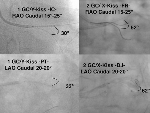
The second finding was “ the X-kiss configuration” . This pattern was observed at three levels: the GCs (expected with a dual wrist access) and at the unexpected levels of the coronary wires and balloons (, ), )). The X-kiss configuration is particularly informative about the potential benefit of addressing distal LMCA stenosis through double wrists double GC. The LMCA anatomy indeed appears more often as a “T” than as a “V-branching” system. Using one GC for each of the side branches allows the catheter’s tip, the wire and the balloon catheter to lie coaxially with that vessel, which led to the observed X figure. A V-kiss pattern is more likely to result from the use of only one GC, particularly if the left main stem is short (). Due to the hemodynamic shear stress (Citation15) and the fractal geometry of arterial coronary bifurcation (Citation16), the burden of atherosclerosis is more extensive at the outer walls of vessels bifurcations. This has been confirmed by pathological findings (Citation17) and intravascular ultrasound studies (Citation18,Citation19). The same shear stress also plays a role during the healing process after stenting.
Figure 6. X versus Y-kiss at the wires level: 1 Guiding Catheter versus 2 Guiding Catheters in LAO and RAO caudal views. Y-kiss wires views are extracted from records of previous single access TRA cases of distal LMCA PCI.
During balloon inflation, the X-kiss configuration could potentially translate into a different distribution of forces against the atherosclerotic plaques particularly at the outer and lateral sites: there is a potential for a different carina shift during balloon angioplasty.
The extensively studied (Citation20,Citation21) after stenting conventional KB technique produces a significant over-expansion of the stent at the MV just in front of the SB (elliptic shape) and corrects only partially the strut apposition (Citation22). It is possible that in the same way as an X-Kiss balloon inflation before stenting may result in a better plaque disruption at its most developed site, the X-Kiss inflation after stenting may alter differently the stent(s) geometry at the SB origin with a better stent coverage as compared to the actual single GC KB technique. It may result in a more anatomically correct reconstruction. These possibilities should be confirmed by IVUS or OCT imaging or by bench study. The wires and balloons did not exhibit the X-kiss pattern in case 4 and 5 (LMCA equivalent disease and proximal LAD artery/1st diagonal artery) perhaps due to the long length of the LM stem or to the simultaneous double wiring of the LAD artery (case 4). In such cases, the location of atherosclerotic plaques is similarly handled by the way of conventional single GC kissing balloon and kissing stenting but at the expense of a larger vascular access.
Conclusion
We describe the use of a “slender” double wrist vascular access with double 5F GCs for the treatment of severe distal unprotected LMCA disease and of LM equivalent or proximal LAD/diagonal artery disease. For distal LMCA disease, an X-kiss configuration resulted from the wiring and ballooning of each branch of the LMCA with a different GC. This unique pattern could potentially lead to a more anatomically respectful rebuilding of the LMCA by the kissing balloon inflations performed before and after stenting. The use of smaller sites for vascular entry at the expense of starting simultaneously from both wrists may result in a “bigger and better” solution than the current practice of using a unique large-bore GC. Moreover, the described technique may extend the benefits of LMCA TRA PCI for patients previously recused due to small vascular access.
References
- Windecker S, 2014 ESC/EACTS Guidelines on myocardial revascularization. Eur Heart J. 2014 Aug;35(37):2541–619. doi:10.1093/eurheartj/ehu278. AQ5
- Teirstein PS, Price MJ. Left main percutaneous coronary intervention. J Am Coll Cardiol. 2012;60:1605–12. doi:10.1016/j.jacc.2012.01.085.
- Jolly SS, Mehta SR. Coronary intervention: radialartery access comes of age. Lancet. 2015 Jun 20;385. doi:10.1016/S0140-6736(15)60507-4.
- Sunil VR, Kedev S. Approaching the post-femoral era for coronary angiography and intervention. JACC. 2015;8(4):524–26.
- Dangoisse V, Guédès A, Gabriel L, Jamart J, Chenu P, Marchandise B, Schroeder E. Full conversion from transfemoral to transradial approach for percutaneous coronary interventions results in a similar success rate and a rapid reduction of in-hospital cardiac and vascular major events. EuroIntervention. 2013 Jul;9(3):345–52. doi:10.4244/EIJV9I3A56.
- Patel T, Shah S, Pancholy S. “Combo” technique for the use of 7F guide catheter system during transradial approach. Circ Cardiovasc Interv. 2015;86:1033–40. doi:10.1002/ccd.v86.6.
- Dangoisse V. Slender TRA-PCI are back-up improving techniques dependent. J Cardiol Curr Res. 2015;3(6):00126. doi:10.15406/jccr2015.03.00126.
- Kiemeneij F, Yoshimachi F, Matsukage T, Amoroso G, Fraser D, Claessen BE, Saito S. Focus on maximal miniaturisation of transradial coronary access materials and techniques by the Slender Club Japan and Europe: an overview and classification. EuroIntervention. 2015 Feb;10(10):1178–86. doi:10.4244/EIJY14M09_09.
- Darderont O, Leymarie JL, Lefevre T, Albiero R, Mortier P, Louvard Y. Technical aspects of the provisional side branch stunting strategy. EuroIntervention. 2015;11:V86–V90. doi:10.4244/EIJV11SVA19.
- Chen S-L, Zhang Y, Xu B, Ye F, Zhang J, Tian N, Liu Z, Qian X, Ding S, Li F, et al. Five-year clinical follow-up of unprotected left main bifurcation lesion stenting: one-stent versus two-stent techniques versus double-kissing crush technique. EuroIntervention. 2012;8:803–14. doi:10.4244/EIJV8I7A123.
- Guedes A & Dangoisse V, Gabriel L, et al. Low rate of conversion to transfemoral approach when attempting both radial arteries for coronary angiography and percutaneous coronary intervention: a study of 1,826 consecutive procedures. J Invasive Cardiol. 2010 Sept;22(9):391–97.
- Sgueglia Gregory A, Bernard C. Kissing Balloon inflation in percutaneous coronary interventions. J Am Coll Cardiol Intv. 2012;5:503–11.
- Chou S-H, Lin C-P, Lin Y-C, Kuo C-T, Lin M-S, Chang C-J. Double guiding catheters for complex percutaneous coronary intervention. Tex Heart Inst J. 2012;39(1):112–15.
- Eichhöfer J, Sastry S, Fraser D. Transradial conventional crush stenting with double guiding catheter technique. EuroPCR; 2008. pcronline.com/EuroIntervention.pcronline.com/Lectures/2008.
- Malek AM, Alper SL, Sligo I. Hemodynamic shear stress and its role in atherosclerosis. Jama. 1999;282:2035–42. doi:10.1001/jama.282.21.2035.
- Finit G, Gilard M, Perrenot B, Rioufol G, Motreff P, Gavit L, Prost R. Fractal geometry of arterial coronary bifurcations: a quantitative coronary angiography and intravascular ultrasound analysis. EuroIntervention. 2007;3:490–98. doi:10.4244/EIJV3I4A87.
- Nakazawa G, Yazdani SK, Finn AV, Vorpahl M, Kolodgie FD, Renu V. Patological findings at bifurcation lesions. The impact of flow distribution on atherosclerosis and arterial healing after stent implantation. J Am Coll Cardiol. 2010;55(16):1679-87.
- Oviedo C, Machara A, Mintz GS, Araki H, So-Yeon C, Tsujita K, Kubo T, Doi H, Trample B, Lanky AJ, et al. Intravascular ultrasound classification of plaque distribution in left main coronary artery bifurcations. Where is the plaque really located? Circ Cardiovasc Interv. 2010;3:105–12. doi:10.1161/CIRCINTERVENTIONS.109.906016.
- Medina A, Martin P, de Lezo JS, Novoa J, Melian F, Hernandez E, de Lezo JS, Pan M, Burgos L, Amador C, et al. Ultrasound study of the prevalence of plaque at the carina in lesions that affect the coronary bifurcation: implication for treatment with provisional stent. Rev Esp Cardiol. 2011;64(1):43–50. doi:10.1016/j.recesp.2010.07.006.
- Foin N, Sayan S, Allegria E, Petardo R, Sukh N, Francis DP, Di MC, Davies JE. Maximal expansion capacity with current DES platforms: a critical factor for stent selection in the treatment of left main bifurcation? EuroIntervention. 2013;8:1315–25. doi:10.4244/EIJV8I11A200.
- Guérin P, Pilet P, Finet G, Gouëffic Y, N’Guyen J-M, Crochet D, Bijou D, Pacaud P, Loirand G. Drug-eluting stents in bifurcations, bench study of strut deformation and coating lesions. Circ Cardiovasc Interv. 2010;3:120–26. doi:10.1161/CIRCINTERVENTIONS.108.846089.
- Foin N, Secco GG, Ghilencea L, Krams R, di Mario C. Final proximal post-dilatation is necessary after kissing balloon in bifurcation stenting. EuroIntervention. 2011;7:597–604. doi:10.4244/EIJV7I5A96.