
ABSTRACT
Cancer is one of the most common health problems responsible for outnumbered deaths worldwide. Nanomedicine plays an important role in developing alternative and more effective treatment strategies for cancer theranostics. However, the toxicity, high cost and nanoparticles (NPs) production complexity are some of the major issues that obstruct the use of existing nanomedicine. Recently, the green synthesis of biogenic NPs from plants and microbial sources has become an emerging field due to their safer, eco-friendly, simple, fast, energy efficient, low-cost and less toxic nature. Interestingly, NPs play a key role in diagnosis of tumor at the initial stage by allowing cellular visualization. Furthermore, prospective applications of green NPs include magnetically responsive drug delivery, anti-cancer activity, photo-thermal therapy and bio-imaging. The present review provides perspective on the use of anti-cancer green bionanomaterials with a focus on their present status and future prospects in the theranostics of cancer.
GRAPHICAL ABSTRACT
1. Cancer: a global menace
According to the National Cancer Institute (NCI), United States (U.S), cancer contains over more than 100 types Institute (Citation1). Besides, it is anticipated that the incidence of cancer by 2030 will reach around 21.7 million and there would be about 13 million deaths owing to aging and population growth. The latest report published in June 2016 by iMShealth (Institute for Healthcare Informatics) anticipates the global cancer treatment market to reach $150 Billion by 2020 Informatics (Citation2). Nevertheless, the tumor load in the future is likely to be considerably larger due to lifestyle adoption, leading to an increased risk of developing tumor, such as physical inactivity, cigarette smoking and unhealthy diet in the economically developing countries’ society (Citation3). Notably, cancer causes one in seven deaths globally, causing more deaths than malaria, tuberculosis and AIDS Society (Citation3 , Citation4). Impressively, the largest proportion of cancer cases is in developing countries, accounting for around 57% of new cases and 65% of cancer-related deaths. In 2012, cancer caused premature deaths of about 4.3 million people and the number of premature cancer deaths are presumed to increase by 44% between 2012 and 2030 Organization (Citation5). By 2025, it is estimated that in the industrialized world only 20–25% of the people will be aged over 65 years, and of those 50–60% who die of tumor will be aged over 75 years (Citation6). On 1 January 2016, over 15.5 million of the population of the United States had a previous history of cancer disease. By 1 January, 2026, this number is estimated to increase to 20.3 million. Moreover, the high prevalent cancers in 2016 that affects women are uterine corpus (757,190), breast (3,560,570), and colon and rectum cancer (727,350). For men, prostate (3,306,760), melanoma (614,460), and colon and rectum (724,690) cancers are most common (Citation7). Additionally, nearly 189,910 new cancer patients and about 69,410 cancer-related deaths were estimated among black people in 2016 in the United States, involving 95,920 cases among women and 93,990 cases in men. Although black people have higher death rates due to cancer than whites, the discrepancy has lessened for all types of cancer combined and for prostate and lung cancers (in males only).
Radiotherapy, chemotherapy and surgery are some of the cancer treatments which are used to improve a patient’s life. Besides, one of the major problems in the cancer treatment process is the side-effects owing to conventional treatment strategies (Citation8–10). Recent cancer research findings have enhanced understanding about carcinogenesis, the metastatic cascade and genetic factors that influence tumor growth and development. Overall, despite some progress in cancer control, incidence and death rates are increasing for cancer types. Recently, the applications of nanobiotechnology have revealed novel strategies for the treatment and diagnosis of cancer. Thereby, the current review article aims to highlight significant applications of biosynthesized green nanomaterials in cancer theranostics and the hurdles in their way to clinical trials.
2. Green bionanomaterials: an insight
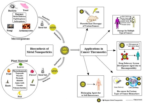
Green bionanomaterials involving metals such as gold, silver, copper, titanium, zinc and iron prepared from different bio-sources, and have been reported for various biomedical applications (Citation11). In addition, metal NPs are used in drug delivery, gene delivery, medicine, cell labeling, sensors, food packaging, wound dressings, etc. However, some applications of metal NPs are still under development such as magnetically responsive drug delivery, photo-thermal therapy and photo-imaging () (Citation12–14). Although fabrication of NPs has attracted great interest for various physical and chemical processes, but there is an urgent need to explore alternative routes owing to some unsatisfying conditions of these methods such as the need for high temperature in the thermo-reductive process or intensive energy in the laser ablation process (Citation15). Moreover, the fabrication of NPs by a chemical process may require toxic substrates, generates harmful wastes or requires large amounts of energy and even has low productivity (Citation16). Hence, natural products and resources may ultimately prove to be more efficient and cost-effective. Remarkably, many plant extracts, algae and an extensive range of microorganisms containing bacteria, actinomycetes, fungi and yeast are reported to be proficient in synthesizing bionanomaterials, Priester et al. (Citation17), Barabadi et al. (Citation18), Honary et al. (Citation19), Rahimi et al. (Citation20), Salunke et al. (Citation11), Ovais et al. (Citation21), Mukherjee et al. (Citation22), Patra et al. (Citation23) and Ovais et al. (Citation24). Interestingly, isolated chloroplasts were reported to be able to catalyze the biosynthesis of bionanomaterials. More interestingly, microalgae were designed for ecofriendly, scalable and permanent photobioreactors for sustainable and continuous fabrication of valuable bionanomaterials (Citation15). Notably, the exact mechanism of biosynthesis of metal NPs is not well understood yet (Citation25). However, it is believed that some enzymes play a role in bio-reducing metal ions to form metal NPs (Citation26, Citation27). shows the schematic mechanism of microbial-mediated synthesis of metal NPs (Citation11). The negatively charged bacterial cell wall interacts electrostatically with metal ions having positive charge. In addition, the bioreduction process may be catalyzed by enzymes inside the cells or on the cell surface. Salunke et al. (Citation28) suggested the role of Saccharomyces cerevisiae cell wall components, some proteins and alcoholic compounds in the synthesis and stabilization of MnO2 NPs (Citation28). The mechanism of extracellular microbial-mediated biofabrication of metal NPs is basically due to microbial nitrate reductase responsible for reduction of metal ions into metallic NPs (Citation11). All the mentioned mechanisms lead to extracellular or intracellular fabrication of metal NPs. In case of phytosynthesis of metal NPs, it is believed that phytochemicals such as proteins, flavonoids, polyphenols, alkaloids, saponins, phenols, essential oils and polyols which are present in the plants extract play a main role in bio-reducing the metal ions, converting them to metal NPs and also capping of the synthesized NPs for stabilization (Citation13, Citation29–31). The plant-mediated mechanism of biogenic silver NPs’ (AgNPs’) production is illustrated in .
Figure 1. Biological synthesis and applications of metal NPs in biomedical and environmental fields (Adapted with permission from Singh et al. (Citation12)).
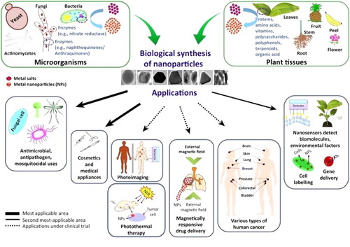
Figure 2. Schematic mechanism of microbial-mediated synthesis of metal nanoparticles (Adapted with permission from Salunke et al. (Citation11)).
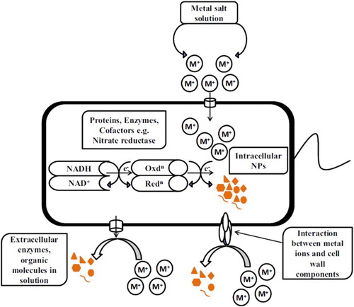
Figure 3. Plant-mediated mechanism of biogenic silver nanoparticles’ (AgNPs’) production: a step forward for cancer nanomedicine (Adapted with permission from Ovais et al. (Citation21)).
2.1. Anti-cancer activity of green nanomaterials: A mechanistic approach
In vitro anti-cancer activity of metal bionanoparticles has been confirmed against different cancer cell lines, i.e. MDA-MB-231 and MCF-7 (human breast adenocarcinoma) (Citation32, Citation33), A549 (human lung adenocarcinoma) (Citation34, Citation35), HCT116 (human colon colorectal carcinoma) (Citation36), HeLa (human cervical cancer) (Citation37, Citation10, Citation38), NCI-H460 (non-small cell lung cancer) (Citation39), U87 (glioblastoma multiforme cell) (Citation40), PANC-1 (pancreatic adenocarcinoma) (Citation41), MG-63 (osteosarcoma cell) (Citation42, Citation43), AGS (human gastric carcinoma) (Citation44, Citation45), SMMC-7721 (human hepatoma cells) (Citation46), LS174T (human colon adenocarcinoma cell) (Citation46), HT-29 (human colorectal adenocarcinoma) (Citation47), Varsha B (Citation48), Caco-2 (human epithelial colorectal adenocarcinoma) (Citation47, Citation49), HCT 15 (human colon adenocarcinoma) (Citation50, Citation51), PC-3 (human prostate carcinoma) (Citation52, Citation53), U937 (human histiocytic lymphoma cell) (Citation54), COLO205 (human colon adenocarcinoma) (Citation41, Citation54), B16F10 (mouse melanoma) (Citation54, Citation55), 4T1 (mouse breast cancer) (Citation56), CRL-1451 (mouse lung adenocarcinoma) (Citation56), EAC (ehrlich ascites carcinoma) (Citation57), CT-26 (mouse colon adenocarcinoma) (Citation56), WEHI-3B (mouse leukemia) (Citation56), CEM-ss (human T acute lymphoblastic leukemia) (Citation58), CaOV-3 (ovarian adenocarcinoma) (Citation41), Jurkat (human T acute lymphoblastic leukemia) (Citation58, Citation59), HL-60 (human acute myeloid leukemia) (Citation41, Citation60), K562 (human chronic myelogenous leukemia) (Citation58, Citation61), A431 (human vulvar squamous cell carcinoma) (Citation62), HNGC-2 (human adult glioma tissue) (Citation55), ECV-304 (human urinary bladder carcinoma) (Citation55), RAW254.7 (mouse leukemia) (Citation49), SiHa (human cervical squamous cell carcinoma) (Citation63), DL (dalton’s lymphoma) (Citation64), HepG2 (hepatic cancer) (Citation65–Citation67), HEp-2 (human larynx carcinoma) (Citation68, Citation69), ZR-75-1 (human caucasian breast carcinoma) (Citation70), DAUDI (human burkitt’s lymphoma) (Citation70), T47D (human breast cancer) (Citation71), KB (human oral cancer) (Citation72, Citation73), C26 (murine colon carcinoma) (Citation74), LoVo (human colon adenocarcinoma) (Citation75), LoVo/DX (multidrug resistance human colon adenocarcinoma sub-line) (Citation75). The anti-cancer activity of various metal bionanoparticles from different bio-sources has been listed in and from studies conducted in recent years by several research groups. Based on the enlisted studies, metal NPs exhibit a dose-dependent cytotoxic activity on various cancer cell lines. Moreover, biocompatibility testing of metal NPs has been done on different normal cell lines such as HaCaT (human keratinocyte) (Citation76, Citation77), HMEC (human mammary epithelial cell) (Citation33), HDFa (human normal skin dermal fibroblast) (Citation39), HEK 293 (normal human embryonic kidney cell) (Citation40, Citation78), Vero (african green monkey kidney cell) (Citation79, Citation80), RWPE-1 (non-malignant human prostate epithelial cell) (Citation81), PBMC (human peripheral blood mononuclear cell) (Citation82), HL-7702 (normal human liver cell) (Citation46), 3T3 (normal mice fibroblast cell) (Citation83, Citation84), HUVEC (human umbilical vein endothelial cell) (Citation55), Rat L6 (rat skeletal muscle cell) (Citation85), MDCK (canine cocker spaniel kidney cell) (Citation86), CV-1 (monkey African green kidney fibroblast) (Citation87), WI-38 (human caucasian fetal lung) (Citation87), HEK-293 (human embryo kidney) (Citation88), BHK21 (hamster syrian kidney) (Citation89), MRC-5 (normal human lung fibroblast cell) (Citation41, Citation45), Raw 264.7 (Murine macrophage cell lines) (Citation35), 3T3-L1 (mouse embryo) (Citation90), PLs (human normal peripheral lymphocytes) (Citation34), hFOB (human fetal osteoblast progenitor cell) (Citation91), MCF10A (human breast epithelial cell) (Citation71), H9c2 (rat cardiac myoblast) (Citation92), NIH3T3 (mouse fibroblast) (Citation93) and C2C12 (mouse muscle myoblast) (Citation94).
Table 1. Phyto-sources for synthesis of bionanoparticles and their anticancer activity in various cell lines for the past 5 years.
Table 2. Microbial sources for synthesis of bionanoparticles and their anticancer activity in various cell lines for the past 5 years.
According to the literature, several studies reported nontoxicity and biocompatibility of some metal NPs in normal cell lines, but high toxicity in cancer cell lines. For instance, Anand et al. (Citation95) reported notable antitumor activity of phyto-synthesized palladium NPs against A549, but no toxicity was found in human normal peripheral lymphocytes cells (Citation95). Besides, Du et al. (Citation96) reported plant-mediated synthesis and anti-cancer activity of AgNPs against Hela and A549 in the range of 1–5 µg/mL, but no significant cytotoxic effect was observed up to 10 µg/mL in normal HaCaT and also ≤ 2 µg/mL in Raw 264.7 (Citation96). Similarly, Uma Suganya et al. (Citation33) reported substantial in vitro anti-cancer activity of biosynthesized AuNPs (2–10 μg/mL) against MDA-MB-231, but no significant toxicity was found in normal HMEC in the range of 5–80 μg/mL, indicating considerable biocompatibility of NPs in normal cells (Citation33). Also, Kummara et al. (Citation97) showed very high cytotoxicity of green synthesized AgNPs against NCI-H460 at 240 ppm, but no cytotoxicity was found in normal HDFa (Citation97). Furthermore, Singh et al. (Citation98) reported not only remarkable cytotoxicity of phyto-fabricated AgNPs against A549 and HeLa above 5 μg/mL, but also no significant toxicity in normal RAW 264.7 (Citation98). Further work showed antitumor effects of bio-fabricated AgNPs (0.02–50 µg/mL) against MCF-7; however, no cytotoxic effect was found in normal human blood mononuclear cells (Citation99). In addition, Namvar et al. (Citation58) reported biocompatibility of biological AuNPs in normal human blood mononuclear cells up to 100 µg/mL, but substantial cytotoxicity against cancer cell lines including CEM-ss, Jurkat, HL-60 and K562 (Citation58). Likewise, Venkatesan et al. (Citation76) showed biocompatibility of biosynthesized AuNPs in normal HaCaT in the range of 10–50 µg/mL (Citation76). Furthermore, Yang et al. (Citation87) reported no cytotoxicity of plant-mediated synthesized AuNPs (10–160 μg/mL) in normal CV-1, WI-38 (Citation87). Although these studies depicted biocompatibility of metal NPs, some studies are not in agreement. Specifically, Majeed et al. (Citation100) reported fungus-mediated synthesis of AgNPs by using Penicillium decumbens (MTCC-2494) and assessed significant cytotoxic effects by MTT assay in the range of 20–120 μg/mL against A-549 while 50% survival was demonstrated in the normal Vero cell line (Citation100). In other study reported by Baharara et al. (Citation101), as evidenced by MTT assay, biosynthesized AuNPs inhibit proliferation of HeLa cells (IC50:100 µg/mL) and normal bone marrow mesenchymal stem cells (IC50:300 µg/mL). However, cytotoxicity of AuNPs in bone marrow were lower than cancerous Hela cells (Citation101). Likewise, Xia et al. (Citation46) affirmed stronger cytotoxic effects of biogenic AgNPs (IC50: 27.75 µg/mL) against human cancerous hepatoma SMMC-7721 cells but showed lower cytotoxic against human normal liver (HL-7702) cells (IC50: 81.39 µg/mL) (Citation46). Furthermore, Ma et al. (Citation102) reported that the biosynthesized AgNPs, using a cell-free filtrate of the fungus strain Penicillium aculeatum Su1 as a reducing agent, presented higher biocompatibility toward human bronchial epithelial cells and high cytotoxicity in a dose-dependent manner with an IC50 of 48.73 μg/mL toward A549 cells. Additionally, Kasithevar (Citation13) reported a simple and rapid synthesis of AgNPs using aqueous leaf extract of Alysicarpus monilifer mostly spherical in shape with a mean size of 15 ± 2 nm. Their study showed no cytotoxicity of AgNPs against Vero cell lines at the concentration of 200 µg/mL after 72 h of incubation. In contrast, Valli Nachiyar et al. (Citation103) reported microbial-mediated synthesis of titanium dioxide (TiO2) NPs. In their study, TiO2 NPs were found to be more toxic against normal HaCaT (IC50: 55 μg/mL) than cancerous HEp2 cell lines (IC50:172 μg/mL) (Citation103). Moreover, Lima et al. (Citation104) reported genotoxic effects of microbial biosynthesized spherical AgNPs (Average size: 40.3 ± 3.5 nm) at concentrations of 5.0 and 10.0 μg/mL (Citation104). In a study, natural anti-cancer flavone Chrysin (ChR), (5, 7-Dihydroxyflavone ChR), was used to synthesize AgNPs and AuNPs in a greener route without toxic additives. ChR strongly reduces Ag+ and Au3+ into their nano-forms with uniform size, shape and surface chemistry. In vitro anti-cancer results revealed that the prepared NPs exhibit enhanced cytotoxicity than ChR against treated two different breast carcinoma cell lines (MDA-MB-231 and MDA-MB-468) (Citation105). Besides, Rajendran et al. (Citation106) reported the highly stable flavonoid apigenin conjugated to gold nanoparticles (ap-AuNPs) are formed when apigenin reacts with Au3+ under appropriate conditions. The ap-AuNPs are also found to exhibit toxicity toward cancerous A431 cell lines, while being nontoxic toward normal epidermoid cells (HaCat). Additionally, Sahu et al. (Citation107) investigated the role of pure aqueous solution of plant secondary metabolites, namely hesperidin, naringin and diosmin, in the biosynthesis of AgNPs. The secondary metabolites have the polyhydroxy group which may be responsible for their role in the reduction of metal ions into NPs. In this study, the cytotoxicity of the synthesized AgNPs was investigated on the cancerous HL-60 cell line. The result represented that AgNPs synthesized using naringin as reducing agent had higher stability and better cytotoxic activity (Citation107). Interestingly, a fish-intestine-associated bacterial strain was a potential source of exopolysaccharide (EPS) production reported with a significant ability to reduce iron-based materials and convert them to iron oxide nanoparticles (FeONPs). EPS was extracted from a spore-forming strain of Bacillus subtilis isolated from the gut microbiome of the freshwater fish Oreochromis mossambicus. In this study, the in vitro cytotoxicity effects of free EPS and EPS-stabilized FeONPs were probed in the human epidermoid carcinoma cell line A431. The IC50 values of EPS and EPS-stabilized FeONPs were found to be 350.18 and 62.946 mg/mL, respectively (Citation108). In a study, AgNPs were biosynthesized by an aqueous leaf extract of Erythrina suberosa (Roxb.). Following that, the cytotoxicity of AgNPs was compared with plant extract and AgNO3 against the A-431 osteosarcoma cell line. The IC50 values were determined to be around 106.15, 74.02 and 136.73 μg/mL for leaf extract, AgNPs and AgNO3, respectively, indicating excellent anti-cancer activity of AgNPs among all (Citation109). Notably, the exact mechanism of metal NPs’ anti-cancer activity is not fully understood yet. However, it is believed that reactive oxygen species (ROS) generation, Sub-G1 arrest in cell, up-regulation of p53 protein and caspase-3 expression, inhibition of VEGF-induced activities are the major proposed anti-cancer mechanisms (Citation21, Citation110). Recent advances in the proposed mechanisms for anti-cancer activity shown by colloidal biogenic AgNPs are shown in . Importantly, the ROS leads to activation of caspase-3 which is responsible for cell apoptosis by arresting the cell cycle at the G2/M phase (Citation111). Besides, increased oxidative stress leads to oxidation of glutathione (GSH) to glutathione disulfide (GSSG) through the oxidation process. Notably, GSH is known as an antioxidant that prevents cells from ROS damages, consequently resulting in remarkably much MNPs’ cytotoxicity and loss of GSH (Citation24). Furthermore, AgNPs were shown to downregulate the activity of a recognized enzyme involved in DNA damage repair named DNA-dependent protein kinase (Citation112). Coccini et al. (Citation113) showed that the AgNP-induced oxidative stress genes involved Gpx1, SOD, FMO2 and GAPDH in different organs indicating AgNP-induced toxicity. Jeyaraj et al. (Citation114) evaluated the caspase-mediated apoptotic cell death on treatment of biosynthesized AgNPs in the HeLa cell line. AgNPs exhibited the downregulation of Bcl-2 gene and, conversely, upregulation of the Bax gene. This regulation triggered the cascade and regulates the caspases 3, 8 and 9 which are responsible for apoptotic cell death (Citation114). More interestingly, some studies reported neither cancerous nor normal cells showed metal NPs’ mediated cytotoxicity. Specifically, Patra et al. (Citation55) reported phytosynthesis of AuNPs and AgNPs and assessed their lack of cytotoxic effect in the range of 0.3–2.5 µM on different cancer and normal cell lines. In another study, the in vitro cytotoxicity of microbial biosynthesized AuNPs (0.01–1000 μg/mL) on normal 3T3-L1, H9c2 and cancerous HepG2 cell lines showed the nontoxic and biocompatible nature of biosynthesized AuNPs (Citation92).
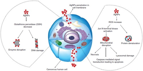
Figure 4. Recent advances in proposed mechanisms for anticancer activity shown by colloidal biogenic silver nanoparticles (AgNPs) (Adapted with permission from Ovais et al. (Citation21)).
The cytotoxicity of NPs may depend on parameters such as particle size, surface area and surface reactivity (Citation115). Upon understanding the mechanism of metallic NPs’ synthesis from plants and microbes, strategies can be designed for optimum synthesis of NPs of the desired shape and size. Eventually, based on heterogeneity of bio-sources, various factors such as temperature, synthesis conditions, reaction time, substrate concentrations and pH can have significant impacts on the rate of synthesis reaction.
2.2. Biocompatible nanomaterials for cancer diagnosis
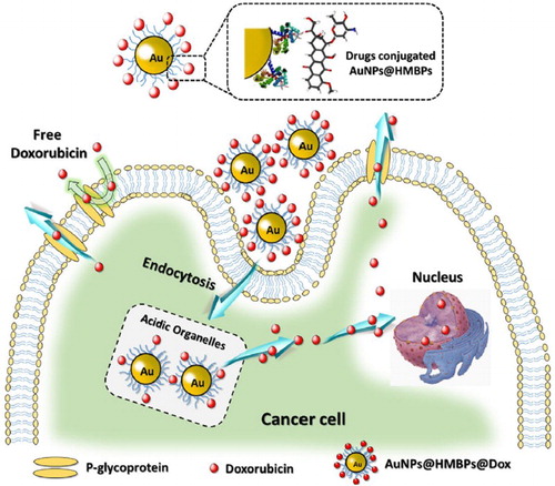
Recent nano-medical developments helped the progress of NPs for diagnostic and therapeutic (theranostics) applications. Notably, NPs can play a vital role in cancer diagnosis at an early stage by allowing visualization of cancer cells. Cancer diagnostic instruments used routinely in preclinical research and clinical practice involve MRI, CT, ultrasound, optical imaging, positron emission tomography, photo-acoustic imaging and single-photon emission CT (SPECT). In this regard, these instruments differ based on their underlying physical principles, sensitivity and specificity to contrast agents, tissue contrast, spatial resolution, quantitativeness and tissue penetration (Citation116). Remarkably, alpha fetal protein (AFP) is a cancer biomarker in clinical diagnosis associated with the disease progression and therapeutic responses of liver cancer. Xuan et al. (Citation117) reported a plasmonic ELISA strategy by means of alkaline phosphatase-mediated growth of AgNPs for the colorimetric detection of serum AFP. This plasmonic ELISA provided high sensitivity displaying high performance in cancer diagnosis and therapeutic monitoring. This plasmonic assay is based on the in situ generation of AgNPs, leading to rapid color with various degrees of yellow, which can be easily performed on currently available instruments in clinical laboratories. Surprisingly, cancer detection has been widely investigated by applying metal NPs for marking tumor cells to find tumor-target fluorescence bio-imaging as an excellent fluorescent probe (Citation118). Specifically, Ge et al., (Citation118) reported biosynthesis of fluorescent Au/Ce nanoclusters (NCs) (1.2–2.2 nm) as highly sensitive bio-imaging agents. As demonstrated in , the Au/Ce NCs show significant fluorescence in the HeLa cancer cells (A–C). Moreover, D depicted the variations of fluorescence intensity between cancerous and normal cell types. Besides, the same results were obtained in HepG2 in the same situation. In contrast, the control group including L02 cells showed almost no intracellular fluorescence. Furthermore, according to the acceptable in vitro results, Au/Ce NCs were also investigated for in vivo bio-imaging in a tumor mice model of cervical carcinoma. As shown in , fluorescence was observed around the tumor after 24 h of subcutaneous Au/Ce NCs injection (Citation118). This is in agreement with the study of Wang et al., 2013, which demonstrated green synthesis of silver NCs employing glutathione and showed in vivo fluorescent tumor imaging through living animal tumors (Citation119). Furthermore, Mukherjee et al. (Citation120) reported green synthesis of monodispersed AgNPs (20–60 nm) by using Olax scandens leaf extract, with spherical shape and high stability for several days. In this study the fluorescence properties of biogenic AgNPs were observed through two cell lines (A549 & B16F10) employing a fluorescence microscopy. The untreated cells used as control and those treated with chemically fabricated AgNPs did not exhibit fluorescence, while those cancerous cells that were treated with biological synthesized AgNPs exhibited very strong fluorescence indicating the internalization of AgNPs by A549 & B16F10 cells (Citation120). Overall, based on the aforementioned, the use of nanotechnology will be the best strategy for cancer diagnosis due to their self-fluorescence ability. One of the major drawbacks of current cancer treatment strategies is the lack of targeted drug delivery, resulting in systemic toxicity (Citation8 , Citation121). Recently, magnetically targeted NPs have overcome this significant disadvantage of non-specificity as carriers for FDA-approved anti-cancer drugs. The basic principle is the loading of a particular anti-cancer drug on to a magnetic NP carrier like biocompatible super-paramagnetic iron oxide nanoparticles followed by injecting into the blood stream. Thereupon, with the application of external magnetic fields (high-gradient), the particular drug delivery system (DDS) is targeted specifically at cancerous cells via changes in physiological conditions (osmolality, enzymatic activity and temperature). Indeed, by applying this strategy the associated side-effects along with the amount of the drug delivered are reduced, ultimately leading to reduced systemic toxicity (Citation122 , Citation123). Seo et al. (Citation124). have reported a novel study of biosynthesizing AuNPs via employing heavy metal binding proteins (HMBPs) of genetically engineered Escherichia coli acting as reducing, stabilizing and capping agent. The synthesized NPs were spherical in nature having a size of 5–20 nm in diameter. Furthermore, the group loaded doxorubicin (Dox) on the AuNPs@HMBPs DDS for treating the cancerous HeLa cells. The process of AuNPs@HMBPs’ preparation along with Dox-loaded AuNPs@HMBPs’ cytotoxic evaluation are illustrated in (Citation124).
Figure 5. Fluorescence images of HeLa cancer cells which were incubated in the absence of Au/Ce NCs (A), in the presence of 50 μmol/L (B) and 150 μmol/L (C) Au/Ce NCs solutions for 24 h. (D) The fluorescence intensity variations along cross-sections a (in A), b (in B) or c (in C). Fluorescence images were collected by applying fluorescence excitation wavelength at 488 nm (Adapted with permission from Ge et al. (Citation118)).
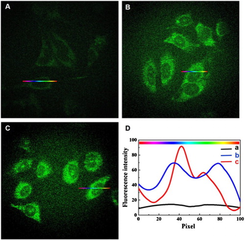
Figure 6. Tumor nude mice models of cervical carcinoma in vivo imaging. (A) In vivo fluorescence imaging 24 h after a subcutaneous injection of 5 mmol/L Au/Ce NCs solution near the tumor. (B) In vivo fluorescence imaging 24 h after an intravenous injection of 5 mmol/L Au/Ce NCs solution through the tail. (C) Control nude mice without tumor after intravenous injection equivalent to PBS through the tail. Fluorescent Au/Ce NCs were observed inside the tumors using a 455 nm excitation wavelength (Adapted with permission from Ge et al. (Citation118)).
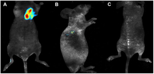
Figure 7. Biosynthesis of AuNPs by employing heavy metal binding proteins (HMBPs) in recombinant E. coli and subsequent conjugation of doxorubicin. Doxorubicin-loaded AuNPs complexes can be easily fragmented to release doxorubicin from AuNPs to defuse through the cancer cell (Adapted with permission from Seo et al. (Citation124)).
3. Hurdles for green nanomaterials as future cancer nanomedicine
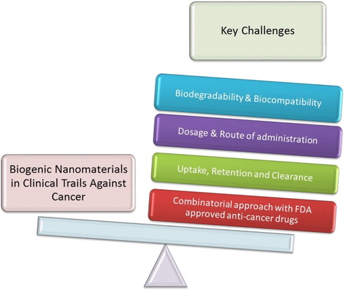
A wide range of applications of metal nanoparticle therapeutics in the future is doubtless though there are still many challenges to be overcome and eventually routine clinical practice. If we talk about phytosynthesis, many amino acids, polysaccharides, flavonoids, alkaloids, vitamins, etc. exist in the metal NPs’ medium method, which play a role in NPs’ function, even if their surfaces are washed and purified since the residuals would stick to the NPs’ surface. Likewise, in microbial synthesis, there are a number of microorganisms involving saprophytes and even pathogenic microbes such as E. coli introduced as a bio-source for preparation of metal NPs which would cause some hazards according to clinical studies. As an example, Aspergillus niger, Aspergillus flavus, Fusarium solani, etc. have been employed to biosynthesize metal NPs (Citation26, Citation125Citation126 Citation127 Citation128–Citation129). Importantly, another limitation is the protein corona effect, which occurs via adsorption of proteins on the colloidal NPs’ surfaces when the nanoparticle enters the biological system (Citation130). This adsorption actually compromises the ultimate dependability of NPs in the in vivo system (Citation131). Overall, nanotechnology-based products are highly expensive probably due to difficulties in the manufacturing and preserving process; thereby its progress would cost a high amount of money. The key hurdles faced by researchers for entrance of biosynthesized NPs into the clinical phase have been graphically illustrated in (Citation132Citation133 Citation135 – Citation134).
Figure 8. Major challenges for scientists before biosynthesized nanomaterials enter clinical trials for cancer theranostics.
By and large, many challenges and issues came in the way of scaling up nanoparticles/nanoconjugates’ production to industrial scale. NPs produced using routine top-down or bottom-up approaches in the lab vary greatly from those obtained from commercial producers. These approaches include sonication, emulsification, organic solvents evaporation and use, homogenization, centrifugation, elevated temperature, crosslinking and lyophilization (Citation35). Practically, a slight change in the optimized conditions of NPs’ synthesis can render the NPs biologically inactive, less stable and highly impure. Hence, a robust production process should be used at the lab-scale for NPs with an extraordinary reproducibility rate to be viable for large-scale commercial production.
3.1. Diffusion and penetration
In general, diffusive resistance of various tissues (intestine, vagina, nose mucosal membranes and lungs) poses a major barrier in nanomedicine delivery. Researchers have scientifically proved that the sedimentation and diffusion velocities of NPs highly affect their penetration and cellular uptake. Specifically, it is also reported that the cellular uptake and penetration are independent of surface coating, size, density, initial dose concentration and morphology of NPs (Citation136). In addition, Cho et al. (Citation137) reported that the shape dependency of AuNPs for their penetration through the cells could vary depending on their surface functional group. Wang et al. (Citation138) studied endocytosis of AuNPs with different sizes (45, 70 and 110 nm) in the human cancerous cell lines (CL1-0 and HeLa). This study revealed that the optimal size for the penetration into cells was around 45 nm. Interestingly, Chithrani et al. (Citation139) evaluated the effect of spherical and rod-shaped AuNPs on cellular uptake in the HeLa cell line. This study depicted that spherical-shaped AuNPs had a higher cellular uptake in comparison to rod-shaped AuNPs because of variable biophysical properties such receptor diffusion kinetics. More interestingly, it was suggested that the surface coating of NPs impact on rate of uptake (Citation140). For instance, Sur et al. (Citation141) investigated cytotoxicity and cellular penetration of modified AgNPs with glucose, lactose, etc. in normal (L929) and cancerous (A549) cell lines. In this study, no differences were revealed in the cellular penetration of lactose- and glucose-modified AgNPs in L929, though there was a substantial growth in the cellular internalization of lactose-modified AgNPs into the A549, could be explained by the role of the chemical nature of the ligand in cellular uptake.
3.2. Toxicological and immunological aspect
Mukherjee and coworkers experimentally proved the nontoxic nature of these biogenic NPs in C57BL6/J female mice via various biochemical parameters along with a histopathology study of different organs. Even after successive injections of biogenic AuNPs (1 or 10 mg/kg/day) for seven days in healthy mice, no toxicity was observed (Citation142). Moreover, it is interesting to note that in another study the same group reported that mice treated with chemically synthesized AuNPs showed broken alveolar walls with hyperplasic sinusoids as compared to the untreated or green synthesized AuNPs-treated mice (Citation143). On the other hand, various chronic and acute toxicities of liver and nephrons were reported to be associated with zinc, platinum and cerium oxide NPs (Citation144 Citation145 –Citation146). Hence, it is very crucial to screen the potential nanoparticles/nanomaterials for various toxicity studies including their pharmacokinetics, pharmacodynamics and responses to immune system before moving on to clinical trials (Citation147 –Citation148 Citation149 Citation150 ).
3.3. Biodegradability, metabolic kinetics and clearance
Biodegradability is very vital aspect to be considered before exploiting AuNPs in vivo. Polymeric NPs are naturally biodegradable and are cleared form the body. However, in case of metallic NPs biodegradability is slow and poses serious challenges in terms of toxicity (Citation50 , Citation147, Citation151). Although detailed knowledge regarding the mechanism of metallic NPs’ biodegradability and clearance is still unclear, few recent studies validate the gradual excretion of metal NPs in feces and urine even up to 14 days (Citation147 , Citation152). Furthermore, the studies shed light on the positively charged AuNPs, which may encounter a negative charge repulsion from the glomerular basement membrane in nephrons and pass out via urine. In another study reported by Rengan and coworkers, liposomal AuNPs were demonstrated for their biodegradability and as a potential DDS for cancer therapy. Moreover, metabolic degradation of the delivery system was noticed in liver and hepatocytes followed by its easy excretion via the renal route (Citation147). All the studies discussed in detail that in vivo biodistribution, metabolic kinetics, clearance and ultimate dependability of NPs are determined by their shape, size and morphology.
4. Conclusions and future prospects
Green nanomaterials are currently in a highly investigative phase for the treatment and diagnosis of cancer but the ultimate dependability is yet to be decided in clinical trials. A number of new possibilities have come into account in relation to the use of green nanomaterials, owing to their biocompatibility and effectiveness. Moreover, many types of cancers that do not have cures today may be cured by these green nanomaterials in the future. Additionally, thorough understanding of key physiological barriers in vivo is the key to effectively deliver NPs into the tumor. Furthermore, the knowledge regarding the safety of nanomaterials is not sufficient and comprehensive acute and chronic toxicity in clinical studies should be observed to identify the hazards associated with the use of NPs. Considering the above discussion, it is expected that green nanomaterials could emerge as future cancer therapeutics and diagnostics agents in the near future.
Acknowledgements
We gratefully acknowledge Dr Sudip Mukherjee (Post.doc Researcher, Department of Bioengineering, Rice University, Houston, TX, USA) for his valuable suggestions and comments over the content of the manuscript.
Disclosure statement
No potential conflict of interest was reported by the authors.
Notes on contributors
Mr. Hamed Barabadi graduated as a Doctor of Pharmacy from Mazandaran University of Medical Sciences, Sari, Iran, 2014. He is currently working on his Ph.D. in Pharmaceutical Biotechnology, School of Pharmacy, Shahid Beheshti University of Medical Sciences, Tehran, Iran. His main research interests are in nanobiotechnology with an emphasis on nanomedicine, nanobiomaterials and bioprocessing.
Mr. Muhammad Ovais is pursuing a Ph.D. degree in Nanomedicine from CAS Key Laboratory for Biomedical Effects of Nanomaterials and Nanosafety & CAS Center for Excellence in Nanoscience, National Center for Nanoscience and Technology, Beijing, People’s Republic of China. He has completed his MPhil-Biotechnology degree in 2017 from Quaid-i-Azam University, Pakistan and earned his BS-Biotechnology degree in 2015 with distinction from the University of Peshawar, Pakistan. His research interest lies in the development novel nano-delivery systems for cancer theranostics. He owns to his credit 19 original research and review articles in prestigious journals with a cumulative impact factor of above 50.
Prof. Dr. Zabta khan Shinwari is a Tenured Professor at Department of Biotechnology, Quaid-i-Azam University and Secretary General of Pakistan Academy of Sciences. He did his Post-doc from Japan International Research Center for Agricultural Sciences and Ph.D. from Kyoto University. His research interest lies in Medicinal plants, Molecular Systematics, Cancer Nanomedicine, and dual-use education. He is UNESCO Laureate and Avicenna Gold Medalist for Ethics in Science, 2015. Dr. Zabta owns to his credit 355 scientific publications with cumulative citations of 5842, having h-index of 37 and i10-index of 129. Moreover, he remained Vice Chancellor of many Universities in Pakistan and has won numerous national and international scientific projects. He also has to his credit highest National Civil Awarded, “Tamgha-e-Imtiaz”.
Dr. Muthupandian Saravanan graduated in Microbiology from Madurai Kamaraj University and earned his doctorate with specialization in Medical Microbiology and Nanomedicine from Sathyabama University, India. Thereafter, He did his Post Doctoral Research through Israel Government research fellowship (2011–2012) in Institute of Drug research, Faculty of Medicine, Hebrew University of Jerusalem, Israel, focusing his research on Nano-biomaterials & their Biomedical Applications. Prior to post-doc, he worked as an Assistant Professor, Department of Biotechnology, SRM University, Chennai, India. Presently, he is an Associate Professor (under UNDP) at Department of Medical Microbiology and Immunology, Institute of Bio-medical Sciences, College of Health Science, Mekelle University, Ethiopia. Dr. Saravanan owns to his credit 60 publications with cumulative citations of 1045, having h-index of 16 and i10-index of 17. He has participated in more than 40 national and international conferences and also published in more than 30 international peer-reviewed journals. He was a recipient of many fellowships and awards – notably, DST-SERC Young Scientist Award in 2012, International Fellowship “Advanced Course on Diagnostics” Sponsored by LSH&TM & Fondation Mérieux, in France 2013, International Fellowship “Pertussis: biology, epidemiology and prevention” meeting Sponsored by Fondation Mérieux & WHO in France 2014, International Union of Microbiological Societies (IUMS) travel grant in 2015 to Canada, International Fellowship “Advanced Course on Antibiotics” (AdCAb) Sponsored by Institute of Pasteur and Fondation Mérieux France, 2016. His research interest lies in Microbial Drug Resistance, Nano-biotechnology, and Nanomedicine to combat Antimicrobial resistance (AMR) and Cancer.
ORCID
Muthupandian Saravanan http://orcid.org/0000-0002-1480-3555
References
- Institute, N.C. “What is cancer?” https://www.cancer.gov/about-cancer/understanding/what-is-cancer (accessed Nov 6)
- Informatics, I.I.f.H. http://www.imshealth.com/en/thought-leadership/ims-institute
- Society, T.A.C. Global Cancer Facts & Figures. 3rd Edition. http://www.cancer.org/Research/CancerFactsStatistics/global-cancer-facts-figures-3rd-edition (accessed Nov 6)
- Jemal, A.; Bray, F.; Center, M.M.; Ferlay, J.; Ward, E.; Forman, D. Global Cancer Statistics. CA: A Cancer J. Clinicians 2011, 61 (2), 69–90.
- Organization, W.H.: “Cancer Control: A Global Snaptshot in 2015”. http://www.who.int/cancer/cancer-snapshot-2015/en/ (accessed Nov 6)
- Tanneberger, S. Palliative Care in Advanced Cancer. In ESMO Handbook of Advanced Cancer Care; Catane, R., Cherny, N., Kloke, M., Tanneberger, S., Schrijvers, D., Eds.; Taylor & Francis, 2006; pp 5–9.
- Miller, K.D.; Siegel, R.L.; Lin, C.C.; Mariotto, A.B.; Kramer, J.L.; Rowland, J.H.; Stein, K.D.; Alteri, R.; Jemal, A. Cancer Treatment and Survivorship Statistics, 2016. CA: A Cancer J. Clinicians 2016, 46 (2), 231–239.
- Rao, P.V.; Nallappan, D.; Madhavi, K.; Rahman, S.; Jun Wei, L.; Gan, S.H. Phytochemicals and Biogenic Metallic Nanoparticles as Anticancer Agents. Oxid. Med. Cell Longev. 2016, 2016 (33), 12–27.
- Lokina, S.; Stephen, A.; Kaviyarasan, V.; Arulvasu, C.; Narayanan, V. Cytotoxicity and Antimicrobial Activities of Green Synthesized Silver Nanoparticles. Eur. J. Med. Chem. 2014, 76 (11), 256–263.
- Anwar, A.; Ovais, M.; Khan, A.; Raza, A. Docetaxel Loaded Solid Lipid Nanoparticles: A Novel Drug Delivery System. IET Nanobiotechnol. 2017, 11 (6), 1–34.
- Salunke, B.K.; Sawant, S.S.; Lee, S.-I.; Kim, B.S. Microorganisms as Efficient Biosystem for the Synthesis of Metal Nanoparticles: Current Scenario and Future Possibilities. World J. Microbiol. Biotechnol. 2016, 32 (5), 1–16.
- Singh, P.; Kim, Y.-J.; Zhang, D.; Yang, D.-C. Biological Synthesis of Nanoparticles From Plants and Microorganisms. Trends Biotechnol. 2016, 34 (7), 588–599.
- Kasithevar, M.; Saravanan, M.; Prakash, P.; Kumar, H.; Ovais, M.; Barabadi, H.; Shinwari, Z.K. Green Synthesis of Silver Nanoparticles Using Alysicarpus Monilifer Leaf Extract and its Antibacterial Activity Against MRSA and CoNS Isolates in HIV Patients. J. Interdiscip. Nanomed. 2017, 2 (2), 131–141.
- Khalil, A.T.; Ovais, M.; Ullah, I.; Ali, M.; Shinwari, Z.K.; Hassan, D.; Maaza, M. Sageretia Thea (Osbeck.) Modulated Biosynthesis of NiO Nanoparticles and Their in Vitro Pharmacognostic, Antioxidant and Cytotoxic Potential. Artif. Cells Nanomed. Biotechnol. 2017. DOI: 10.1080/21691401.2017.1345928
- Dahoumane, S.A.; Mechouet, M.; Wijesekera, K.; Filipe, C.D.M.; Sicard, C.; Bazylinski, D.A.; Jeffryes, C. Algae-mediated Biosynthesis of Inorganic Nanomaterials as A Promising Route in Nanobiotechnology - A Review. Green Chem. 2017, 19 (3), 552–587.
- Barabadi, H.a.S.H. Biofabrication of Gold and Silver Nanoparticles for Pharmaceutical Applications. Pharm. Biomed. Res. 2016, 2 (1), 1–7.
- Priester, J.H.; Van De Werfhorst, L.C.; Ge, Y.; Adeleye, A.S.; Tomar, S.; Tom, L.M.; Piceno, Y.M.; Andersen, G.L.; Holden, P.A. Effects of TiO2 and Ag Nanoparticles on Polyhydroxybutyrate Biosynthesis By Activated Sludge Bacteria. Environ. Sci. Technol. 2014, 48 (24), 14712–14720.
- Barabadi, H.; Honary, S.; Mohammadi, M.A.; Ahmadpour, E.; Rahimi, M.T.; Alizadeh, A.; Naghibi, F.; Saravanan, M. Green Chemical Synthesis of Gold Nanoparticles by Using Penicillium Aculeatum and Their Scolicidal Activity Against Hydatid Cyst Protoscolices of Echinococcus Granulosus. Environ. Sci. Pollut. Res. 2017, 24 (6), 5800–5810.
- Honary, S.; Barabadi, H.; Ebrahimi, P.; Naghibi, F.; Alizadeh, A. Development and Optimization of Biometal Nanoparticles by Using Mathematical Methodology: A Microbial Approach. J. Nano Res. 2015, 30 (7), 333–341.
- Rahimi, M.T.; Ahmadpour, E.; Esboei, B.R.; Spotin, A.; Koshki, M.H.K.; Alizadeh, A.; Honary, S.; Barabadi, H.; Mohammadi, M.A. Scolicidal Activity of Biosynthesized Silver Nanoparticles Against Echinococcus Granulosus Protoscolices. Int. J. Surg. 2015, 19 (10), 128–133.
- Ovais, M.; Khalil, A.T.; Raza, A.; Khan, M.A.; Ahmad, I.; Islam, N.U.; Saravanan, M.; Ubaid, M.F.; Ali, M.; Shinwari, Z.K. Green Synthesis of Silver Nanoparticles via Plant Extracts: Beginning A new era in Cancer Theranostics. Nanomedicine 2016, 11 (23), 3157–3177.
- Mukherjee, S.; Dasari, M.; Priyamvada, S.; Kotcherlakota, R.; Bollu, V.S.; Patra, C.R. A Green Chemistry Approach for the Synthesis of Gold Nanoconjugates That Induce the Inhibition of Cancer Cell Proliferation Through Induction of Oxidative Stress and Their in Vivo Toxicity Study. J. Mater. Chem. B. 2015, 3 (18), 3820–3830.
- Patra, C.R.; Mukherjee, S.; Kotcherlakota, R. Biosynthesized Silver Nanoparticles: A Step Forward for Cancer Theranostics? Nanomedicine 2014, 9 (10), 1445–1448.
- Ovais, M.; Raza, A.; Naz, S.; Islam, N.U.; Khalil, A.T.; Ali, S.; Khan, M.A.; Shinwari, Z.K. Current State and Prospects of the Phytosynthesized Colloidal Gold Nanoparticles and Their Applications in Cancer Theranostics. Appl. Microbiol. Biotechnol. 2017, 101 (9), 1–15.
- Honary, S.; Barabadi, H.; Gharaei-Fathabad, E.; Naghibi, F. Green Synthesis of Silver Nanoparticles Induced by the Fungus Penicillium Citrinum. Trop. J. Pharm. Res. 2013, 12 (1), 7–11.
- Barabadi, H.; Honary, S.; Ebrahimi, P.; Mohammadi, M.A.; Alizadeh, A.; Naghibi, F. Microbial Mediated Preparation, Characterization and Optimization of Gold Nanoparticles. Braz. J. Microbiol. 2014, 45 (4), 1493–1501.
- Subbaiya, R.; Saravanan, M.; Priya, A.R.; Shankar, K.; Selvam, M.; Ovais, M.; Balajee, R.; Barabadi, H. Biomimetic Synthesis of Silver Nanoparticles From Streptomyces Atrovirens and Their Potential Anticancer Activity Against Human Breast Cancer Cells. IET Nanobiotechnol. 2017. DOI: 10.1049/iet-bt.2016.0222
- Salunke, B.K.; Sawant, S.S.; Lee, S.-I.; Kim, B.S. Comparative Study of MnO2 Nanoparticle Synthesis by Marine Bacterium Saccharophagus Degradans and Yeast Saccharomyces Cerevisiae. Appl. Microbiol. Biotechnol. 2015, 99 (13), 5419–5427.
- Rajan, R.; Chandran, K.; Harper, S.L.; Yun, S.-I.; Kalaichelvan, P.T. Plant Extract Synthesized Silver Nanoparticles: An Ongoing Source of Novel Biocompatible Materials. Ind Crops Prod 2015, 70 (6), 356–373.
- Khalil, A.T.; Ovais, M.; Ullah, I.; Ali, M.; Khan Shinwari, Z.; Maaza, M. Biosynthesis of Iron Oxide (Fe2O3) Nanoparticles via Aqueous Extracts of Sageretia Thea (Osbeck.) and Their Pharmacognostic Properties. Green Chem. Lett. Rev. 2017, 10 (4), 186–201.
- Khalil, A.T.; Ovais, M.; Ullah, I.; Ali, M.; Shinwari, Z.K.; Maaza, M. Physical Properties, Biological Applications and Biocompatibility Studies on Biosynthesized Single Phase Cobalt Oxide (Co 3 O 4) Nanoparticles via Sageretia Thea (Osbeck.). Arab. J. Chem. 2017. DOI: 10.1016/j.arabjc.2017.07.004
- Sivaraj, R.; Rahman, P.K.; Rajiv, P.; Narendhran, S.; Venckatesh, R. Biosynthesis and Characterization of Acalypha Indica Mediated Copper Oxide Nanoparticles and Evaluation of its Antimicrobial and Anticancer Activity. Spectrochim. Acta, Part A 2014, 129 (17), 255–258.
- KS, U.S.; Govindaraju, K.; Kumar, G.; Prabhu, D.; Arulvasu, C.; Karthick, V.; Changmai, N. Anti-proliferative Effect of Biogenic Gold Nanoparticles Against Breast Cancer Cell Lines (MDA-MB-231 & MCF-7). Appl. Surf. Sci. 2016, 371 (12), 415–424.
- Gengan, R.; Anand, K.; Phulukdaree, A.; Chuturgoon, A. A549 Lung Cell Line Activity of Biosynthesized Silver Nanoparticles Using Albizia Adianthifolia Leaf. Colloids Surf. B 2013, 105 (34), 87–91.
- Singh, H.; Du, J.; Yi, T.-H. Green and Rapid Synthesis of Silver Nanoparticles Using Borago Officinalis Leaf Extract: Anticancer and Antibacterial Activities. Artif. Cells Nanomed Biotechnol. 2016, 45 (7), 1310–1316.
- Jena, S.; Singh, R.K.; Panigrahi, B.; Suar, M.; Mandal, D. Photo-bioreduction of Ag+ Ions Towards The Generation of Multifunctional Silver Nanoparticles: Mechanistic Perspective and Therapeutic Potential. J. Photochem. Photobiol, B 2016, 164 (43), 306–313.
- Rajan, A.; Vilas, V.; Philip, D. Studies on Catalytic, Antioxidant, Antibacterial and Anticancer Activities of Biogenic Gold Nanoparticles. J Mol Liq 2015, 212 (31), 331–339.
- Prabakaran, K.; Ragavendran, C.; Natarajan, D. Mycosynthesis of Silver Nanoparticles From Beauveria Bassiana and its Larvicidal, Antibacterial, and Cytotoxic Effect on Human Cervical Cancer (HeLa) Cells. RSC Adv. 2016, 6 (51), 44972–44986.
- Kummara, S.; Patil, M.B.; Uriah, T. Synthesis, Characterization, Biocompatible and Anticancer Activity of Green and Chemically Synthesized Silver Nanoparticles – A Comparative Study. Biomed. Pharmacother. 2016, 84 (6), 10–21.
- Mishra, P.; Ray, S.; Sinha, S.; Das, B.; Khan, M.I.; Behera, S.K.; Yun, S.-I.; Tripathy, S.K.; Mishra, A. Facile bio-Synthesis of Gold Nanoparticles by Using Extract of Hibiscus Sabdariffa and Evaluation of its Cytotoxicity Against U87 Glioblastoma Cells Under Hyperglycemic Condition. Biochem. Eng. J. 2016, 105 (6), 264–272.
- Namvar, F.; Azizi, S.; Rahman, H.S.; Mohamad, R.; Rasedee, A.; Soltani, M.; Rahim, R.A. Green Synthesis, Characterization, and Anticancer Activity of Hyaluronan/Zinc Oxide Nanocomposite. Onco Targets Ther. 2016, 9 (12), 4549–4559.
- Nayak, D.; Ashe, S.; Rauta, P.R.; Kumari, M.; Nayak, B. Bark Extract Mediated Green Synthesis of Silver Nanoparticles: Evaluation of Antimicrobial Activity and Antiproliferative Response Against Osteosarcoma. Mater. Sci. Eng: C 2016, 58 (11), 44–52.
- Chatterjee, A.; Loganathan, A.; Niroshinee, V.; Abraham, J. Biosynthesis of Lanthanum Nanoparticles Using Green Gram Seeds and Their Effect on Microorganisms. Res. J. Pharmac, Biol. Chem. Sci. 2016, 7 (2), 1462–1470.
- 4i4) Salehi, S.; Shandiz, S.A.S.; Ghanbar, F.; Darvish, M.R.; Ardestani, M.S.; Mirzaie, A.; Jafari, M. Phytosynthesis of Silver Nanoparticles Using Artemisia Marschalliana Sprengel Aerial Part Extract and Assessment of Their Antioxidant, Anticancer, and Antibacterial Properties. Int. J. Nanomed. 2016, 11 (12), 1835–1846.
- Rashmezad, M.A.; Ali Asgary, E.; Tafvizi, F.; Shandiz, S.; Ataollah, S.; Mirzaie, A. Comparative Study on Cytotoxicity Effect of Biological and Commercial Synthesized Nanosilver on Human Gastric Carcinoma and Normal Lung Fibroblast Cell Lines. Tehran Univ. Med. J. TUMS Publ. 2015, 72 (12), 799–807.
- Xia, Q.H.; Ma, Y.J.; Wang, J.W. Biosynthesis of Silver Nanoparticles Using Taxus Yunnanensis Callus and Their Antibacterial Activity and Cytotoxicity in Human Cancer Cells. Nanomaterials 2016, 6 (9), 160–100.
- Salari, Z.; Ameri, A.; Forootanfar, H.; Adeli-Sardou, M.; Jafari, M.; Mehrabani, M.; Shakibaie, M. Microwave-assisted Biosynthesis of Zinc Nanoparticles and Their Cytotoxic and Antioxidant Activity. J. Trace Elem. Med. Biol. 2017, 39 (16), 116–123.
- Varsha, B.; Rajasekar, A.; Kathiravan, G. Xylorious Biogenic Synthesis of Silver Nanoparticle and Their Cytotoxicity Effects Against HT-29 Cell Line. Res. J. Pharm, Biol. Chem. Sci. 2016, 7 (5), 1578–1583.
- Premasudha, P.; Venkataramana, M.; Abirami, M.; Vanathi, P.; Krishna, K.; Rajendran, R. Biological Synthesis and Characterization of Silver Nanoparticles Using Eclipta Alba Leaf Extract and Evaluation of its Cytotoxic and Antimicrobial Potential. Bull. Mater. Sci. 2015, 38 (4), 965–973.
- Manikandan, R.; Manikandan, B.; Raman, T.; Arunagirinathan, K.; Prabhu, N.M.; Basu, M.J.; Perumal, M.; Palanisamy, S.; Munusamy, A. Biosynthesis of Silver Nanoparticles Using Ethanolic Petals Extract of Rosa Indica and Characterization of its Antibacterial, Anticancer and Anti-Inflammatory Activities. Spectrochim. Acta, Part A 2015, 138 (7), 120–129.
- Prabhu, D.; Arulvasu, C.; Babu, G.; Manikandan, R.; Srinivasan, P. Biologically Synthesized Green Silver Nanoparticles From Leaf Extract of Vitex Negundo L. Induce Growth-Inhibitory Effect on Human Colon Cancer Cell Line HCT15. Process Biochem. 2013, 48 (2), 317–324.
- Prasad, P.R.; Kanchi, S.; Naidoo, E. In-vitro Evaluation of Copper Nanoparticles Cytotoxicity on Prostate Cancer Cell Lines and Their Antioxidant, Sensing and Catalytic Activity: One-pot Green Approach. J. Photochem. Photobiol, B 2016, 11 (2), 122–130.
- He, Y.; Du, Z.; Ma, S.; Cheng, S.; Jiang, S.; Liu, Y.; Li, D.; Huang, H.; Zhang, K.; Zheng, X. Biosynthesis, Antibacterial Activity and Anticancer Effects Against Prostate Cancer (PC-3) Cells of Silver Nanoparticles Using Dimocarpus Longan Lour. Nanoscale Res. Lett. 2016, 11 (1), 1–10.
- Nalavothula, R.; Alwala, J.; Nagati, V.B.; Manthurpadigya, P.R. Biosynthesis of Silver Nanoparticles Using Impatiens Balsamina Leaf Extracts and its Characterization and Cytotoxic Studies Using Human Cell Lines. Int. J. ChemTech Res. 2015, 7 (5), 2460–2468.
- Patra, S.; Mukherjee, S.; Barui, A.K.; Ganguly, A.; Sreedhar, B.; Patra, C.R. Green Synthesis, Characterization of Gold and Silver Nanoparticles and Their Potential Application for Cancer Therapeutics. Mater. Sci. Eng: C 2015, 53, 298–309.
- Namvar, F.; Rahman, H.S.; Mohamad, R.; Azizi, S.; Tahir, P.M.; Chartrand, M.S.; Yeap, S.K. Cytotoxic Effects of Biosynthesized Zinc Oxide Nanoparticles on Murine Cell Lines. Evidence-Based Complementary Altern. Med. 2015, 17 (15), 743–751.
- Raman, R.P.; Parthiban, S.; Srinithya, B.; Kumar, V.V.; Anthony, S.P.; Sivasubramanian, A.; Muthuraman, M.S. Biogenic Silver Nanoparticles Synthesis Using the Extract of the Medicinal Plant Clerodendron Serratum and its in-Vitro Antiproliferative Activity. Mater. Lett. 2015, 160 (67), 400–403.
- Namvar, F.; Rahman, H.S.; Mohamad, R.; Rasedee, A.; Yeap, S.K.; Chartrand, M.S.; Azizi, S.; Tahir, P.M. Apoptosis Induction in Human Leukemia Cell Lines by Gold Nanoparticles Synthesized Using the Green Biosynthetic Approach. J. Nanomater. 2015, 21 (8), 1–10.
- Namvar, F.; Rahman, H.S.; Mohamad, R.; Baharara, J.; Mahdavi, M.; Amini, E.; Chartrand, M.S.; Yeap, S.K. Cytotoxic Effect of Magnetic Iron Oxide Nanoparticles Synthesized via Seaweed Aqueous Extract. Int. J. Nanomed. 2014, 9 (1), 2479–2488.
- Sulaiman, G.M.; Mohammad, A.A.; Abdul-Wahed, H.E.; Ismail, M.M. Biosynthesis, Antimicrobial and Cytotoxic Effects of Silver Nanoparticles Using Rosmarinus Officinalis Extract. Digest J. Nanomater. Biostruct. 2013, 8 (1), 34–41.
- Dorosti, N.; Jamshidi, F. Plant-mediated Gold Nanoparticles by Dracocephalum Kotschyi as Anticholinesterase Agent: Synthesis, Characterization, and Evaluation of Anticancer and Antibacterial Activity. J. Appl. Biomed. 2016, 11 (7), 123–131.
- Nayak, D.; Pradhan, S.; Ashe, S.; Rauta, P.R.; Nayak, B. Biologically Synthesised Silver Nanoparticles From Three Diverse Family of Plant Extracts and Their Anticancer Activity Against Epidermoid A431 Carcinoma. J. Colloid Interface Sci. 2015, 457 (13), 329–338.
- Jha, A.K.; Prasad, K. Green Syntheis Of Silver Nanoparticles And Its Activity On SiHa Cervical Cancer Cell Line. Adv. Mater. Lett. 2014, 5 (12), 501–505.
- Mittal, A.K.; Bhaumik, J.; Kumar, S.; Banerjee, U.C. Biosynthesis of Silver Nanoparticles: Elucidation of Prospective Mechanism and Therapeutic Potential. J. Colloid Interface Sci. 2014, 415 (11), 39–47.
- Kumar, B.; Smita, K.; Seqqat, R.; Benalcazar, K.; Grijalva, M.; Cumbal, L. In Vitro Evaluation of Silver Nanoparticles Cytotoxicity on Hepatic Cancer (Hep-G2) Cell Line and Their Antioxidant Activity: Green Approach for Fabrication and Application. J. Photochem. Photobiol, B 2016, 159 (11), 8–13.
- Roopan, S.M.; Kumar, S.H.S.; Madhumitha, G.; Suthindhiran, K. Biogenic-production of SnO2 Nanoparticles and its Cytotoxic Effect Against Hepatocellular Carcinoma Cell Line (HepG2). Appl. Biochem. Biotechnol. 2015, 175 (3), 1567–1575.
- Khalil, A.T.; Ovais, M.; Ullah, I.; Ali, M.; Shinwari, Z.K.; Khamlich, S.; Maaza, M. Sageretia Thea (Osbeck.) Mediated Synthesis of Zinc Oxide Nanoparticles and its Biological Applications. Nanomedicine 2017, 12 (15), 1767–1789.
- Renugadevi, K.; Inbakandan, D.; Bavanilatha, M.; Poornima, V. Cissus Quadrangularis Assisted Biosynthesis of Silver Nanoparticles with Antimicrobial and Anticancer Potentials. Int. J. Pharm. Bio Sci. 2012, 3 (3), 437–445.
- Devi, J.S.; Bhimba, B.V.; Peter, D.M. Production of Biogenic Silver Nanoparticles Using Sargassum Longifolium and its Applications. Indian J. Geo-Mar. Sci. 2013, 42 (1), 125–130.
- Ahmad Siddiqui, E.; Ahmad, A.; Julius, A.; Syed, A.; Khan, S.; Kharat, M.; Pai, K.; Kadoo, N.; Gupta, V. Biosynthesis of Anti-Proliferative Gold Nanoparticles Using Endophytic Fusarium Oxysporum Strain Isolated From Neem (A. Indica) Leaves. Curr. Top. Med. Chem. 2016, 16 (18), 2036–2042.
- Ortega, F.G.; Fernández-Baldo, M.A.; Fernández, J.G.; Serrano, M.J.; Sanz, M.I.; Diaz-Mochón, J.J.; Lorente, J.A.; Raba, J. Study of Antitumor Activity in Breast Cell Lines Using Silver Nanoparticles Produced by Yeast. Int. J. Nanomed. 2015, 10 (5), 2021–2031.
- Inbakandan, D.; Kumar, C.; Bavanilatha, M.; Ravindra, D.N.; Kirubagaran, R.; Khan, S.A. Ultrasonic-assisted Green Synthesis of Flower Like Silver Nanocolloids Using Marine Sponge Extract and its Effect on Oral Biofilm Bacteria and Oral Cancer Cell Lines. Microb. Pathog. 2016, 99 (22), 135–141.
- Bhakya, S.; Muthukrishnan, S.; Sukumaran, M.; Grijalva, M.; Cumbal, L.; Benjamin, J.F.; Kumar, T.S.; Rao, M. Antimicrobial, Antioxidant and Anticancer Activity of Biogenic Silver Nanoparticles – an Experimental Report. RSC Adv. 2016, 6 (84), 81436–81446.
- Potara, M.; Bawaskar, M.; Simon, T.; Gaikwad, S.; Licarete, E.; Ingle, A.; Banciu, M.; Vulpoi, A.; Astilean, S.; Rai, M. Biosynthesized Silver Nanoparticles Performing as Biogenic SERS-Nanotags for Investigation of C26 Colon Carcinoma Cells. Colloids Surf. B 2015, 133 (11), 296–303.
- Maliszewska, I. Microbial Mediated Synthesis of Gold Nanoparticles: Preparation, Characterization and Cytotoxicity Studies. Dig J. Nanomater. Bios 2013, 8 (11), 1123–1131.
- Venkatesan, J.; Manivasagan, P.; Kim, S.-K.; Kirthi, A.V.; Marimuthu, S.; Rahuman, A.A. Marine Algae-Mediated Synthesis of Gold Nanoparticles Using A Novel Ecklonia Cava. Bioprocess Biosyst. Eng. 2014, 37 (8), 1591–1597.
- Wang, C.; Mathiyalagan, R.; Kim, Y.J.; Castro-Aceituno, V.; Singh, P.; Ahn, S.; Wang, D.; Yang, D.C. Rapid Green Synthesis of Silver and Gold Nanoparticles Using Dendropanax Morbifera Leaf Extract and Their Anticancer Activities. Int. J. Nanomed. 2016, 11 (6), 3691–3699.
- Wani, K.; Choudhari, A.; Chikate, R.; Kaul-Ghanekar, R. Synthesis and Characterization of Gold Nanoparticles Using Ficus Religiosa Extract. Carbon Sci. Technol. 2013, 5 (1), 203–210.
- Prasannaraj, G.; Sahi, S.V.; Ravikumar, S.; Venkatachalam, P. Enhanced Cytotoxicity of Biomolecules Loaded Metallic Silver Nanoparticles Against Human Liver (HepG2) and Prostate (PC3) Cancer Cell Lines. J. Nanosci. Nanotechnol. 2016, 16 (5), 4948–4959.
- Raj, P.; Khusro, A. In-vitro Anticancer and Antioxidant Activity of Gold Nanoparticles Conjugate with Tabernaemontana Divaricata Flower SMs Against MCF -7 Breast Cancer Cells. Korean Chem. Eng. Res. 2016, 54 (1), 75–80.
- Saikia, I.; Sonowal, S.; Pal, M.; Boruah, P.K.; Das, M.R.; Tamuly, C. Biosynthesis of Gold Decorated Reduced Graphene Oxide and its Biological Activities. Mater. Lett. 2016, 178 (14), 239–242.
- Tippayawat, P.; Phromviyo, N.; Boueroy, P.; Chompoosor, A. Green Synthesis of Silver Nanoparticles in Aloe Vera Plant Extract Prepared by A Hydrothermal Method and Their Synergistic Antibacterial Activity. PeerJ. Preprints 2016, 4 (56), 1912–1920.
- Arokiyaraj, S.; Arasu, M.V.; Vincent, S.; Prakash, N.U.; Choi, S.H.; Oh, Y.-K.; Choi, K.C.; Kim, K.H. Rapid Green Synthesis of Silver Nanoparticles From Chrysanthemum Indicum L and its Antibacterial and Cytotoxic Effects: an in Vitro Study. Int. J. Nanomed. 2014, 9 (13), 379–388.
- Singh, M.; Saurav, K.; Majouga, A.; Kumari, M.; Kumar, M.; Manikandan, S.; Kumaraguru, A. The Cytotoxicity and Cellular Stress by Temperature-Fabricated Polyshaped Gold Nanoparticles Using Marine Macroalgae, Padina Gymnospora. Biotechnol. Appl. Biochem. 2015, 62 (3), 424–432.
- Swamy, M.K.; Akhtar, M.S.; Mohanty, S.K.; Sinniah, U.R. Synthesis and Characterization of Silver Nanoparticles Using Fruit Extract of Momordica Cymbalaria and Assessment of Their in Vitro Antimicrobial, Antioxidant and Cytotoxicity Activities. Spectrochim. Acta, Part A 2015, 151 (54), 939–944.
- Dharmatti, R.; Phadke, C.; Mewada, A.; Thakur, M.; Pandey, S.; Sharon, M. Biogenic Gold Nano-Triangles: Cargos for Anticancer Drug Delivery. Mater. Sci. Eng: C 2014, 44 (28), 92–98.
- Yang, N.; WeiHong, L.; Hao, L. Biosynthesis of Au Nanoparticles Using Agricultural Waste Mango Peel Extract and its in Vitro Cytotoxic Effect on two Normal Cells. Mater. Lett. 2014, 134 (6), 67–70.
- Arunkumar, P.; Vedagiri, H.; Premkumar, K. Rapid Bioreduction of Trivalent Aurum Using Banana Stem Powder and its Cytotoxicity Against MCF-7 and HEK-293 Cell Lines. J. Nanopart. Res. 2013, 15 (3), 1–8.
- Harne, S.; Sharma, A.; Dhaygude, M.; Joglekar, S.; Kodam, K.; Hudlikar, M. Novel Route for Rapid Biosynthesis of Copper Nanoparticles Using Aqueous Extract of Calotropis Procera L. Latex and Their Cytotoxicity on Tumor Cells. Colloids Surf. B 2012, 95 (23), 284–288.
- Kalpana, D.; Pichiah, P.T.; Sankarganesh, A.; Park, W.S.; Lee, S.M.; Wahab, R.; Cha, Y.S.; Lee, Y.S. Biogenesis of Gold Nanoparticles Using Plant Powders and Assessment of in Vitro Cytotoxicity in 3T3-L1 Cell Line. J. Pharm. Innov 2013, 8 (4), 265–275.
- Tiwari, M.; Jain, P.; Hariharapura, R.C.; Narayanan, K.; Bhat, U.; Udupa, N.; Rao, J.V. Biosynthesis of Copper Nanoparticles Using Copper-Resistant Bacillus Cereus, A Soil Isolate. Process. Biochem. 2016, 51 (10), 1348–1356.
- Kalpana, D.; Srikanth, K.; Pichiah, P.T.; Cha, Y.S.; Lee, Y.S. Synthesis, Characterization and In Vitro Cytotoxicity of Gold Nanoparticles Using Cultural Filtrate of Low Shear Modeled Microgravity and Normal Gravity Cultured K. Pneumoniae. Macromol. Res. 2014, 22 (5), 487–493.
- Syed, A.; Saraswati, S.; Kundu, G.C.; Ahmad, A. Biological Synthesis of Silver Nanoparticles Using the Fungus Humicola sp. and Evaluation of Their Cytoxicity Using Normal and Cancer Cell Lines. Spectrochim. Acta, Part A 2013, 114 (55), 144–147.
- Mishra, A.; Tripathy, S.K.; Wahab, R.; Jeong, S.-H.; Hwang, I.; Yang, Y.-B.; Kim, Y.-S.; Shin, H.-S.; Yun, S.-I. Microbial Synthesis of Gold Nanoparticles Using the Fungus Penicillium Brevicompactum and Their Cytotoxic Effects Against Mouse Mayo Blast Cancer C2C12 Cells. Appl. Microbiol. Biotechnol. 2011, 92 (3), 617–630.
- Anand, K.; Tiloke, C.; Phulukdaree, A.; Ranjan, B.; Chuturgoon, A.; Singh, S.; Gengan, R. Biosynthesis of Palladium Nanoparticles by Using Moringa Oleifera Flower Extract and Their Catalytic and Biological Properties. J. Photochem. Photobiol, B 2016, 165 (34), 87–95.
- Du, J.; Singh, H.; Yi, T.-H. Antibacterial, Anti-Biofilm and Anticancer Potentials of Green Synthesized Silver Nanoparticles Using Benzoin gum (Styrax Benzoin) Extract. Bioprocess Biosyst. Eng. 2016, 39 (12), 1923–1931.
- Kummara, S.; Patil, M.B.; Uriah, T. Synthesis, Characterization, Biocompatible and Anticancer Activity of Green and Chemically Synthesized Silver Nanoparticles – A Comparative Study. Biomed. Pharmacotherapy 2016, 84 (12), 10–21.
- Singh, H.; Du, J.; Yi, T.-H. Green and Rapid Synthesis of Silver Nanoparticles Using Borago Officinalis Leaf Extract: Anticancer and Antibacterial Activities. Artif. Cells Nanomed. Biotechnol. 2016, 15 (11), 1–7.
- Heydari, R.; Rashidipour, M. Green Synthesis of Silver Nanoparticles Using Extract of oak Fruit Hull (Jaft): Synthesis and in Vitro Cytotoxic Effect on MCF-7 Cells. Int. J. Breast Cancer 2015, 33 (16), 45–54.
- Majeed, S.; bin Abdullah, M.S.; Dash, G.K.; Ansari, M.T.; Nanda, A. Biochemical Synthesis of Silver Nanoprticles Using Filamentous Fungi Penicillium Decumbens (MTCC-2494) and its Efficacy Against A-549 Lung Cancer Cell Line. Chin. J. Nat. Med. 2016, 14 (8), 615–620.
- Baharara, J.; Ramezani, T.; Divsalar, A.; Mousavi, M.; Seyedarabi, A. Induction of Apoptosis by Green Synthesized Gold Nanoparticles Through Activation of Caspase-3 and 9 in Human Cervical Cancer Cells. Avicenna J. Med. Biotechnol. 2016, 8 (2), 75–83.
- Ma, L.; Su, W.; Liu, J.X.; Zeng, X.X.; Huang, Z.; Li, W.; Liu, Z.C.; Tang, J.X. Optimization for Extracellular Biosynthesis of Silver Nanoparticles by Penicillium Aculeatum Su1 and Their Antimicrobial Activity and Cytotoxic Effect Compared with Silver Ions. Mater. Sci. Eng. C – Mater. Biol. Appl. 2017, 77, 963–971.
- Valli Nachiyar, C.; Vijayalakshmi, R.; Nivedha, K.; Bavanilatha, M.; Swetha, S. Moraxella Osloensis Mediated Synthesis of TiO2 Nanoparticles. Int. J. Pharmacy Pharmac. Sci. 2016, 8 (5), 397–400.
- Lima, R.; Feitosa, L.; Ballottin, D.; Marcato, P.; Tasic, L.; Durán, N. In Cytotoxicity and genotoxicity of biogenic silver nanoparticles, Journal of Physics: Conference Series, 2013; IOP Publishing: 2013; p 012020.
- Sathishkumar, G.; Bharti, R.; Jha, P.K.; Selvakumar, M.; Dey, G.; Jha, R.; Jeyaraj, M.; Mandal, M.; Sivaramakrishnan, S. Dietary Flavone Chrysin (5,7-Dihydroxyflavone ChR) Functionalized Highly-Stable Metal Nanoformulations for Improved Anticancer Applications. RSC Adv. 2015, 5 (109), 89869–89878.
- Rajendran, I.; Dhandapani, H.; Anantanarayanan, R.; Rajaram, R. Apigenin Mediated Gold Nanoparticle Synthesis and Their Anti-Cancer Effect on Human Epidermoid Carcinoma (A431) Cells. RSC Adv. 2015, 5 (63), 51055–51066.
- Sahu, N.; Soni, D.; Chandrashekhar, B.; Satpute, D.B.; Saravanadevi, S.; Sarangi, B.K.; Pandey, R.A. Synthesis of Silver Nanoparticles Using Flavonoids: Hesperidin, Naringin and Diosmin, and Their Antibacterial Effects and Cytotoxicity. Int. Nano Lett. 2016, 6 (3), 173–181.
- Vignesh, V.; Sathiyanarayanan, G.; Sathishkumar, G.; Parthiban, K.; Sathish-Kumar, K.; Thirumurugan, R. Formulation of Iron Oxide Nanoparticles Using Exopolysaccharide: Evaluation of Their Antibacterial and Anticancer Activities. RSC Adv. 2015, 5 (35), 27794–27804.
- Mohanta, Y.K.; Panda, S.K.; Jayabalan, R.; Sharma, N.; Bastia, A.K.; Mohanta, T.K. Antimicrobial, Antioxidant and Cytotoxic Activity of Silver Nanoparticles Synthesized by Leaf Extract of Erythrina Suberosa (Roxb.). Front. Mol. Biosci. 2017, 4 (18), 1–14.
- Hackenberg, S.; Scherzed, A.; Kessler, M.; Hummel, S.; Technau, A.; Froelich, K.; Ginzkey, C.; Koehler, C.; Hagen, R.; Kleinsasser, N. Silver Nanoparticles: Evaluation of DNA Damage, Toxicity and Functional Impairment in Human Mesenchymal Stem Cells. Toxicol. Lett. 2011, 201 (1), 27–33.
- Rai, M.; Ingle, A.P.; Birla, S.; Yadav, A.; Santos, C.A.D. Strategic Role of Selected Noble Metal Nanoparticles in Medicine. Crit. Rev. Microbiol. 2016, 42 (5), 696–719.
- Coulter, J.; Hyland, W.; Nicol, J.; Currell, F. Radiosensitising Nanoparticles as Novel Cancer Therapeutics – Pipe Dream or Realistic Prospect? Clin Oncol 2013, 25 (10), 593–603.
- Coccini, T.; Gornati, R.; Rossi, F.; Signoretto, E.; Vanetti, I.; Bernardini, G.; Manzo, L. Gene Expression Changes in rat Liver and Testes After Lung Instillation of A low Dose of Silver Nanoparticles. J. Nanomed Nanotechnol. 2014, 5 (5), 1–12.
- Jeyaraj, M.; Rajesh, M.; Arun, R.; MubarakAli, D.; Sathishkumar, G.; Sivanandhan, G.; Dev, G.K.; Manickavasagam, M.; Premkumar, K.; Thajuddin, N. An Investigation on the Cytotoxicity and Caspase-Mediated Apoptotic Effect of Biologically Synthesized Silver Nanoparticles Using Podophyllum Hexandrum on Human Cervical Carcinoma Cells. Colloids Surf. B 2013, 102 (41), 708–717.
- Lima, R.; Seabra, A.B.; Durán, N. Silver Nanoparticles: A Brief Review of Cytotoxicity and Genotoxicity of Chemically and Biogenically Synthesized Nanoparticles. J. Appl. Toxicol. 2012, 32 (11), 867–879.
- Baetke, S.C.; Lammers, T.; Kiessling, F. Applications of Nanoparticles for Diagnosis and Therapy of Cancer. Br. J. Radiol. 2015, 88 (1054), 2015–2023.
- Xuan, Z.H.; Li, M.M.; Rong, P.F.; Wang, W.; Li, Y.J.; Liu, D.B. Plasmonic ELISA Based on the Controlled Growth of Silver Nanoparticles. Nanoscale 2016, 8 (39), 17271–17277.
- Ge, W.; Zhang, Y.; Ye, J.; Chen, D.; Rehman, F.U.; Li, Q.; Chen, Y.; Jiang, H.; Wang, X. Facile Synthesis of Fluorescent Au/Ce Nanoclusters for High-Sensitive Bioimaging. J Nanobiotechnol. 2015, 13 (1), 1–8.
- Gao, S.; Chen, D.; Li, Q.; Ye, J.; Jiang, H.; Amatore, C.; Wang, X. Near-infrared Fluorescence Imaging of Cancer Cells and Tumors Through Specific Biosynthesis of Silver Nanoclusters. Sci. Rep. 2014, 4 (12), 1–6.
- Mukherjee, S.; Chowdhury, D.; Kotcherlakota, R.; Patra, S.; Vinothkumar, B.; Bhadra, M.P.; Sreedhar, B.; Patra, C.R. Potential Theranostics Application of Bio-Synthesized Silver Nanoparticles (4-in-1 System). Theranostics. 2014, 4 (3), 316–335.
- Krukiewicz, K.; Zak, J.K. Biomaterial-based Regional Chemotherapy: Local Anticancer Drug Delivery to Enhance Chemotherapy and Minimize its Side-Effects. Mater. Sci. Eng: C 2016, 62 (12), 927–942.
- Fathi Karkan, S.; Mohammadhosseini, M.; Panahi, Y.; Milani, M.; Zarghami, N.; Akbarzadeh, A.; Abasi, E.; Hosseini, A.; Davaran, S. Magnetic Nanoparticles in Cancer Diagnosis and Treatment: A Review. Artif. Cells Nanomed. Biotechnol. 2016, 55 (7), 1–5.
- Wadajkar, A.S.; Menon, J.U.; Kadapure, T.; Tran, R.T.; Yang, J.; Nguyen, K.T. Design and Application of Magnetic-Based Theranostic Nanoparticle Systems. Recent Pat. Biomed. Eng. 2013, 6 (1), 47–57.
- Seo, J.M.; Kim, E.B.; Hyun, M.S.; Kim, B.B.; Park, T.J. Self-assembly of Biogenic Gold Nanoparticles and Their use to Enhance Drug Delivery Into Cells. Colloids Surf. B 2015, 135 (21), 27–34.
- El-Aziz, A.; Al-Othman, M.; Mahmoud, M.; Metwaly, H. Biosynthesis of Silver Nanoparticles Using Fusarium Solani and its Impact on Grain Borne Fungi. Dig J. Nanomater. Biostruct. 2015, 10 (2), 655–662.
- Ghazwani, A.A. Biosynthesis of Silver Nanoparticles by Aspergillus Niger, Fusarium Oxysporum and Alternaria Solani. Afr. J. Biotechnol. 2015, 14 (26), 2170–2174.
- Shakouri, V.; Salouti, M.; Mohammadi, B.; Zonooz, N.F. Procedure Optimization for Increasing Biosynthesis Rate of Gold Nanoparticles by Aspergillus Flavus Supernatant. Synth. React. Inorg, Met-Org, Nano-Met. Chem. 2016, 46 (10), 1468–1472.
- Samson, R.A.; Houbraken, J.; Summerbell, R.C.; Flannigan, B.; Miller, J.D. Common and Important Species of Fungi and Actinomycetes in Indoor Environments. Microorganisms Home Indoor Work Environ: Divers, Health Impacts, Invest. Control. 2016, 43 (12), 285–473.
- Schuster, E.; Dunn-Coleman, N.; Frisvad, J.; Van Dijck, P. On the Safety of Aspergillus Niger – A Review. Appl. Microbiol. Biotechnol. 2002, 59 (4-5), 426–435.
- Del Pino, P.; Pelaz, B.; Zhang, Q.; Maffre, P.; Nienhaus, G.U.; Parak, W.J. Protein Corona Formation Around Nanoparticles – From the Past to the Future. Mater Horiz 2014, 1 (3), 301–313.
- Vroman, L.; Adams, A.; Fischer, G.; Munoz, P. Interaction of High Molecular Weight Kininogen, Factor XII, and Fibrinogen in Plasma at Interfaces. Blood 1980, 55 (1), 156–159.
- Wason, M.S.; Zhao, J. Cerium Oxide Nanoparticles: Potential Applications for Cancer and Other Diseases. Am. J. Transl. Res. 2013, 5 (2), 126–131.
- Bao, C.; Conde, J.; Polo, E.; Del Pino, P.; Moros, M.; Baptista, P.V.; Grazu, V.; Cui, D.; De La Fuente, J.M. A Promising Road with Challenges: Where are Gold Nanoparticles in Translational Research? Nanomedicine 2014, 9 (15), 2353–2370.
- Arvizo, R.; Bhattacharya, R.; Mukherjee, P. Gold Nanoparticles: Opportunities and Challenges in Nanomedicine. Expert Opin. Drug Deliv. 2010, 7 (6), 753–763.
- Desai, N. Challenges in Development of Nanoparticle-Based Therapeutics. AAPS J. 2012, 14 (2), 282–295.
- Cho, E.C.; Zhang, Q.; Xia, Y. The Effect of Sedimentation and Diffusion on Cellular Uptake of Gold Nanoparticles. Nat. Nanotechnol. 2011, 6 (6), 385–391.
- Cho, E.C.; Au, L.; Zhang, Q.; Xia, Y. The Effects of Size, Shape, and Surface Functional Group of Gold Nanostructures on Their Adsorption and Internalization by Cells. Small 2010, 6 (4), 517–522.
- Wang, S.-H.; Lee, C.-W.; Chiou, A.; Wei, P.-K. Size-dependent Endocytosis of Gold Nanoparticles Studied by Three-Dimensional Mapping of Plasmonic Scattering Images. J. Nanobiotechnol. 2010, 8 (1), 33–41.
- Chithrani, B.D.; Ghazani, A.A.; Chan, W.C. Determining the Size and Shape Dependence of Gold Nanoparticle Uptake Into Mammalian Cells. Nano Lett. 2006, 6 (4), 662–668.
- Arvizo, R.R.; Bhattacharyya, S.; Kudgus, R.A.; Giri, K.; Bhattacharya, R.; Mukherjee, P. Intrinsic Therapeutic Applications of Noble Metal Nanoparticles: Past, Present and Future. Chem. Soc. Rev. 2012, 41 (7), 2943–2970.
- Sur, I.; Cam, D.; Kahraman, M.; Baysal, A.; Culha, M. Interaction of Multi-Functional Silver Nanoparticles with Living Cells. Nanotechnology 2010, 21 (17), 1751–1759.
- Mukherjee, S.; Vinothkumar, B.; Prashanthi, S.; Bangal, P.R.; Sreedhar, B.; Patra, C.R. Potential Therapeutic and Diagnostic Applications of one-Step in Situ Biosynthesized Gold Nanoconjugates (2-in-1 System) in Cancer Treatment. RSC Adv. 2013, 3 (7), 2318–2329.
- Mukherjee, S.; Sau, S.; Madhuri, D.; Bollu, V.S.; Madhusudana, K.; Sreedhar, B.; Banerjee, R.; Patra, C.R. Green Synthesis and Characterization of Monodispersed Gold Nanoparticles: Toxicity Study, Delivery of Doxorubicin and its bio-Distribution in Mouse Model. J. Biomed. Nanotechnol. 2016, 12 (1), 165–181.
- Aalapati, S.; Ganapathy, S.; Manapuram, S.; Anumolu, G.; Prakya, B.M. Toxicity and bio-Accumulation of Inhaled Cerium Oxide Nanoparticles in CD1 Mice. Nanotoxicology 2014, 8 (7), 786–798.
- Triboulet, S.; Aude-Garcia, C.; Armand, L.; Collin-Faure, V.; Chevallet, M.; Diemer, H.; Gerdil, A.; Proamer, F.; Strub, J.-M.; Habert, A. Comparative Proteomic Analysis of the Molecular Responses of Mouse Macrophages to Titanium Dioxide and Copper Oxide Nanoparticles Unravels Some Toxic Mechanisms for Copper Oxide Nanoparticles in Macrophages. PloS one. 2015, 10 (4), 1–22.
- Elder, A.; Yang, H.; Gwiazda, R.; Teng, X.; Thurston, S.; He, H.; Oberdörster, G. Testing Nanomaterials of Unknown Toxicity: an Example Based on Platinum Nanoparticles of Different Shapes. Adv. Mater. 2007, 19 (20), 3124–3129.
- Rengan, A.K.; Bukhari, A.B.; Pradhan, A.; Malhotra, R.; Banerjee, R.; Srivastava, R.; De, A. In Vivo Analysis of Biodegradable Liposome Gold Nanoparticles as Efficient Agents for Photothermal Therapy of Cancer. Nano Lett. 2015, 15 (2), 842–848.
- Patra, C.R.; Moneim, S.S.A.; Wang, E.; Dutta, S.; Patra, S.; Eshed, M.; Mukherjee, P.; Gedanken, A.; Shah, V.H.; Mukhopadhyay, D. In Vivo Toxicity Studies of Europium Hydroxide Nanorods in Mice. Toxicol. Appl. Pharmacol. 2009, 240 (1), 88–98.
- Lasagna-Reeves, C.; Gonzalez-Romero, D.; Barria, M.; Olmedo, I.; Clos, A.; Ramanujam, V.S.; Urayama, A.; Vergara, L.; Kogan, M.J.; Soto, C. Bioaccumulation and Toxicity of Gold Nanoparticles After Repeated Administration in Mice. Biochem. Biophys. Res. Commun. 2010, 393 (4), 649–655.
- Cassano, D.; Martir, D.R.; Signore, G.; Piazza, V.; Voliani, V. Biodegradable Hollow Silica Nanospheres Containing Gold Nanoparticle Arrays. Chem. Commun 2015, 51 (49), 9939–9941.
- Park, J.-H.; Gu, L.; Von Maltzahn, G.; Ruoslahti, E.; Bhatia, S.N.; Sailor, M.J. Biodegradable Luminescent Porous Silicon Nanoparticles for in Vivo Applications. Nat. Mater. 2009, 8 (4), 331–336.
- Mukherjee, S.; Patra, C.R. Therapeutic Application of Anti-Angiogenic Nanomaterials in Cancers. Nanoscale. 2016, 8 (25), 12444–12470.
- Baharara, J.; Namvar, F.; Ramezani, T.; Mousavi, M.; Mohamad, R. Silver Nanoparticles Biosynthesized Using Achillea Biebersteinii Flower Extract: Apoptosis Induction in MCF-7 Cells via Caspase Activation and Regulation of Bax and Bcl-2 Gene Expression. Molecules 2015, 20 (2), 2693–2706.
- Farah, M.A.; Ali, M.A.; Chen, S.-M.; Li, Y.; Al-Hemaid, F.M.; Abou-Tarboush, F.M.; Al-Anazi, K.M.; Lee, J. Silver Nanoparticles Synthesized From Adenium Obesum Leaf Extract Induced DNA Damage, Apoptosis and Autophagy via Generation of Reactive Oxygen Species. Colloids Surf. B 2016, 141 (5), 158–169.
- Vinmathi, V.; JACOB, S.J.P. A Green and Facile Approach for the Synthesis of Silver Nanoparticles Using Aqueous Extract of Ailanthus Excelsa Leaves, Evaluation of its Antibacterial and Anticancer Efficacy. Bull. Mater. Sci. 2015, 38 (3), 625–628.
- Firdhouse, M.J.; Lalitha, P. Biosynthesis of Silver Nanoparticles Using the Extract of Alternanthera Sessilis – Antiproliferative Effect Against Prostate Cancer Cells. Cancer Nanotechnol. 2013, 4 (6), 137–143.
- Sathishkumar, P.; Vennila, K.; Jayakumar, R.; Yusoff, A.R.M.; Hadibarata, T.; Palvannan, T. Phyto-synthesis of Silver Nanoparticles Using Alternanthera Tenella Leaf Extract: An Effective Inhibitor for the Migration of Human Breast Adenocarcinoma (MCF-7) Cells. Bioprocess Biosyst. Eng. 2016, 39 (4), 651–659.
- Balasubramani, G.; Ramkumar, R.; Krishnaveni, N.; Pazhanimuthu, A.; Natarajan, T.; Sowmiya, R.; Perumal, P. Structural) of Characterization, Antioxidant and Anticancer Properties of Gold Nanoparticles Synthesized From Leaf Extract (Decoction Antigonon Leptopus Hook. & Arn. J. Trace Elem. Med. Biol. 2015, 30 (24), 83–89.
- Kalaiarasi, K.; Prasannaraj, G.; Sahi, S.V.; Venkatachalam, P. Phytofabrication of Biomolecule-Coated Metallic Silver Nanoparticles Using Leaf Extracts of in Vitro-Raised Bamboo Species and its Anticancer Activity Against Human PC3 Cell Lines. Turk. J. Biol. 2015, 39 (2), 223–232.
- Caroling, G.; Tiwari, S.K.; Ranjitham, A.M.; Suja, R. Biosynthesis of Silver Nanoparticles Using Aqueous Broccoli Extract-Characterization and Study of Antimicrobial, Cytotoxic Effects. Asian J. Pharmac. Clin. Res. 2013, 1 (1), 165–172.
- Bhuvaneswari, R.; Xavier, R.J.; Arumugam, M. Biofabrication and its in Vitro Toxicity Mechanism of Silver Nanoparticles Using Bruguiera Cylindrica Leaf Extract. Karbala Int. J. Modern Sci. 2015, 1 (2), 129–134.
- Ashokkumar, T.; Prabhu, D.; Geetha, R.; Govindaraju, K.; Manikandan, R.; Arulvasu, C.; Singaravelu, G. Apoptosis in Liver Cancer (HepG2) Cells Induced by Functionalized Gold Nanoparticles. Colloids Surf. B 2014, 123 (31), 549–556.
- Remya, R.; Rajasree, S.R.; Aranganathan, L.; Suman, T. An Investigation on Cytotoxic Effect of Bioactive AgNPs Synthesized Using Cassia Fistula Flower Extract on Breast Cancer Cell MCF-7. Biotechnol. Rep. 2015, 8 (5), 110–115.
- Ranjitham, A.; Suja, R.; Caroling, G.; Tiwari, S. In Vitro Evaluation of Antioxidant, Antimicrobial, Anticancer Activities and Characterisation of Brassica Oleracea. var. Bortrytis. L Synthesized Silver Nanoparticles. Int. J. Pharm. Pharm. Sci. 2013, 5 (4), 239–251.
- Ebrahiminezhad, A.; Bagheri, M.; Taghizadeh, S.-M.; Berenjian, A.; Ghasemi, Y. Biomimetic Synthesis of Silver Nanoparticles Using Microalgal Secretory Carbohydrates as A Novel Anticancer and Antimicrobial. Adv. Nat. Sci: Nanosci. Nanotechnol. 2016, 7 (1), 1–8.
- Anju Varghese, R.; Anandhi, P.; Arunadevi, R.; Boovisha, A.; Sounthari, P.; Saranya, J.; Parameswari, K.; Chitra, S. Satin Leaf (Chrysophyllum Oliviforme) Extract Mediated Green Synthesis of Silver Nanoparticles: Antioxidant and Anticancer Activities. J. Pharmac. Sci. Res. 2015, 7 (6), 266–273.
- Chanthini, A.B.; Balasubramani, G.; Ramkumar, R.; Sowmiya, R.; Balakumaran, M.D.; Kalaichelvan, P.T.; Perumal, P. Structural Characterization, Antioxidant and in Vitro Cytotoxic Properties of Seagrass, Cymodocea Serrulata (R. Br.) Asch. & Magnus Mediated Silver Nanoparticles. J. Photochem. Photobiol, B 2015, 153 (22), 145–152.
- Palaniappan, P.; Sathishkumar, G.; Sankar, R. Fabrication of Nano-Silver Particles Using Cymodocea Serrulata and its Cytotoxicity Effect Against Human Lung Cancer A549 Cells Line. Spectrochim. Acta, Part A 2015, 138 (22), 885–890.
- Gajendran, B.; Chinnasamy, A.; Durai, P.; Raman, J.; Ramar, M. Biosynthesis and Characterization of Silver Nanoparticles From Datura Inoxia and its Apoptotic Effect on Human Breast Cancer Cell Line MCF7. Mater. Lett. 2014, 122 (33), 98–102.
- Abbasi, T.; Anuradha, J.; Ganaie, S.U.; Abbasi, S.A. Biomimetic Synthesis of Nanoparticles Using Aqueous Extracts of Plants (Botanical Species). J. Nano Res. 2015, 31 (6), 138–202.
- Ghosh, S.; Nitnavare, R.; Dewle, A.; Tomar, G.B.; Chippalkatti, R.; More, P.; Kitture, R.; Kale, S.; Bellare, J.; Chopade, B.A. Novel Platinum–Palladium Bimetallic Nanoparticles Synthesized by Dioscorea Bulbifera: Anticancer and Antioxidant Activities. Int. J. Nanomed. 2015, 10 (34), 7477–7490.
- Sre, P.R.; Reka, M.; Poovazhagi, R.; Kumar, M.A.; Murugesan, K. Antibacterial and Cytotoxic Effect of Biologically Synthesized Silver Nanoparticles Using Aqueous Root Extract of Erythrina Indica lam. Spectrochim. Acta, Part A 2015, 135 (44), 1137–1144.
- Kumar, B.; Smita, K.; Cumbal, L.; Camacho, J.; Hernández-Gallegos, E.; de Guadalupe Chávez-López, M.; Grijalva, M.; Andrade, K. One pot Phytosynthesis of Gold Nanoparticles Using Genipa Americana Fruit Extract and its Biological Applications. Mater. Sci. Eng: C 2016, 62 (7), 725–731.
- Thakore, S.; Rathore, P.S.; Jadeja, R.N.; Thounaojam, M.; Devkar, R.V. Sunflower oil Mediated Biomimetic Synthesis and Cytotoxicity of Monodisperse Hexagonal Silver Nanoparticles. Mater. Sci. Eng: C 2014, 44 (5), 209–215.
- Dipankar, C.; Murugan, S. The Green Synthesis, Characterization and Evaluation of the Biological Activities of Silver Nanoparticles Synthesized From Iresine Herbstii Leaf Aqueous Extracts. Colloids Surf B 2012, 98 (11), 112–119.
- Kudle, K.R.; Donda, M.R.; Merugu, R.; Prashanthi, Y.; Rudra, M.P. Investigation on the Cytotoxicity of Green Synthesis and Characterization of Silver Nanoparticles Using Justicia Adhatoda Leaves on Human Epitheloid Carcinoma Cells and Evaluation of Their Antibacterial Activity. Int. J. Drug Dev. Res 2014, 6 (1), 0975–9344.
- Gandhiraj, V.; Sathish Kumar, K.; Madhusudhanan, J.; Sandhya, J. Antitumor Activity of Biosynthesized Silver Nano Particles From Leaves of Momordica Charantia Against MCF-7 Cell Line. Int. J. Chem. Tech. Res. 2015, 8 (7), 351–362.
- Suman, T.; Rajasree, S.R.; Kanchana, A.; Elizabeth, S.B. Biosynthesis, Characterization and Cytotoxic Effect of Plant Mediated Silver Nanoparticles Using Morinda Citrifolia Root Extract. Colloids Surf. B: Biointerfaces. 2013, 106 (32), 74–78.
- Sankar, R.; Karthik, A.; Prabu, A.; Karthik, S.; Shivashangari, K.S.; Ravikumar, V. Origanum Vulgare Mediated Biosynthesis of Silver Nanoparticles for its Antibacterial and Anticancer Activity. Colloids Surf. B 2013, 108 (11), 80–84.
- Mittal, A.K.; Tripathy, D.; Choudhary, A.; Aili, P.K.; Chatterjee, A.; Singh, I.P.; Banerjee, U.C. Bio-synthesis of Silver Nanoparticles Using Potentilla Fulgens Wall. ex Hook. and its Therapeutic Evaluation as Anticancer and Antimicrobial Agent. Mater. Sci. Eng: C 2015, 53 (15), 120–127.
- Reddy, P.R.; Ganesh, S.D.; Saha, N.; Zandraa, O.; Sáha, P. Ecofriendly Synthesis of Silver Nanoparticles From Garden Rhubarb (Rheum Rhabarbarum). J. Nanotechnol. 2016, 16 (3), 76–84.
- Jafarirad, S.; Mehrabi, M.; Divband, B.; Kosari-Nasab, M. Biofabrication of Zinc Oxide Nanoparticles Using Fruit Extract of Rosa Canina and Their Toxic Potential Against Bacteria: A Mechanistic Approach. Mater. Sci. Eng: C 2016, 59 (14), 296–302.
- Dhas, T.S.; Kumar, V.G.; Karthick, V.; Govindaraju, K.; Narayana, T.S. Biosynthesis of Gold Nanoparticles Using Sargassum Swartzii and its Cytotoxicity Effect on HeLa Cells. Spectrochim. Acta, Part A 2014, 133 (14), 102–106.
- Jeyaraj, M.; Sathishkumar, G.; Sivanandhan, G.; MubarakAli, D.; Rajesh, M.; Arun, R.; Kapildev, G.; Manickavasagam, M.; Thajuddin, N.; Premkumar, K. Biogenic Silver Nanoparticles for Cancer Treatment: an Experimental Report. Colloids Surf. B: Biointerfaces 2013, 106 (18), 86–92.
- Gorbe, M.; Bhat, R.; Aznar, E.; Sancenón, F.; Marcos, M.D.; Herraiz, F.J.; Prohens, J.; Venkataraman, A.; Martínez-Máñez, R. Rapid Biosynthesis of Silver Nanoparticles Using Pepino (Solanum Muricatum) Leaf Extract and Their Cytotoxicity on HeLa Cells. Materials (Basel). 2016, 9 (5), 1–15.
- Ramar, M.; Manikandan, B.; Marimuthu, P.N.; Raman, T.; Mahalingam, A.; Subramanian, P.; Karthick, S.; Munusamy, A. Synthesis of Silver Nanoparticles Using Solanum Trilobatum Fruits Extract and its Antibacterial, Cytotoxic Activity Against Human Breast Cancer Cell Line MCF 7. Spectrochim. Acta, Part A 2015, 140 (29), 223–228.
- Sathishkumar, M.; Pavagadhi, S.; Mahadevan, A.; Balasubramanian, R. Biosynthesis of Gold Nanoparticles and Related Cytotoxicity Evaluation Using A549 Cells. Ecotoxicol. Environ. Saf. 2015, 114 (11), 232–240.
- Rajasekharreddy, P.; Rani, P.U. Biofabrication of Ag Nanoparticles Using Sterculia Foetida L. Seed Extract and Their Toxic Potential Against Mosquito Vectors and HeLa Cancer Cells. Mater. Sci. Eng: C 2014, 39 (7), 203–212.
- Thampi, N.; Shalini, J.V. Bio-Prospecting the in-Vitro Antioxidant and Anti-Cancer Activities of Silver Nanoparticles Synthesized From the Leaves of Syzygium Samarangense. Int. J. Pharmacy Pharm. Sci. 2015, 7 (7), 269–274.
- Khalifa, K.; Hamouda, R.; Hamza, D.H.A. In Vitro Antitumor Activity of Silver Nanoparticles Biosynthesized by Marine Algae. Dig J. Nanomater. Biostruct. 2016, 11 (1), 213–221.
- Chatterjee, A.; Nishanthini, D.; Sandhiya, N.; Abraham, J. Biosynthesis of Titanium Dioxide Nanoparticles Using Vigna Radiata. Asian J. Pharm. Clin. Res. 2016, 28 (11), 85–88.
- Azizi, S.; Mohamad, R.; Rahim, R.A.; Moghaddam, A.B.; Moniri, M.; Ariff, A.; Saad, W.Z.; Namvab, F. ZnO–Ag Core Shell Nanocomposite Formed by Green Method Using Essential Oil of Wild Ginger and Their Bactericidal and Cytotoxic Effects. Appl. Surf. Sci. 2016, 384 (44), 517–524.
- Osman, M.; Eid, M.; Khattab, O.; El-Hallouty, S.; Mahmoud, D.; Manawaty, M.-E. In Vitro Cytotoxicity of Biosynthesized Ag/CS NP Against MCF7, PC3 and A549 Cancer Cell Lines. Int. J.Pharm. Tech Res. 2015, 8 (5), 1011–1017.
- Sulaiman, G.M.; Hussien, H.T.; Saleem, M.M. Biosynthesis of Silver Nanoparticles Synthesized by Aspergillus Flavus and Their Antioxidant, Antimicrobial and Cytotoxicity Properties. Bull Mater Sci 2015, 38 (3), 639–644.
- Anand, B.G.; Thomas, C.N.; Prakash, S.; Kumar, C.S. Biosynthesis of Silver Nano-Particles by Marine Sediment Fungi for A Dose Dependent Cytotoxicity Against HEp2 Cell Lines. Biocatal Agric Biotechnol. 2015, 4 (2), 150–157.
- Roy, S.; Das, T.K.; Maiti, G.P.; Basu, U. Microbial Biosynthesis of Nontoxic Gold Nanoparticles. Mater. Sci. Eng: B 2016, 203 (33), 41–51.
- Sunkar, S.; Nachiyar, C.V.; Lerensha, R.; Renugadevi, K. Biogenesis of TiO2 Nanoparticles Using Endophytic Bacillus Cereus. J. Nanopart. Res. 2014, 16 (11), 1–11.
- Pourali, P.; Yahyaei, B. Biological Production of Silver Nanoparticles by Soil Isolated Bacteria and Preliminary Study of Their Cytotoxicity and Cutaneous Wound Healing Efficiency in rat. J. Trace Elem. Med. Biol. 2016, 34 (12), 22–31.
- Murugan, M.; Anthony, K.J.P.; Jeyaraj, M.; Rathinam, N.K.; Gurunathan, S. Biofabrication of Gold Nanoparticles and its Biocompatibility in Human Breast Adenocarcinoma Cells (MCF-7). J. Ind. Eng. Chem. 2014, 20 (4), 1713–1719.
- Forootanfar, H.; Adeli-Sardou, M.; Nikkhoo, M.; Mehrabani, M.; Amir-Heidari, B.; Shahverdi, A.R.; Shakibaie, M. Antioxidant and Cytotoxic Effect of Biologically Synthesized Selenium Nanoparticles in Comparison to Selenium Dioxide. J. Trace Elem. Med. Biol. 2014, 28 (1), 75–79.
- Kanakalakshmi, A.; Janaki, V.; Shanthi, K.; Kamala-Kannan, S. Biosynthesis of Cr (III) Nanoparticles From Electroplating Wastewater Using Chromium-Resistant Bacillus Subtilis and its Cytotoxicity and Antibacterial Activity. Artif. Cells Nanomed. Biotechnol. 2017, 6 (19), 1–6.
- Nachiyar, V.; Sunkar, S.; Prakash, P. Biological Synthesis of Gold Nanoparticles Using Endophytic Fungi. Der Pharma Chem. 2015, 7 (55), 31–38.
- Rajeshkumar, S. Anticancer Activity of eco-Friendly Gold Nanoparticles Against Lung and Liver Cancer Cells. J. Genet. Eng. Biotechnol. 2016, 14 (1), 195–202.
- Rajeshkumar, S.; Malarkodi, C.; Vanaja, M.; Annadurai, G. Anticancer and Enhanced Antimicrobial Activity of Biosynthesizd Silver Nanoparticles Against Clinical Pathogens. J. Mol. Struct. 2016, 1116 (55), 165–173.
- Iram, S.; Khan, S.; Ansary, A.A.; Arshad, M.; Siddiqui, S.; Ahmad, E.; Khan, R.H.; Khan, M.S. Biogenic Terbium Oxide Nanoparticles as the Vanguard Against Osteosarcoma. Spectrochim Acta, Part A 2016, 168 (6), 123–131.
- Husseiny, S.M.; Salah, T.A.; Anter, H.A. Biosynthesis of Size Controlled Silver Nanoparticles by Fusarium Oxysporum, Their Antibacterial and Antitumor Activities. Beni-Suef Univ. J. Basic Appl. Sci. 2015, 4 (3), 225–231.
- Srivastava, P.; Braganca, J.M.; Kowshik, M. In Vivo Synthesis of Selenium Nanoparticles by Halococcus Salifodinae BK18 and Their Anti-Proliferative Properties Against HeLa Cell Line. Biotechnol. Prog. 2014, 30 (6), 1480–1487.
- Syed, A.; Raja, R.; Kundu, G.C.; Gambhir, S.; Ahmad, A. Extracellular Biosynthesis of Monodispersed Gold Nanoparticles, Their Characterization, Cytotoxicity Assay, Biodistribution and Conjugation with the Anticancer Drug Doxorubicin. J. Nanomed. Nanotechnol. 2012, 44 (3), 123–131.
- Manivasagan, P.; Alam, M.S.; Kang, K.-H.; Kwak, M.; Kim, S.-K. Extracellular Synthesis of Gold Bionanoparticles by Nocardiopsis sp. and Evaluation of its Antimicrobial, Antioxidant and Cytotoxic Activities. Bioprocess Biosyst. Eng. 2015, 38 (6), 1167–1177.
- Manivasagan, P.; Oh, J. Production of A Novel Fucoidanase for the Green Synthesis of Gold Nanoparticles by Streptomyces sp. and its Cytotoxic Effect on HeLa Cells. Mar. Drugs 2015, 13 (11), 6818–6837.
- Manivasagan, P.; Venkatesan, J.; Senthilkumar, K.; Sivakumar, K.; Kim, S.-K. Biosynthesis, Antimicrobial and Cytotoxic Effect of Silver Nanoparticles Using A Novel Nocardiopsis sp. MBRC-1. Biomed. Res. Int. 2013, 31 (16), 1–9.
- Rathod, D.; Golinska, P.; Wypij, M.; Dahm, H.; Rai, M. A New Report of Nocardiopsis Valliformis Strain OT1 From Alkaline Lonar Crater of India and its use in Synthesis of Silver Nanoparticles with Special Reference to Evaluation of Antibacterial Activity and Cytotoxicity. Med. Microbiol. Immunol. 2016, 205 (5), 435–447.
- Raman, J.; Reddy, G.R.; Lakshmanan, H.; Selvaraj, V.; Gajendran, B.; Nanjian, R.; Chinnasamy, A.; Sabaratnam, V. Mycosynthesis and Characterization of Silver Nanoparticles From Pleurotus Djamor var. Roseus and Their in Vitro Cytotoxicity Effect on PC3 Cells. Process Biochem. 2015, 50 (1), 140–147.
- Yehia, R.S.; Al-Sheikh, H. Biosynthesis and Characterization of Silver Nanoparticles Produced by Pleurotus Ostreatus and Their Anticandidal and Anticancer Activities. World J. Microbiol. Biotechnol. 2014, 30 (11), 2797–2803.
- Ramalingam, V.; Rajaram, R.; PremKumar, C.; Santhanam, P.; Dhinesh, P.; Vinothkumar, S.; Kaleshkumar, K. Biosynthesis of Silver Nanoparticles From Deep sea Bacterium Pseudomonas Aeruginosa JQ989348 for Antimicrobial, Antibiofilm, and Cytotoxic Activity. J.. Basic Microbiol. 2014, 54 (9), 928–936.
- Arun, G.; Eyini, M.; Gunasekaran, P. Green Synthesis of Silver Nanoparticles Using the Mushroom Fungus Schizophyllum Commune and its Biomedical Applications. Biotechnol. Bioprocess Eng. 2014, 19 (6), 1083–1090.
- Oves, M.; Khan, M.S.; Zaidi, A.; Ahmed, A.S.; Ahmed, F.; Ahmad, E.; Sherwani, A.; Owais, M.; Azam, A. Antibacterial and Cytotoxic Efficacy of Extracellular Silver Nanoparticles Biofabricated From Chromium Reducing Novel OS4 Strain of Stenotrophomonas Maltophilia. PloS one 2013, 8 (3), 1–14.
- Shanmugasundaram, T.; Radhakrishnan, M.; Gopikrishnan, V.; Pazhanimurugan, R.; Balagurunathan, R. A Study of the Bactericidal, Anti-Biofouling, Cytotoxic and Antioxidant Properties of Actinobacterially Synthesised Silver Nanoparticles. Colloids Surf. B 2013, 111 (21), 680–687.
- Abd-Elnaby, H.M.; Abo-Elala, G.M.; Abdel-Raouf, U.M.; Hamed, M.M. Antibacterial and Anticancer Activity of Extracellular Synthesized Silver Nanoparticles From Marine Streptomyces Rochei MHM13. Egypt. J. Aquatic Res. 2016, 42 (3), 301–312.
- Kulandaivelu, B.;Gothandam, K. Cytotoxic Effect on Cancerous Cell Lines by Biologically Synthesized Silver Nanoparticles. Braz. Arch. Biol. Technol. 2016, 59 (33), 111–119.