
ABSTRACT
Spermatogenesis is a complex process of proliferation and differentiation during male germ cell development whereby undifferentiated spermatogonial germ cells evolve into maturing spermatozoa. In this developmental process the interactions between different cell types are finely regulated, hence any disruption in these relationships leads to male infertility. The twitcher mouse, the murine model of Krabbe disease, is characterized by deficiency of galactosylceramidase, an enzyme also involved in the metabolism of the galactosyl-alkyl-acyl-glycerol, the precursor of sulfogalactosyl-alkyl-acyl-glycerol, the most abundant glycolipid in spermatozoa. Twitcher mice are sterile due to alterations of spermatogenesis resulting in the production of spermatozoa with abnormally swollen acrosomes and bent flagella, mainly at the midpiece–principal piece junction. The current study employs light, fluorescence, and electron microscopy to examine the defective spermiogenesis leading to the morphological abnormalities of mature sperm. This study reveals that alterations in germ cell development can be initially detected at the stage VIII and IX of spermatogenesis. The disrupted spermatogenetic process leads to a reduced number of elongating spermatids and spermatozoa in these mutant animals. Electron microscopy analysis demonstrates major acrosomal and chromatin condensation defects in the mutants. In addition, in twitcher mice, the epididymal architecture is impaired, with stereocilia of caput and corpus broken, detached and completely spread out into the lumen. These findings indicate that seminolipid expression is crucial for proper development of spermatocytes and spermatids and for their normal differentiation into mature spermatozoa.
Abbreviations: GALC: galactosylceramidase; GalAAG: galactosyl-alkyl-acyl-glycerol; SGalAAG: sulfogalactosylalkylacylglycerol; PND: postnatal day; PAS: periodic acid-Schiff stain; TEM: transmission electron microscopy; SEM: scanning electron microscopy; PFA: paraformaldheyde
Introduction
Male infertility affects 1 in 25 men in the Western world and is the cause of considerable social and financial burden [De Kretser and Baker Citation1999; Holden et al. Citation2005]. The establishment of male fertility in humans is not completed until puberty. Since the great majority of genes and processes involved in sperm production appear to be conserved between mice and men, mice are considered excellent models to study human infertility. An additional advantage of the mouse is that the key time points for the appearance of particular types of germ cells are well defined [Bellvé et al. Citation1977; Russell et al. Citation1990] and that full fertility is completed by 5–7 weeks of age. Many factors are involved in the alteration of the semen parameters at all stages of spermatogenesis contributing to human infertility, but few of them can be modeled in vitro or in cell culture. Mouse models, natural occurring or obtained by in vitro mutagenesis, provide a powerful method to analyze several of these steps giving new insights into the origin of male infertility [Jamsai and O’Bryan Citation2011; Michaelis et al. Citation2013].
The general importance of lysosomal hydrolases in male fertility is well documented by the observation that both the nervous system and the reproductive system are impaired in the knockout mice for the sphingomyelinase, arilsulphatase A, or h-hexosaminidase [Butler et al. Citation2002; Trasler et al. Citation1998; Xu et al. Citation2011]. Furthermore, the consensus suggests that sperm acrosome is a modified lysosome containing several acidic hydrolases, including h-hexosaminidase, h-glucuronidase, α-mannosidase, and h-galactosidase [Yanagimachi et al. Citation1994; Tulsiani et al. Citation1998a]. These data support the hypothesis that lysosomal enzymes may exert pleiotropic effects specifically on spermiogenesis.
Galactosylceramidase (GALC) is a lysosomal enzyme which hydrolyzes the terminal galactose from galactosylceramide, a typical component of the myelin membrane. The failure to adequately degrade this glycosphingolipid results in the characteristic pathological findings observed in tissue from humans and animals affected by globoid cell leukodystrophy [Kobayashi et al. Citation1980; Wenger et al. Citation2001]. This enzyme also catalyzes the hydrolysis of the terminal galactose from galactosyl-alkyl-acyl-glycerol (GalAAG), the precursor of seminolipid (sulfogalactosylalkylacylglycerol; SGalAAG), a glycolipid expressed on the membrane of germ cells [Ishizuka Citation1997]. In the twitcher mouse, a natural occurring mouse model of globoid cell leukodystrophy, the deficiency of GALC results in accumulation of GalAAG in the testis.
Previously published data [Luddi et al. Citation2005] demonstrated in this mutant the importance of GALC in normal sperm maturation and function. This study indicated that affected spermatozoa have abnormally swollen acrosomes and angulation of the flagellum mainly at the midpiece–principal piece junction. Electron microscopy analysis of sperms obtained from vas deferens showed multiple folding of the principal piece, redundant acrosomal membrane detached from the nucleus and folded over. Disorganization and abnormal arrangements of the axoneme components were also detected.
In a more recent publication, by assessing gene expression levels of key hormones and their receptors, both in brain and testis, we showed a significant impairment of gonadotropin-releasing hormone, luteinizing hormone, follicle stimulating hormone, and androgen receptor expression, highlighting a pivotal role of the central nervous system in twitcher mouse infertility [Puggioni et al. Citation2015]. To our knowledge, a systematic evaluation of twitcher spermatogenesis has never been attempted. Thus the aim of this work was to investigate the GalAAG physiological function and the morphological consequences of GalAAG impairment in spermatogenesis by studying the twitcher male reproductive system and to identify at what stage of spermiogenesis the morphological abnormalities appear.
Results
Localization of the increased GalAAG
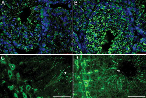
The localization of GalAAG in the testis of wild type and twitcher mice was analyzed by immunofluorescence. Positive staining revealed that GalAAG is mainly localized in Sertoli cells and in spermatid tail (). This localization is retained at later stages of sperm cell maturation, in fact, utilizing the same antibody, positive staining was detected in the middle piece of isolated sperm cells [Luddi et al. Citation2005]. The quantitative increase of GalAAG, previously demonstrated by lipid analysis [Luddi et al. Citation2005] could also be detected in immunocytochemistry, as indicated by the more intense fluorescence signal observed in the twitcher testis (,) compared to that of the wild type mice (,).
Figure 1. Immunofluorescence localization of galactosyl-alkyl-acyl-glycerol (GalAAG) in the testis of wild type (A,C) and twitcher mice (B,D) of tubules from stage VII-VIII. GalAAG (green) accumulation in Sertoli cell cytoplasm (asterisk) and spermatid tails (arrow head) is evident. Nuclei are stained in blue. Scale bar 25 μm.
Submicroscopic analysis of twitcher and wild type testes
Examination of periodic acid-Schiff stain (PAS)-stained cross-section of the twitcher and wild type seminiferous tubules shows that the morphogenetic processes proceed normally to step 4-5. In fact, the seminiferous epithelium architecture as well as the formation of the acrosome appears normal; the intercellular relationships are also maintained in the mutant (,). The stage was established based on the observation that in round spermatids the angle subtended by the stained acrosome is over 40° and extends up to 95°.
Figure 2. Light and transmission electron microscopy of twitcher and wild type seminiferous tubules at stage 4-5. Periodic acid-Schiff staining (PAS) highlights normal morphology of round spermatids both in wild type (A) and in twitcher (B). The acrosomes (arrow heads) are shown in red in the online version. Electron microscopy micrographs confirm that round spermatids at step 4-5 does not show differences between wild type (C) and twitcher (D); the acrosomal vesicle (asterisk) is tightly adherent to nuclear membrane and present a dense granule. BL: basal lamina; N: nucleus; M: mitochondria. Scale bar 20 µm (A,B), 2 µm (C,D).
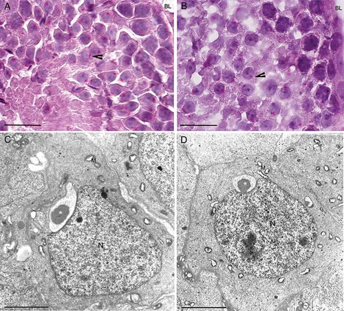
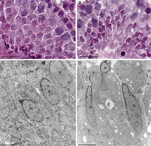
Electron microscopy analysis on ultrathin sections of the seminiferous tubules from twitcher and wild type mice was performed focusing on round spermatids starting from step 5. shows round spermatids with normal morphology at step 4-5 both in wild type (panel C) and twitcher (panel D) mice. The acrosomal vesicle, with the typical round profile, shows a well conformed granule in contact with the nucleus. It is important to point out that the negligible differences in the size of acrosomal vesicle observed in these round spermatids are most likely due to the slightly different level of the sections. No other obvious differences were detected analyzing stages 3 to 7 by transmission electron microscopy (TEM; data not shown). The earliest alterations in twitcher spermatid differentiation were found at steps 8-9. At step 8 the acrosomal complex in twitcher round spermatids appears less defined () than the wild type (). At the ultrastructural level in the twitcher mouse the development of the acrosome seems to be delayed () compared to wild type (). At step 9 these differences are more pronounced and the stained acrosomes appear morphologically abnormal (,). At the ultramicroscopic level (,) the sub-acrosomal space in twitcher spermatids appears enlarged; acrosomal projections in the anterior region of the nucleus are frequently detected and swollen acrosomes can also be found.
Figure 3. Light and transmission electron microscopy of twitcher and wild type tubule at stage 8. Periodic acid-Schiff staining (PAS) of round spermatids from wild type (A) and twitcher (B) mice shows an elongating acrosome cup-shaped. The acrosomes (asterisk) are shown in red in the online version. Electron microscopy micrographs showing spermatids from wild type (C) and twitcher mouse (D): the ruffling of the outer acrosomal membrane is evident only in gametes from mutant mice (arrowheads). BL: basal lamina; N: nucleus; M: mitochondria. Scale bar 100 µm (A,B), 3 µm (C,D).
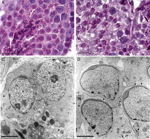
Figure 4. Light and transmission electron microscopy of twitcher and wild type tubule at stage 9. Periodic acid-Schiff staining (PAS) highlights the elongating acrosomes in wild type (A) and in twitcher (B), where altered acrosomal architecture is evident. The acrosomes (asterisk) are shown in red in the online version. Electron microscopy micrographs showing spermatids from wild type (C) and twitcher mouse (D). At ultrastructural level the sub-acrosomal space in twitcher spermatids appears enlarged (arrow heads) and swollen acrosomes and acrosomal projections in the anterior region of the nucleus are frequently detected (arrow). BL: basal lamina; N: nucleus; M: mitochondria. Scale bar 100 µm (A,B), 3 µm (C,D).
The analysis at step 16 of the spermatogenetic wave progression confirms that in twitcher spermatids, abnormally swollen acrosomes, often detached from the nucleus and folded over, () are present in comparison with wild type spermatids (). These morphological abnormalities were previously demonstrated in sperms from vas deferens [Luddi et al. Citation2005].
Figure 5. Electron microscopy micrographs of elongated spermatids at step 16 from wild type (A) and from twitcher mice (B). In wild type testis, spermatids have well conformed nucleus and acrosome. In panel B, the twitcher elongated spermatids reveal major structural acrosomal abnormalities. Indeed, the acrosome is redundant, swollen, and detached from the nucleus (arrow heads). N: nucleus; M: mitochondria. Scale bar 2 µm (A,B).
Analysis of twitcher and wild type epididymis
The high expression levels of lysosomal enzymes reported in the epididymis [Tulsiani et al. Citation1998b] indicated an essential role for these enzymes in remodeling the acrosomal membrane during the epididymal transit. Based on the altered acrosome morphology observed in twitcher sperm from deferens, a morphologic analysis of epididymis was carried out both in twitcher and in wild type mice.
Ultrastructural analysis at the TEM shows a completely different picture in the epididymis of twitcher compared to wild type mice. In the corpus epididymidis, the major cell types, including principal, apical, and clear cells are well represented, and their relative position is maintained both in wild type and twitcher epithelium. In the wild type mice the stereocilia were attached to the columnar epithelial cells and many sperms were present into the lumen (,), whereas in twitcher mice the lumen appeared filled with shedded stereocilia, and few sperms were present (,). In regard to epididimal sperm morphology, normal sperm head and tail interacting with epididymal stereocilia are visible in wild type epididymis (); in twitcher mice abnormally shaped sperms with large residual bodies and cytoplasmic droplets are interspersed among the broken stereocilia (,).
Figure 6. Transmission electron microscopy of corpus epididymidis from wild type (A,C) and from twitcher (B,D) mouse. The epithelium appears well structured both in wild type and in twitcher mice (A,B), but the apical region shows several differences: well shaped stereocilia are present in wild type mice (C) whereas, in twitcher mice, these structures are broken, detached, and completely spread out into the lumen (D). Sc: stereocilia. Scale bar: A,B, 10 µm; C,D, 2 µm.
Figure 7. Transmission electron microscopy of sperm in the epididymal lumen at the level of cauda from wild type (A) and twitcher mouse (B,C). In twitcher epididymal lumen, misshaped sperms with structural acrosomal abnormalities (asterisk), large residual bodies, and cytoplasmic droplets are clearly visible. N: nucleus; Ax: axoneme; Sc: stereocilia. Scale bar 2 µm.
Abnormality of epididymal architecture was also confirmed by scanning electron microscopy (SEM) (). Well organized stereocilia project into the lumen of the tubules of the wild type mice, whereas in the mutants the epididymal lumen results were larger and empty for the loss of well structured stereocilia.
Figure 8. Scanning electron microscopy of epididymis from wild type (A) and twitcher mouse (B). In wild type mice well developed stereocilia project into the lumen of the tubule, while in the mutants the epididymal epithelium appears disorganized, the lumen larger and empty for the loss of stereocilia. Scale bar 20 µm.
Discussion
Alterations in sperm maturation and acrosome morphology, in addition to a significant accumulation of undegraded GalAAG and minor changes in the SGalAAG concentration, were previously reported in the testes of the twitcher mouse [Luddi et al. Citation2005]. Using an immunohistochemical approach, we have focused our attention on the differentiation of twitcher germ cells from pachytene spermatocyte stage onwards to carefully assess at what stage morphologic abnormalities occur.
In mammalian testes and spermatozoa the seminolipid, SGalAAG, is the most abundant glycolipid and comprises about 3% of total lipids. The synthesis of SGalAAG from GalAAG increases rapidly in the early phase of spermatocyte development and it is maintained in the later germ cell stages [Goto-Inoue et al. Citation2009]. The absence of mature spermatozoa in knockout mice, deficient in the enzyme which synthesize these lipids, has clearly demonstrated that a proper balance of seminolipid and its precursor are essential for normal spermatogenesis [Fujimoto et al. Citation2000; Honke et al. Citation2002]. Our data, not only confirm that GalAAG is expressed in spermatocytes as well as in Sertoli cells and spermatid tails, but also show increased expression of this lipid in the twitcher Sertoli cells compared to wild type mice. It has been suggested that the interaction between Sertoli cells and spermatocytes is essential for spermatocytes differentiation and that any alteration in their metabolism would result in impairment of this relationship [Honke et al. Citation2002].
The PAS analysis of twitcher and wild type mice tubules presented in this study shows that the alterations of sperm maturation and acrosome morphology are first detectable at stage VIII of the seminiferous epithelium cycle, clearly demonstrating that these abnormalities are not caused by post-testicular degeneration. At this stage, preleptotene and pachytene spermatocytes are well shaped, but there are evident pathological alterations in spermatids at step 8. Obviously, elongated spermatids at step 16, also present at this stage, show clear alterations in the acrosome architecture and re-absorption of the cytoplasmic droplets.
In the transition between steps 7-8, many important processes take place, in particular the nucleus reaches the cell surface, and the inner membrane surface of the acrosome is in contact with the nuclear membrane, while the outer membrane surface of the acrosome approaches the plasma membrane. It can be hypothesized that the alteration in glycolipid composition observed in the twitcher testes could affect membrane fluidity and composition, thus interfering with proper interaction between acrosome and nuclear or plasma membrane and/or Sertoli cells. All these interactions have a pivotal role, allowing the proper crosstalk between cells involved in spermiohistogenesis. It has been demonstrated that seminolipids present in the lipid rafts contribute to the sperm cell membrane shape and stability and that they are also involved in recognition processes and cell–cell adhesion [Bou Khalil et al. Citation2006]. The abnormal GalAAG and SGalAAG accumulation observed in twitcher testes could interfere with lipid raft composition, thus explaining morphological abnormalities observed in twitcher spermatids and mature sperms.
We have previously demonstrated that the altered lipid metabolism, due to GALC deficiency, is not the only cause of male infertility. Our data indicated that the severe involvement of the CNS causes a derangement of the hormone gene expression profile and interferes with the crosstalk between the nervous system and the male gonads, thus contributing to the infertility of the twitcher mouse [Puggioni et al. Citation2015]. In a previous study, we have also shown that the number of apoptotic cells, mainly condensing spermatids, was significantly increased in the twitcher compared to the wild-type mice [Luddi et al. Citation2005]. This increase could, indeed, be correlated to the unbalanced hormonal profile. The data of Zhu et al. [Citation1997] show that in the rat reduced levels of testosterone induce apoptosis of germ cells. It is interesting to note, that in the present study we report that the abnormalities of sperm maturation and acrosome morphology were first identified at stage VIII, coincident with the androgen-dependent stages (VII and VIII) [Zhu et al. Citation1997]. While we have not measured the level of intratesticular testosterone, we have demonstrated a significant reduction of lipid droplets in the Leydig cells of twitcher mice [Puggioni et al. Citation2015]. Based on the established correlation between the synthesis of testosterone and the number of lipid droplets present in Leydig cells [Chigurupati et al. Citation2008], a reduction in testosterone production could be reasonably hypothesized in the affected mice thus explaining the increased number of apoptotic cells observed in twitcher testis [Luddi et al. Citation2005].
The high levels of expression of lysosomal enzymes measured in the epididymis of wild type mice [Belmonte et al. 1995], the hairpin morphology, and the presence of large cytoplasmic droplets in twitcher sperm previously described [Luddi et al. Citation2005] suggested that the epidydimal maturation could also be impaired in the mutant mice. TEM and SEM data demonstrate that the epididymal architecture is, indeed, impaired in twitcher mice, with stereocilia of caput and corpus broken, detached, and completely spread into the lumen. It may be hypothesized that this structural impairment should be a consequence of substrate accumulation, causing the massive macrophage activation and the considerable inflammation feature of this pathology. This is in agreement with several publications reporting that the presence of detached stereocilia in the lumen of epididymis is associated to direct or indirect evidence of infection and to the presence of varicocele in many of the analyzed cases, strongly suggesting a cause-effect relationship between these conditions [Comiter et al. Citation1995; González-Jiménez and Villanueva-Díaz Citation2006].
In summary, the present data add a significant advance to the current knowledge on the importance of these glycolipids during sperm maturation and differentiation. In fact, changes in the concentration of glycolipids can induce a modification in the arrangement and fluidity of the plasma membrane of germ cells. These changes could adversely affect the lateral diffusion of lipids and their movements in the different micro-regions throughout the remodeling of the sperm membrane during maturation and transition in the epididymis.
The reported hormonal imbalance [Puggioni et al. Citation2015], due to severe degeneration of the central nervous system, associated to the described morphological and biochemical abnormalities, begin to delineate a clearer picture of the factors interfering with spermatogenesis in the twitcher mice. It is well known that in other lysosomal storage diseases, genetic disorders caused by lysosomal enzyme deficiency, the degeneration of the central nervous system is often associated to defects of spermatogenesis. It is reasonable to hypothesize that the altered hypothalamic-pituitary-testicular axis plays an important role not only in the Twitcher mouse infertility but also in other lysosomal diseases where brain injury is associated to defects of the spermatogenesis.
Materials and methods
Animals
Twitcher heterozygote breeding pairs (C57BL/6J) were purchased from Jackson Laboratory (Bar Harbor, ME, USA) and maintained in our institution under controlled conditions (22 ± 2°C, 55 ± 5% humidity, 12 h light/dark cycle) with water and food ad libitum. The genotypes of all newborn mice were determined by a rapid PCR-based-method using DNA isolated from clipped tail, modified from Sakai and collaborators [Sakai et al. Citation1996]. All the animals were treated according to the recommendations of the European Convention for the Protection of Vertebrate Animals used for experimental and other scientific purposes and approved by the Institutional Animal care and Use Committee.
Tissue preparation
The mice were sacrificed by cervical dislocation on postnatal day (PND) 35 (n = 6 wild type and n = 6 twitcher). The testes were removed immediately and one of each pair was fixed by immersion in Bouin’s after cutting tunica albuginea at the poles of the testes. The other was processed for electron microscopy. The epididymis was processed accordingly for electron microscopy.
Staging of tubules
In order to monitor single stage of the spermatogenesis, three different pairs of mice were used. Each pair consisted of one mutant and one wild-type mouse of the same age; all mice were 35 days of age. Testis from each mouse was removed, cryoprotected, and frozen. Cryostat sections (12 μm) were stained with PAS to identify the stage of spermatid development. Briefly, cryostat sections were hydrated in distilled water. The slides were immersed in a solution of periodic acid 0.5% for 8 min at room temperature and then washed in running water for 10 min. The slides were then immersed in Schiff reagent for 35 min at room temperature, washed for 15 min in running water, double stained in Mayer’s hematoxylin solution for 30 s, washed in running water for several min, dehydrated, and mounted in DPX mountant for histology (Sigma-Aldrich, Milan, Italy).
For staging, one testis cross section per mouse was analyzed under the light microscope. The total number of tubules in each section was counted, and all tubules were assigned a stage based on the criteria outlined by Russell and collaborators [Russell et al. Citation1990].
TEM
Testis from twitcher and wild type mice were removed from mice upon perfusion and fixed for 2 h at 4°C in Karnovsky fixative (paraformaldehyde 1%, glutaraldehyde 5% in cacodylate buffer 0.2 M, pH 7.4). After washing in cacodylate buffer, the tissue was cut in small pieces (not to exceed 1 mm cubed), immersed in the same fixative for 2 h at 4°C, rinsed, and postfixed in 1% buffered OsO4, dehydrated through a graded ethanol changes (50% ethanol - 10 min, 70% ethanol - 10 min, 80% ethanol - 10 min, 95% ethanol with 2 changes within 10 min and then with 3 changes within 15 min of 100% ethanol from a newly opened bottle). The samples were then treated with Propylene oxide for 10 min, 1:1 propylene oxide\resin for minimum of 1 h, 1:2 propylene oxide\resin for 1 h, 100% Epon for over 2-6 h, and finally embedded in Epon Araldite. Ultrathin sections were cut using a Supernova Reichert-Jung (Wien) ultramicrotome, collected on copper grids, stained with uranyl acetate and lead citrate, and examined in a transmission electron microscope Philips 301 (Philips Electronic Instruments Co., Pittsburgh, PA, USA). Magnification was calibrated in each section.
SEM
Epididymis from twitcher and wild type mice were fixed for 1 h in Karnovsky fixative, washed 3 times in cacodylate buffer, dehydrated in increasing concentrations of ethanol, critical point dried, and observed in a scanning electron microscope Philips 500 (Philips Electronic Instruments Co.).
Immunofluorescence
Testes and epididymis were removed from twitcher and wild type mice after perfusion with 4% paraformaldheyde (PFA). The sections of testis and epididymis sliced with a cryostat (7 μm) were hydrated in distilled water, attached on glass slides, and immunolabeled using an indirect procedure. All incubations were performed in blocker solution containing 5% goat serum, to block unspecific binding sites. Sections were incubated with anti-GalAAG mouse monoclonal antibody, 1:100 overnight at 4°C. Slides were then rinsed 3 times with PBS (pH 7.4) and exposed to secondary antibody FITC-labeled goat anti-mouse IgM antibody (Sigma, St. Louis, MO, USA) for 60 min at room temperature in a dark chamber. Specificity of immunostaining was confirmed by both omission of primary antibody and staining of parallel sections with control antibodies. After washing in PBS, the slides were mounted in Prolong antifade (LifeTechologies, CA, USA), then observed with a Leica DMB 6000 microscope (Leica, Solms, Germany), images captured with an CFTR6500 digital camera (Leica), and analysis of 10 fields was performed with LSA software (Leica) by two independent observers.
Declaration of interest
The authors report no declaration of interest.
Additional information
Notes on contributors
Alice Luddi
Study design, data interpretation, manuscript drafting: LA, GM; Data acquisition and analysis: GM, CL, MC, BG; Critical discussion: EC; Study design, manuscript drafting, and critical discussion: PP. All authors read and approved the final manuscript.
Martina Gori
Study design, data interpretation, manuscript drafting: LA, GM; Data acquisition and analysis: GM, CL, MC, BG; Critical discussion: EC; Study design, manuscript drafting, and critical discussion: PP. All authors read and approved the final manuscript.
Laura Crifasi
Study design, data interpretation, manuscript drafting: LA, GM; Data acquisition and analysis: GM, CL, MC, BG; Critical discussion: EC; Study design, manuscript drafting, and critical discussion: PP. All authors read and approved the final manuscript.
Camilla Marrocco
Study design, data interpretation, manuscript drafting: LA, GM; Data acquisition and analysis: GM, CL, MC, BG; Critical discussion: EC; Study design, manuscript drafting, and critical discussion: PP. All authors read and approved the final manuscript.
Giuseppe Belmonte
Study design, data interpretation, manuscript drafting: LA, GM; Data acquisition and analysis: GM, CL, MC, BG; Critical discussion: EC; Study design, manuscript drafting, and critical discussion: PP. All authors read and approved the final manuscript.
Elvira Costantino-Ceccarini
Study design, data interpretation, manuscript drafting: LA, GM; Data acquisition and analysis: GM, CL, MC, BG; Critical discussion: EC; Study design, manuscript drafting, and critical discussion: PP. All authors read and approved the final manuscript.
Paola Piomboni
Study design, data interpretation, manuscript drafting: LA, GM; Data acquisition and analysis: GM, CL, MC, BG; Critical discussion: EC; Study design, manuscript drafting, and critical discussion: PP. All authors read and approved the final manuscript.
References
- Bellvé, A.R., Cavicchia, J.C., Millette, C.F., O’Brien, D.A., Bhatnagar, Y.M. and Dym, M. (1977) Spermatogenic cells of the prepuberal mouse. Isolation and morphological characterization. J Cell Biol 74:68–85.
- Belmonte, S.A., Romano, P., Sartor, T. and Sosa, M.A. (2002) Compartmentalization of lysosomal enzymes in cauda epididymis of normal and castrated rats. Arch Androl 48:193–201.
- Bou Khalil, M., Chakrabandhu, K., Xu, H., Weerachatyanukul, W., Buhr, M., Berger, T., et al. (2006) Sperm capacitation induces an increase in lipid rafts having zona pellucida binding ability and containing sulfogalactosylglycerolipid. Dev Biol 290:220–235.
- Butler, A., He, X., Gordon, R. E., Wu, H.-S., Gatt, S. and Schuchman, E.H. (2002) Reproductive pathology and sperm physiology in acid sphingomyelinase-deficient mice. Am J Pathol 161:1061–1075.
- Chigurupati, S., Son, T.G., Hyun, D.H., Lathia, J.D., Mughal, M.R., Savell, J., et al. (2008) Lifelong running reduces oxidative stress and degenerative changes in the testes of mice. J Endocrinol 199:333–341.
- Comiter, C.V, Bruning, C.O. and Morgentaler, A. (1995) Detached ciliary tufts in the epididymis: a lesson in applied anatomy. Urology 46:740–742.
- De Kretser, D.M. and Baker, H.W. (1999) Infertility in men: recent advances and continuing controversies. J Clin Endocrinol Metab 84:3443–3450.
- Fujimoto, H., Tadano-Aritomi, K., Tokumasu, A., Ito, K., Hikita, T., Suzuki, K., et al. (2000) Requirement of seminolipid in spermatogenesis revealed by UDP-galactose: Ceramide galactosyltransferase-deficient mice. J Biol Chem 275:22623–22626.
- González-Jiménez, M.A. and Villanueva-Díaz, C.A. (2006) Epididymal stereocilia in semen of infertile men: evidence of chronic epididymitis? Andrologia 38:26–30.
- Goto-Inoue, N., Hayasaka, T., Zaima, N. and Setou, M. (2009) The specific localization of seminolipid molecular species on mouse testis during testicular maturation revealed by imaging mass spectrometry. Glycobiol 19:950–957.
- Holden, C.A., McLachlan, R.I., Pitts, M., Cumming, R., Wittert, G., Agius, P.A., et al. (2005) Men in Australia Telephone Survey (MATeS): a national survey of the reproductive health and concerns of middle-aged and older Australian men. Lancet 366:218–224.
- Honke, K., Hirahara, Y., Dupree, J., Suzuki, K., Popko, B., Fukushima, K., et al. (2002) Paranodal junction formation and spermatogenesis require sulfoglycolipids. Proc Natl Acad Sci U S A 99:4227–4232.
- Ishizuka, I. (1997) Chemistry and functional distribution of sulfoglycolipids. Prog Lipid Res 36:245–319.
- Jamsai, D. and O’Bryan, M.K. (2011) Mouse models in male fertility research. Asian J Androl 13:139–151.
- Kobayashi, T., Yamanaka, T., Jacobs, J.M., Teixeira, F. and Suzuki, K. (1980) The Twitcher mouse: an enzymatically authentic model of human globoid cell leukodystrophy (Krabbe disease). Brain Res 202:479–483.
- Luddi, A., Strazza, M., Carbone, M., Moretti, E. and Costantino-Ceccarini, E. (2005) Galactosylceramidase deficiency causes sperm abnormalities in the mouse model of globoid cell leukodystrophy. Exp Cell Res 304:59–68.
- Michaelis, M., Langhammer, M., Hoeflich, A., Reinsch, N., Schoen, J. and Weitzel, J.M. (2013) Initial characterization of an outbreed mouse model for male factor (in)fertility. Andrology 1:772–778.
- Puggioni, E., Governini, L., Gori, M., Belmonte, G., Piomboni, P., Costantino-Ceccarini, E., et al. (2015) Morphological and molecular characterisation of Twitcher mouse spermatogenesis: an update. Reprod Fertil Dev 2015 Feb 10. doi: 10.1071/RD14279. Epub ahead of print.
- Russell, L.D., Ettlin, R.A., Sinha Hikim, A.P. and Clegg, E.D. (1990) Mammalian spermatogenesis. In Histological and Histopathological Evaluation of the Testis (pp. 1–40), Cache River Press: Clearwater.
- Sakai, N., Inui, K., Tatsumi, N., Fukushima, H., Nishigaki, T. Taniike, M., et al. (1996) Molecular cloning and expression of cDNA for murine galactocerebrosidase and mutation analysis of the twitcher mouse, a model of Krabbe’s disease. J Neurochem 66:1118–1124.
- Trasler, J., Saberi, F., Somani, I. H., Adamali, H. I., Huang, J. Q., Fortunato, S. R., et al. (1998) Characterization of the testis and epididymis in mouse models of human Tay Sachs and Sandhoff diseases and partial determination of accumulated gangliosides. Endocrinol 139:3280–3288.
- Tulsiani, D.R.P., Abou-Haila, A., Loeser, C.R., and Pereira, B.M.J. (1998a) The Biological and Functional Significance of the Sperm Acrosome and Acrosomal Enzymes in Mammalian Fertilization. Exp Cell Res 240:151–164.
- Tulsiani, D.R., Orgebin-Crist, M.C. and Skudlarek, M.D. (1998b) Role of luminal fluid glycosyltransferases and glycosidases in the modification of rat sperm plasma membrane glycoproteins during epididymal maturation. J Reprod Fertil Suppl 53:85–97.
- Wenger, D.A., Suzuki, K., Suzuki, Y. and Suzuki, K. (2001) Galactosylceramide lipidosis: globoidcell leukodystrophy (Krabbedisease). In Metabolic and Molecular Basis of Inherited Disease ed. C.R. Scriver, A.L. Beaudet, W.S. Sly and D. Valle, McGraw- Hill Press, NewYork, pp 3669–3694.
- Xu, H., Kongmanas, K., Kadunganattil, S., Smith, C. E., Rupar, T., Goto-Inoue, N., et al. (2011) Arylsulfatase A deficiency causes seminolipid accumulation and a lysosomal storage disorder in Sertoli cells. J Lipid Res 52:2187–2197.
- Yanagimachi, R., Harper, M.J.K., De Kretser, D.M. and Kerr, J.B. (1994) Mammalian fertilization, gamete and zygote transport, and cytology of the testis. In The Physiology of Reproduction 2nd ed. Ed. E. Knobil, J.D. and Neill, Raven Press, New York, pp 189–206.
- Zhu, L.J., Zong, S.D., Phillips, D.M., Moo-Young, A.J, and Bardin, C.W. (1997) Changes in the distribution of intermediate filaments in rat Sertoli cells during the seminiferous epithelium cycle and postnatal development. Anat Rec 248:391–405.