
ABSTRACT
There are variant rates of oocyte degeneration after intracytoplasmic sperm injection (ICSI) among different patients. Oocyte degeneration after ICSI may reflect the cohort of oocyte quality and subsequent embryo development capacity and clinical outcome. This retrospective study analyzed 255 cycles with at least one degenerated oocyte after ICSI (degeneration group) and 243 cycles with no degenerated oocytes after ICSI (control group). Basic characteristics like female age, body mass index, duration of infertility, hormone (FSH, LH, E2) levels on day 3 of menses, and primary infertility patient rate were similar between the two groups (p > 0.05). Total dose of gonadotropin and length of stimulation were also similar between the two groups (p > 0.05), but the degeneration group exhibited a more exuberant response to ovarian stimulation as reflected by more oocytes retrieved (p < 0.05). The number of 2PN embryos available and high quality embryos were similar between the two groups (p > 0.05), but the high quality embryo rate, early cleavage embryo rate, and available embryo rate were all statistically lower than the control group (p < 0.05). Embryo developmental kinetics seemed to be disturbed and embryo fragmentation rate increased in the degeneration group (p < 0.05). However, there was no statistical difference in the distribution of graded embryos transferred, and there were no statistical differences in the pregnancy rate, implantation rate, and abortion rate between the two groups (p > 0.05). We deduce that the presence of oocyte degeneration after ICSI may be associated with decreased embryo quality with embryo development kinetics disturbed. However, the clinical outcomes may not be affected if the premise that sufficient high quality degeneration group embryos are available for transfer.
Introduction
Oocyte degeneration after intracytoplasmic sperm injection (ICSI) is a common phenomenon during human assisted reproduction. It is often characterized by oocyte lysis after withdrawal of the injection needle or being noted the next day by retracted and/or darkened ooplasm [Rosen et al. Citation2006]. Previous reports suggested that the rate of oocyte degeneration is operator dependent, and a smaller injection needle can minimize the incidence of degeneration [Dumoulin et al. Citation2001; Yavas et al. Citation2001]. However, there is still a variant oocyte degeneration rate in different patients even when the same skilled technician uses small injection needles. A multivariate analysis showed that oocyte degeneration after ICSI is not technician or physician dependent, but reflects inherent oocyte quality [Rosen et al. Citation2006]. As there are differences in oocyte quality at the time of oocyte retrieval [Gu et al. Citation2015], oocyte degeneration after ICSI may reflect the different inherent oocyte quality in different patients.
For a cohort of oocytes retrieved from the same couple in the same ICSI treatment cycle, it is not known whether the presence of oocyte degeneration after ICSI can reflect the sibling oocyte quality and affect the associated embryo development potential and clinical pregnancy outcome. Oocyte quality predominantly determines embryo quality [Mikkelsen and Lindenberg Citation2001; Ten et al. Citation2007], and oocyte dysmorphism (like large first poly body, large periviteline space, refractile bodies, or vacuoles) is associated with a decreased oocyte fertilization rate [Setti et al. Citation2011]. It has been reported that empty zona pellucida may indicate decreased oocyte quality and be related to poor ovarian response [Cinar et al. Citation2011], and a giant oocyte in a cohort of retrieved oocytes is associated with abnormal cleavage in sibling embryos [Machtinger et al. Citation2011]. We presume that the presence of oocyte degeneration after intracytoplasmic injection may reflect oocyte quality of the cohort and affect the subsequent embryo developmental potential and clinical pregnancy outcome.
In this study, we retrospectively analyzed 498 cycles using the prolonged GnRH agonist protocol for ovarian stimulation and performed fresh embryo transfer on day 3 after insemination. A total of 255 cycles presented with at least one oocyte degenerated after injection and were defined as the degeneration group. In comparison, 243 cycles with no degenerated oocyte after ICSI were defined as the control group. Basic characteristics, embryo quality, and clinical outcomes of the two groups were compared and analyzed to test our hypothesis.
Results and discussion
Oocyte degeneration after ICSI commonly occurs during ICSI treatment. It has been reported that the rate of degeneration ranges from 0 to 18% in ICSI [Rosen et al. Citation2006]. In this study, we analyzed 5,130 mature oocytes that underwent ICSI treatment in 498 cycles, and found 476 mature oocytes in 255 cycles degenerated after ICSI treatment. The oocyte degeneration rate is 9.28% and degeneration cycle rate is 51.20%. Two operators both with more than 3 years experience carried out the ICSI treatment in this study, and there was no statistical difference in oocyte degeneration rates between the two operators (9.61% vs. 8.93%, p = 0.401). Little is known about the causes of oocyte degeneration and its potential effect on the cohort of sibling oocyte quality, subsequent embryo development capacity, and clinical pregnancy outcomes in cycles with at least one oocyte degenerated after ICSI. In this study, two groups of ICSI cycles were considered based on whether oocyte degeneration occurred after ICSI. Basic characteristics, including day 3 embryo quality and clinical outcomes of the two groups were compared and analyzed.
As shown in , there was no significant difference in female age, body mass index, duration of infertility, FSH level on day 3 of menses, LH level on day 3 of menses, E2 level on day 3 of menses, and primary infertility patient rate (p > 0.05). There was no significant difference in rates of ejaculated spermatozoa, testicular spermatozoa, and epididymal spermatozoa, there was also no significant difference in the rates of fresh spermatozoa and frozen-thawed spermatozoa used for ICSI (p > 0.05). The quality of the embryo that is transferred predominantly determines the clinical pregnancy outcome [Berger et al. Citation2014]. In this study, there was no significant difference in the transferred embryo number, the distribution of the grades of embryos transferred, and endometrial thickness on the day of hCG; accordingly, there was also no significant difference in the clinical pregnancy rate, implantation rate, and abortion rate between the two groups (as shown in , ). The uncompromised implantation rate in the degeneration group may reflect a similar developmental potential of the transferred embryos between the two groups.
Table 1. Basic characteristics of the two groups.
Table 2. Cycle outcome variables of the two groups.
Figure 1. Distribution of day 3 embryo grades. Day 3 embryos were assessed and divided into four different grades according to the ASEBIR embryo assessment criteria [Alpha Scientists in Reproductive Medicine and ESHRE Special Interest Group of Embryology, et al. Citation2011]. Grade 1 and grade 2 embryos were considered as high quality embryos, grade 1, grade 2, and grade 3 embryos were considered as available embryos, while grade 4 embryos were usually deserted. (A) shows that grade 1 and grade 2 embryo rates decreased while grade 3 and grade 4 embryo rates increased as a function of total day 3 embryos in the degeneration group (p = 0.035). (B) shows there was no significant difference in the distribution transferred embryo grades on day 3 between the two groups (p = 0.537).
![Figure 1. Distribution of day 3 embryo grades. Day 3 embryos were assessed and divided into four different grades according to the ASEBIR embryo assessment criteria [Alpha Scientists in Reproductive Medicine and ESHRE Special Interest Group of Embryology, et al. Citation2011]. Grade 1 and grade 2 embryos were considered as high quality embryos, grade 1, grade 2, and grade 3 embryos were considered as available embryos, while grade 4 embryos were usually deserted. (A) shows that grade 1 and grade 2 embryo rates decreased while grade 3 and grade 4 embryo rates increased as a function of total day 3 embryos in the degeneration group (p = 0.035). (B) shows there was no significant difference in the distribution transferred embryo grades on day 3 between the two groups (p = 0.537).](/cms/asset/86da7ffd-c08c-4d5c-938d-253332d78ff7/iaan_a_1272648_f0001_oc.jpg)
Previous studies have reported heterogeneity in the cohort of oocytes retrieved. Oocyte degeneration after ICSI is likely a function of the inherent oocyte quality. The remaining oocytes within the cohort may be of good quality and not affected [Xia Citation1997; Rosen et al. Citation2006], but embryo quality and morphology parameters like fragmentation were not analyzed. In comparison, as reported here, grade 1 and grade 2 embryo rates decreased while grade 3 and grade 4 embryo rates increased on day 3 after fertilization (, p = 0.035). As shown in , the degeneration group exhibited a more exuberant response to ovarian stimulation as reflected by more oocytes retrieved (p < 0.001). While the degeneration group received more mature oocytes (p < 0.05), the high quality embryo rate, available embryo rate, and early cleavage embryo rate in the degeneration group were all significantly lower than in the control group (p < 0.05). The mature oocyte rate in the degeneration group was significantly higher than in the control group (p < 0.001), but the 2 PN embryo fertilization rate (2PN embryos/MII oocytes) was significantly lower in the degeneration group (p < 0.001). All these results indicate that the presence of oocyte degeneration after intracytoplasmic injection may reflect poor quality of the cohort of oocytes and affect subsequent embryo development.
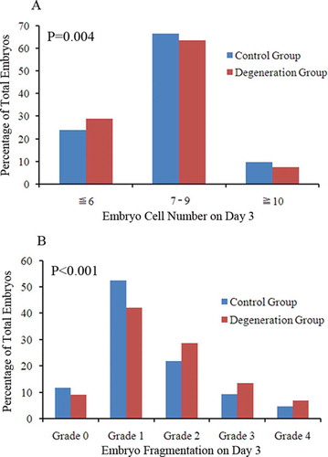
Embryo morphology is greatly associated with embryo development quality and capacity [Munne Citation2006], and embryo morphology assessment is currently the most important factor for the prediction of pregnancy [Alpha Scientists in Reproductive Medicine and ESHRE Special Interest Group of Embryology, et al. Citation2011]. On one hand, it has been shown that embryos with only 2-3 cells and those with more than 8 cells on day 3 have significantly higher rates of aneuploidy compared with those with 7-8 cells [Magli et al. Citation2007]. On the other hand, embryos with 8 cells on day 3 had the highest implantation potential compared with other day 3 embryos with either a lower or higher number of cells [Racowsky et al. Citation2003]. Cellular fragmentation is a sensitive proxy for predicting embryo euploidy [Moayeri et al. Citation2008]. In this study, embryo development kinetics in the degeneration group was perturbed as reflected by a lower proportion of ‘normal’ embryos with 7-9 cells on day 3 (63.43% vs. 66.46%), a lower proportion of ‘accelerated’ embryos with more than 9 cells on day 3 (7.57% vs. 9.71%), and a higher proportion of ‘slow’ embryos with less than 7 cells on day 3 (29.00% vs. 23.83%) (, p = 0.004). ‘Normal’, ‘slow’, and ‘accelerated’ embryo definition followed the Istanbul consensus [Alpha Scientists in Reproductive Medicine and ESHRE Special Interest Group of Embryology, et al. Citation2011]. Embryo fragmentation also statistically increased in the degeneration group as reflected by the decrease of grade 0 and grade 1 embryo rates and increase of grade 2, grade 3, and grade 4 embryo rates on day 3 (, p < 0.001). These results hint that the presence of oocyte degeneration after ICSI may associate with the increase of embryo chromosomal abnormality in the sibling of embryos retrieved in the cohort. However, clinical outcome was not affected as there was no statistical difference between the two groups in clinical pregnancy rate and implantation rate. This observation may reflect the availability of sufficient high quality embryos for transfer in the degeneration group, thereby providing embryos of similar quality for transfer as the control group, and this deduction is compatible with the similar transferred high quality embryo rate between the two groups.
Figure 2. Embryo morphology analysis on day 3. The number of day 3 embryo cells were divided into ‘slow’ embryos with ≤ 6 cells, ‘normal’ embryos with 7-9 cells, and ‘accelerated’ embryos with ≥ 10 cells. Embryo fragmentation grades were divided into five grades: Grade 0 embryos were those with no fragmentation, grade 1 embryos were those with < 10% fragmentation, grade 2 embryos were those with 11%-25% fragmentation, grade 3 embryos were those with 26%-50% fragmentation, and grade 4 embryos were those with > 50% fragmentation. (A) Shows the degeneration group got lower proportions of ‘normal’ embryos and ‘accelerated’ embryo, higher proportion of ‘slow’ embryos compared to the control group (p = 0.004). (B) shows that grade 0 and grade 1 fragmentation rates decreased while grade 2, grade 3, and grade 4 fragmentation rates increased in the degeneration group (p < 0.001).
Theoretically, if degenerated oocytes result from oocyte maturation incompetence or the inadequate microenvironment during oocyte development [Van Hoeck et al. Citation2014; Jahromi et al. Citation2015], the sibling oocytes and associated embryos may also be affected. Ooctye quality predominantly determines embryo quality, poor oocyte quality may cause infertility of women and failure of in vitro fertilization treatment [Marteil et al. Citation2009; Sa et al. Citation2011]. Ooctye meiosis is very sensitive to endogenous and exogenous factors that could result in oocyte chromosomal abnormalities [Plachot Citation2001]. Exposure to certain harmful culture medium may impair oocyte development capacity during in vitro culture [Maside et al. Citation2012; Spricigo et al. Citation2012]. For example, oocytes retrieved from under-oxygenated follicles were developmentally compromised and had a high rate of chromosomal defects [Van Blerkom et al. Citation1997]. It has been shown that ovarian stimulation is also related to oocyte and embryo quality. Mild ovarian stimulation significantly reduced the abnormal and mosaic embryo rate compared to the conventional GnRH agonist protocol [Baart et al. Citation2007]. In this study, all patients used the same GnRH agonist protocol to eliminate this disturbing factor. Kaipia et al. [Citation1997] has reported that oocyte fate may be determined by the granulose cells and follicle microenvironment. Oocytes retrieved from less than optimal follicular environments may result in poor quality oocytes, with their fate being realized at the time of ICSI as the developmental capacity of the embryo. Xia [Citation1997] suggested that oocyte degeneration rates were higher in cases where the total number of oocytes retrieved was greater than 20 and E2 level was > 3000 pg/ml on the day of hCG. Logistic regression analysis showed E2 levels on the day of hCG were independent determinants of oocyte degeneration rate [Rosen et al. Citation2006]. These results suggest that the E2 level is important during oocyte maturation. Marmoset oocyte in vitro maturation showed that different concentrations of estradiol positively affect oocyte maturation and embryo development. Only the optimal concentration of estradiol could produce competent oocytes and embryos, and the optimal concentration range is critical [Tkachenko et al. Citation2015]. In this study, the E2 level on the day of hCG was significantly higher in the degeneration group, and the degeneration group retrieved more oocytes than the control group. The high level of E2 may directly affect oocyte maturation during controlled ovarian stimulation and the subsequent embryo development.
It has been reported that embryos from cycles that did not result in pregnancy had significantly diminished blastocyst development and inner cell mass quality compared with those originated from cycles with successful pregnancy [O’Leary et al. Citation2012]. This may reflect the poor quality of the cohort of embryos in another way. These together strengthen our hypothesis that the presence of oocyte degeneration after ICSI may reflect the sibling oocyte quality and the subsequent embryo development capacity.
In conclusion, we found that the presence of oocyte degeneration after ICSI is associated with the quality of the cohort of oocytes and embryos retrieved. The rate of high quality embryos significantly deceased and embryo cleavage kinetics seem to be perturbed in cycles with at least one oocyte degenerated after ICSI. This may be caused by the less than optimal microenvironment during oocyte maturation. However, additional studies are needed to investigate whether there is any correlation between the presence of oocyte degeneration after ICSI and the increased risk of chromosomal abnormalities in the sibling oocytes and embryos in a cohort.
Materials and methods
Institutional review board approval for this study was obtained from Ethical Committee of Medical College Xiamen University. All subjects enrolled in this study gave written formal consent before participation.
Study participants
Patients aged less than 35 years who underwent ICSI for male factor from 2014 to 2015 in our center were enrolled and retrospectively analyzed. A total of 498 cycles using prolonged GnRH agonist protocol [Ren et al. Citation2014] and performed fresh embryo transfer on day 3 after insemination were included in this study. Among the 498 cycles included, 255 cycles with at least one oocyte degenerated after ICSI were defined as the degeneration group, and 243 cycles with no oocyte degenerated after ICSI were defined as the control group. Basic characteristics, embryo quality, and clinical outcomes of the two groups were compared.
Ovarian stimulation
All patients enrolled used prolonged GnRH agonist protocol as previously described [Ren et al. Citation2014] for ovarian stimulation, domestic urinary human menopausal gonadotropin (hMG, Lizhu Parma, Zhuhai, Guangdong, China) was used for ovarian stimulation, 5,000-10,000 IU human chorionic gonadotropin (hCG, Lizhu Parma) was injected when at least one follicle ≥ 18 mm in mean diameter. Oocyte retrieval was performed using transvaginal ultrasound device 34-36 h after hCG injection.
ICSI and degeneration determination
Cumulus denudation was performed 2-4 h after oocyte retrieval, sperm was immobilized in polyvinylpyrrolidone solution, and ICSI was routinely performed 4-6 h after oocye retrieval by two experienced operators. Oocyte degeneration was determined either at the time during ICSI procedure characterized by oocyte lysis after withdrawal of the injection needle or being noted the next day by retracted and/or darkened ooplasm [Rosen et al. Citation2006].
Embryo culture and assessment
Embryos were cultured at 37C° in a humified atmosphere of 5% CO2 in air and observed every day. Embryo grading system was based on numbers of embryo blastomere, fragmentation, and symmetry [Racowsky et al. Citation2003; Reichman et al. Citation2010; Alpha Scientists in Reproductive Medicine and ESHRE Special Interest Group of Embryology, et al. Citation2011]; day 3 embryos were assessed and divided into four different grades according to the Alpha Scientists in Reproductive Medicine and ESHRE Special Interest Group of Embryology, et al. [Citation2011] embryo assessment criteria. Grade 1 and grade 2 embryos were considered as high quality embryos, grade 1, grade 2, and grad 3 embryos were considered as available embryos, while grade 4 embryos, arrested embryos, and embryos with all blastomere degenerated or lysed were usually deserted. Embryo fragmentation grades were divided into five grades: Grade 0 embryos were those with no fragmentation, grade 1 embryos were those with < 10% fragmentation, grade 2 embryos were those with 11%-25% fragmentation, grade 3 embryos were those with 26%-50% fragmentation, and grade 4 embryos were those with > 50% fragmentation. Embryo symmetry was simply defined as even and uneven.
Embryo transfer and pregnancy determinant
Embryo transfer procedure and pregnancy determinant were described in our previous research [Liu Citation2016].
Statistical analysis
Data analysis was performed using SPSS Version 19.0 statistical software. Continuous variables were analyzed using independent sample t-test or the Mann-Whitney test, dichotomous variables were compared using Chi-square test or Fisher’s exact test. P < 0.05 was considered to be statistically significant in all analysis.
Declaration of interests
This work was partially supported by the National Natural Science Foundation of China (81302454) and Natural Science Foundation of Fujian Province (2015D016). The authors declared no potential conflicts of interest with respect to the research, authorship, and/or publication of this article.
Acknowledgments
The authors thank all the staff, especially the embryologists in our lab for their support in generating this manuscript.
Additional information
Notes on contributors
Lanlan Liu
Conceived and designed this study: LLL, JZR; Performed this study: LLL, PL; Analyzed the data: JLC, XMJ; Wrote the manuscript: LLL, JLC.
Jiali Cai
Conceived and designed this study: LLL, JZR; Performed this study: LLL, PL; Analyzed the data: JLC, XMJ; Wrote the manuscript: LLL, JLC.
Ping Li
Conceived and designed this study: LLL, JZR; Performed this study: LLL, PL; Analyzed the data: JLC, XMJ; Wrote the manuscript: LLL, JLC.
Xiaoming Jiang
Conceived and designed this study: LLL, JZR; Performed this study: LLL, PL; Analyzed the data: JLC, XMJ; Wrote the manuscript: LLL, JLC.
Jianzhi Ren
Conceived and designed this study: LLL, JZR; Performed this study: LLL, PL; Analyzed the data: JLC, XMJ; Wrote the manuscript: LLL, JLC.
References
- Alpha Scientists in Reproductive Medicine and ESHRE Special Interest Group of Embryology, et al. (2011) The Istanbul consensus workshop on embryo assessment: proceedings of an expert meeting. Hum Reprod 26(6): 1270–1283.
- Baart, E.B., Martini, E., Eijkemans, M.J., Van Opstal, D., Beckers, N.G., Verhoeff, A., et al. (2007) Milder ovarian stimulation for in-vitro fertilization reduces aneuploidy in the human preimplantation embryo: a randomized controlled trial. Hum Reprod 22(4): 980–988.
- Berger, D.S., Zapantis, A., Merhi, Z., Younger, J., Polotsky, A.J. and Jindal, S.K. (2014) Embryo quality but not pronuclear score is associated with clinical pregnancy following IVF. J Assist Reprod Genet 31(3): 279–283.
- Cinar, O., Demir, B., Dilbaz, S., Saltek, S., Aydin, S. and Goktolga, U. (2011) Does empty zona pellucida indicate poor ovarian response on intra cytoplasmic sperm injection cycles? Gynecol Endocrinol 28(5): 341–344.
- Dumoulin, J.M., Coonen, E., Bras, M., Bergers-Janssen, J.M., Ignoul-Vanvuchelen, R.C., van Wissen, L.C., et al. (2001) Embryo development and chromosomal anomalies after ICSI: effect of the injection procedure. Hum Reprod 16(2): 306–312.
- Gu, L., Liu, H., Gu, X., Boots, C., Moley, K.H. and Wang, Q. (2015) Metabolic control of oocyte development: linking maternal nutrition and reproductive outcomes. Cell Mol Life Sci 72(2): 251–271.
- Jahromi, B.N., Mosallanezhad, Z., Matloob, N., Davari, M. and Ghobadifar, M.A. (2015) The potential role of granulosa cells in the maturation rate of immature human oocytes and embryo development: A co-culture study. Clin Exp Reprod Med 42(3): 111–117.
- Kaipia, A., Hsu, S.Y. and Hsueh, A. J. (1997) Expression and function of a proapoptotic Bcl-2 family member Bcl-XL/Bcl-2-associated death promoter (BAD) in rat ovary. Endocrinology 138(12): 5497–5504.
- Liu, L., Cai, J., Li, P., Chen, Y., Sha, A., and Ren, J. (2016) Clinical outcome of IVF/ICSI cycles with an arrested embryo on day 3. Int J Clin Exp Med 9(8): 16414–16424.
- Machtinger, R., Politch, J.A., Hornstein, M.D., Ginsburg, E.S. and Racowsky, C. (2011) A giant oocyte in a cohort of retrieved oocytes: does it have any effect on the in vitro fertilization cycle outcome? Fertil Steril 95(2): 573–576.
- Magli, M.C., Gianaroli, L., Ferraretti, A.P., Lappi, M., Ruberti, A., Farfalli V., et al. (2007) Embryo morphology and development are dependent on the chromosomal complement. Fertil Steril 87(3): 534–541.
- Marteil, G., Richard-Parpaillon, L. and Kubiak, J.Z. (2009) Role of oocyte quality in meiotic maturation and embryonic development. Reprod Biol 9(3): 203–224.
- Maside, C., Gil, M.A., Cuello, C., Sanchez-Osorio, J., Parrilla, I., Vazquez, J.M., et al. (2012) Exposure of in vitro-matured porcine oocytes to SYBR-14 and fluorescence impairs their developmental capacity. Anim Reprod Sci 1332: 101–108.
- Mikkelsen, A.L. and Lindenberg, S. (2001) Morphology of in-vitro matured oocytes: impact on fertility potential and embryo quality. Hum Reprod 16(8): 1714–1718.
- Moayeri, S.E., Allen, R.B., Brewster, W.R., Kim, M.H., Porto, M. and Werlin, L.B. (2008) Day-3 embryo morphology predicts euploidy among older subjects. Fertil Steril 89(1): 118–123.
- Munne, S. (2006) Chromosome abnormalities and their relationship to morphology and development of human embryos. Reprod Biomed Online 12(2): 234–253.
- O’Leary, T., Duggal, G., Lierman, S., Van den Abbeel, E., Heindryckx, B. and De Sutter, P. (2012) The influence of patient and cohort parameters on the incidence and developmental potential of embryos with poor quality traits for use in human embryonic stem cell derivation. Hum Reprod 27(6): 1581–1589.
- Plachot, M. (2001) Chromosomal abnormalities in oocytes. Mol Cell Endocrinol 183 Suppl 1: S59–63.
- Racowsky, C., Combelles, C.M., Nureddin, A., Pan, Y., Finn, A., Miles, L., et al. (2003) Day 3 and day 5 morphological predictors of embryo viability. Reprod Biomed Online 6(3): 323–331.
- Reichman, D.E., Jackson, K.V. and Racowsky, C. (2010) Incidence and development of zygotes exhibiting abnormal pronuclear disposition after identification of two pronuclei at fertilization check. Fertil Steril 94: 965–970.
- Ren, J., Sha, A., Han, D., Li, P., Geng, J. and Ma, C. (2014) Does prolonged pituitary down-regulation with gonadotropin-releasing hormone agonist improve the live-birth rate in in vitro fertilization treatment? Fertil Steril 102(1): 75–81.
- Rosen, M.P., Shen, S., Dobson, A.T., Fujimoto, V.Y., McCulloch, C.E. and Cedars, M.I. (2006) Oocyte degeneration after intracytoplasmic sperm injection: a multivariate analysis to assess its importance as a laboratory or clinical marker. Fertil Steril 85(6): 1736–1743.
- Sa, R., Cunha, M., Silva, J., Luís, A., Oliveira, C., Teixeira da Silva, J., et al. (2011) Ultrastructure of tubular smooth endoplasmic reticulum aggregates in human metaphase II oocytes and clinical implications. Fertil Steril 96(1): 143–149 e147.
- Setti, A.S., Figueira, R.C., Braga, D.P., Colturato, S. S., Iaconelli Jr., A. and Borges Jr., E. (2011) Relationship between oocyte abnormal morphology and intracytoplasmic sperm injection outcomes: a meta-analysis. Eur J Obstet Gynecol Reprod Biol 159(2): 364–370.
- Spricigo, J.F., Morais, K.S., Yang, B.S. and Dode, M.A. (2012) Effect of the exposure to methyl-beta-cyclodextrin prior to chilling or vitrification on the viability of bovine immature oocytes. Cryobiology 65(3): 319–325.
- Ten, J., Mendiola, J., Vioque, J., de Juan J. and Bernabeu, R. (2007) Donor oocyte dysmorphisms and their influence on fertilization and embryo quality. Reprod Biomed Online 14(1): 40–48.
- Tkachenko, O.Y., Delimitreva, S., Heistermann, M., Scheerer-Bernhard, J. U., Wedi, E. and Nayudu, P.L. (2015) Critical estradiol dose optimization for oocyte in vitro maturation in the common marmoset. Theriogenology 83(8): 1254–1263.
- Van Blerkom, J., Antczak, M. and Schrader, R. (1997) The developmental potential of the human oocyte is related to the dissolved oxygen content of follicular fluid: association with vascular endothelial growth factor levels and perifollicular blood flow characteristics. Hum Reprod 12(5): 1047–1055.
- Van Hoeck, V., Bols, P.E., Binelli, M. and Leroy, J.L. (2014) Reduced oocyte and embryo quality in response to elevated non-esterified fatty acid concentrations: a possible pathway to subfertility? Anim Reprod Sci 149(1–2): 19–29.
- Xia, P. (1997) Intracytoplasmic sperm injection: correlation of oocyte grade based on polar body, perivitelline space and cytoplasmic inclusions with fertilization rate and embryo quality. Hum Reprod 12(8): 1750–1755.
- Yavas, Y., Roberge, S., Khamsi, F., Shirazi, P., Endman, M.W. and Wong, J.C. (2001) Performing ICSI using an injection pipette with the smallest possible inner diameter and a long taper increases normal fertilization rate, decreases incidence of degeneration and tripronuclear zygotes, and enhances embryo development. J Assist Reprod Genet 18(8): 426–435.