
ABSTRACT
The influence of cumulus cells (CC) on the lipid profile of bovine oocytes matured in two different lipid sources was investigated. Cumulus-oocyte complexes (COC) or denuded oocytes (DO) were matured in tissue culture medium (TCM) supplemented with fetal bovine serum (FBS) or serum substitute supplement (SSS). Lipid profiles of TCM, serum supplements, immature CC and oocyte (IO), and in vitro-matured oocytes from COC and DO were then analyzed by matrix assisted laser desorption ionization mass spectrometry (MALDI-MS) and submitted to partial least squares-discriminant analysis (PLS-DA). The developmental competence of such oocytes was also assessed. Differences in lipid composition were observed between two types of sera and distinctly influenced the lipid profile of CC. As revealed by PLS-DA, the abundance of specific ions corresponding to triacylglycerols (TAG) or phospholipids (PL) were higher in COC compared to DO both supplemented with FBS or SSS and to some extent affected the subsequent DO in vitro embryo development. DO exposed to SSS had however a marked diminished ability to develop to the blastocyst stage. These results indicate a modulation by CC of the oocyte TAG and PL profiles associated with a specific cell response to the serum supplement used for in vitro maturation.
Introduction
In human assisted reproductive technologies (ART), the in vitro maturation (IVM) of immature oocytes presents a great technical advantage over conventional in vitro fertilization offering an alternative to expensive hormonal stimulation protocols, while avoiding the risk of ovarian hyperstimulation in patients affected by polycystic ovary syndrome (PCOS). IVM has also been found as a promising approach for fertility preservation in young cancer patients [Loutradis et al. Citation2006; Gilchrist and Thompson Citation2007]. Oocyte cytoplasmic maturation remains however a key limiting step for ART. What defines the developmental competence of oocyte is still unclear, but it seems that the oocyte quality is associated with the metabolism and metabolic rates of both oocytes and cumulus cells (CC) that involve lipids [Auclair et al. Citation2013; Cetica et al. Citation2002]. Intracellular communications establish metabolic coupling between the oocyte and CC through bidirectional exchange of small molecules including adenosine triphosphate (ATP) as well as ions and also oxidative substrates such as pyruvate and free fatty-acids (FA) [Gilchrist Citation2011; Ouandaogo et al. Citation2011; Sanchez-Lazo et al. Citation2014; Sutton et al. Citation2003]. In humans, lipid profiling of CC has been found to be a discriminant for pregnancy [Montani et al. Citation2012]. Oocyte intracellular lipids might also reflect oocyte quality in humans [Matorras et al. 1998] and in cattle [Kim et al. Citation2001]. Oocyte competence was also correlated with FA composition in follicular fluid in humans [Valckx et al. Citation2014; Jungheim et al. Citation2011]. Since it is known that lipid exchange occurs between cells and extracellular fluid compartments, it is likely that the lipid composition and metabolism of oocytes also play regulatory roles in oocyte quality in late reproductive stages such as embryo development and implantation [Lapage Citation1993].
Apart from energy supply, intracellular lipids have a critical role in membrane function. Mammalian cell membranes are composed of a complex array of glycerophospholipids and sphingolipids that vary in head-group and acyl-chain composition [Homa et al. Citation1986; Ohvo-Rekila et al. Citation2002]. Phosphatidylcholines (PC) and sphingomyelins (SM) constitute more than 50% of membrane PL and exert diverse roles in membrane dynamics, protein regulation, signal transduction, and secretion, acting as structural and bioactive lipids [Ohvo-Rekila et al. Citation2002; Hannun and Obeid Citation2008]. In addition to the vast number of cellular processes that PL are associated with, changes in even a single lipid may exert metabolic ‘ripple’ effects with severe functional consequences [Hannun and Obeid Citation2008]. Recent studies showed that the absence of CC leads to aberrant cytoplasmic maturation of denuded oocytes (DO) compared with oocytes matured as intact cumulus-oocyte complex (COC) [Auclair et al. Citation2013; Luciano et al. Citation2005]. Interestingly, analysis of the proteins involved in lipid metabolism revealed that the level of fatty acid synthase (FAS, a protein involved in lipogenesis, encoded by the FASN gene) was significantly lower in DO than in COCs [Auclair et al. Citation2013].
In ART laboratories, embryos and oocytes are successfully cultured in medium supplemented with serum substitute supplement (SSS). Such SSS is prepared from a purified fraction of serum that should contain no lipid component and was specifically designed as an albumin-rich supplement for human in vitro fertilization (IVF) culture medium [Weathersbee et al. Citation1995; Meintjes et al. Citation2009]. Fetal bovine serum (FBS), the most common serum in bovine oocyte and embryo culture, contains, however, very high levels of lipids as cholesterol, fatty acid (FA), phospholipids (PL), and triacylglycerols (TAG) [Stoll and Spector Citation1984]. Familiarity with the lipid species in serum and with the mechanism for transfer of these lipids to the COC is therefore necessary to understand the mechanism involved in the lipid nutrition of oocytes in culture. Studies of the lipidome of oocytes and embryos by mass spectrometry (MS) have highlighted the importance of lipids in oocyte physiology and promising candidates for supplement culture media [Ferguson and Leese Citation1999; Hagarty et al. Citation2006; Ferreira et al. Citation2010; Marei et al. Citation2010; Van Hoeck et al. Citation2011; Lolicato et al. Citation2015]. Lipid analysis by matrix assisted laser desorption ionization mass spectrometry (MALDI-MS) recently provided important information about embryos [Tata et al. Citation2013; Sudano et al. Citation2012] and oocyte development [Ferreira et al. Citation2010; Aardema et al. Citation2013; Ferreira et al. Citation2015; Silva-Santos et al. Citation2014]. The evaluation of oocyte lipid content has been also used to complete the information about oocyte quality that seems to be closely related to its lipid composition [Auclair et al. Citation2013; Lapa et al. Citation2011; Lolicato et al. Citation2015; Ouandaogo et al. Citation2011; Ford and Tavendale Citation2010]. Apparently, many of the studies performed with oocytes matured in media with high lipid levels provided an inaccurate picture of the CC and oocyte metabolism and interactions of these cells in culture.
To gain a better understanding of CC participation in oocyte lipid regulation, we have herein investigated the consequences of CC removal on the lipid profile of the oocyte exposed to either a lipid-rich (FBS) or a lipid-poor (SSS) source during IVM. Cumulus-enclosed and denuded bovine oocytes were therefore matured in media supplemented with FBS or SSS and subsequently investigated by MALDI-MS. The lipid profiles of basal medium TCM, FBS, and SSS as well as immature CC (iCC) and immature oocytes were also analyzed.
Results
Lipid profile of FBS, SSS, and IVM media
First, the pure FBS or SSS supplements’ lipid profiles were analyzed. The MALDI-MS data () of pure FBS shows the ions of m/z 725.5, 756.6, 760.6, 782.6, 810.6, 834.6, 836.6, 856.6, and 923.7 which were mainly attributed to sodiated and protonated molecules of SM, PC, and TAG (see for tentative assignments). The MALDI-MS of the pure SSS supplement is characterized by the ions of m/z 725.5, 748.6, 756.6, 758.6, 760.6, 780.6, 782.6, 808.6, 836.6, and 881.7 (, ) corresponding again to other SM, PC, and TAG components. The lipid compositions in FBS and SSS were indeed distinct particularly in the case of the higher relative abundance of the ions of m/z 725.5, 836.6, and 856.6, represented by a SM (C16:0) and PC containing long-chain fatty acyl residues as C20:4 and C22:6. The TCM medium was then analyzed and its MALDI-MS is found in the SI (Supplemental ). Lipid species at the range of m/z of 700-1200 in TCM medium were not detected. The lipid profile of the pure TCM was therefore characterized by ions that probably correspond to metabolites. According to the supplier, TCM has some lipids in a slight concentration as cholesterol (0.2 mg/L) and vitamins (0.01 mg/L).
Table 1. Phospholipids (PL) and triacylglycerols (TAG) identified via MALDI(+)-MS in the supplements, in the oocytes, and in the denuded oocytes.
Figure 1. MALDI-MS lipid profiles of serum used as supplements for oocytes IVM, before solubilization to culture medium. Spectra were obtained in the positive mode using 2,5-Dihydroxybenzoic acid (DHB) as matrix. Analyses were performed directly on fetal bovine serum - FBS (A) and synthetic serum substitute – SSS (B).
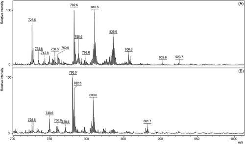
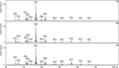
Figure 2. MALDI-MS lipid profiles of pooled cumulus cells from immature and mature oocytes supplemented with sera during 24 h of IVM. (A) iCC: immature cumulus cells; (B) FBS-CC: cumulus cells obtained from FBS-supplemented oocytes after IVM; (C) SSS-CC: cumulus cells obtained from SSS-supplemented oocytes after IVM. FBS: fetal bovine serum; SSS: synthetic serum substitute; IVM: in vitro maturation. Spectra were obtained in the positive mode using 2,5-Dihydroxybenzoic acid (DHB) as matrix.
Lipid profile of cumulus cells collected from GV and MII oocytes
The lipid profile of cumulus cells (CC) was also investigated by MALDI-MS. As shows, the lipid profile of immature cumulus cells (iCC) was characterized by the major ions of m/z 725.5, 734.6, 754.6, 756.6, 760.6, 780.6, 782.6, 784.6, 798.6, 808.6, 810.6, 835.7, 855.7, 881.7, 907.7, and 929.7 corresponding to SM, PC, and TAG (see for assignments). After IVM, the lipid profile of CC obtained from COC matured in FBS and SSS, called FBS-CC and SSS-CC respectively, were also characterized by MALDI-MS ( and ).
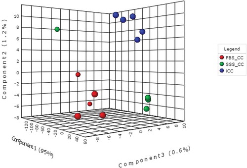
To statistically evaluate the profiles revealed by MALDI-MS and better discriminate differences among the three groups of study (iCC, FBS-CC, SSS-CC) lipid profiles (multivariate data), PLS-DA was applied to the data comprised of the m/z of the ions and the relative intensities of the ion peaks. shows a PLS-DA score plot of samples iCC, FBS-CC, and SSS-CC in which separation of the three clusters of samples was achieved. The variances of PC1, PC2, and PC3 were 95%, 1.2%, and 0.6% respectively, representing 96.8% of the total lipid variability of data. In particular, the VIP plot (Supplemental ) shows that the TAG ions of m/z 881.7 assigned as [POO(52:2) + Na]+, as well as the PC of m/z 760.6 identified as [PC (34:1) + H]+, and 806. 6 [PC (38:6) + H]+ and/or [PC (36:3) + Na]+, present higher abundance in the SSS-CC cluster, contributing to the separation of SSS-CC from the other two groups of samples. The FBS-CC cluster presents however a higher abundance of the ions of m/z 725.5 assigned as [SM (16:0) + Na]+, 809.6 [SM (22:0) + Na]+, and 728.6 assigned as [PC (32:3) + H]+.
Figure 3. Three-dimensional PLS-DA score plot of samples from immature cumulus cells (iCC), FBS-treated cumulus cells (FBS-CC), and SSS-treated cumulus cells (SSS-CC) during IVM. FBS: fetal bovine serum; SSS: synthetic serum substitute; IVM: in vitro maturation. A clear separation between three clusters was achieved. See Supplemental Figure S2 for related variable importance in projection (VIP) plot of the ions.
Lipid profile of immature oocytes and oocytes matured in the presence or absence of cumulus cells
To investigate the influence of CC on the lipid profile of the oocyte during IVM, oocytes were matured in the presence or absence of CC and compared with immature oocytes. Considering that the oocyte and its companion CC take up lipids from the culture medium, we have also investigated whether exposure to poor and enriched-lipid media supplemented with SSS and FBS, respectively, would differentially affect the lipid profiles and development of the oocytes.
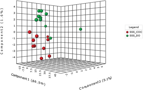
First, the lipid profile of immature oocytes (IO) was analyzed by MALDI-MS. As shows, the spectrum is dominated by the ions of m/z 703.5, 706.6, 725.5, 732.6, 734.6, 754.6, 756.6, 760.6, 782.6, 784.6, 808.6, 810.6, 855.7, and 881.7 corresponding to SM, PC, and TAG (). Oocytes were therefore matured with and without cumulus cells in culture medium supplemented with FBS or SSS and then analyzed by MALDI-MS with subsequent PCA analysis. For simplicity, we will term these oocytes as FBS-DO, FBS-COC, SSS-DO, and SSS-COC. The MALDI-MS of these samples were all characterized again by similar lipid ions corresponding to SM, PC, and TAG (see for assignments) as observed for the IO. We then statistically evaluated the differences in lipid profiles of IO, FBS-COC, and FBS-DO (, vs. 4D). PLS-DA score plot of the IO, FBS-COC, and FBS-DO revealed good separation between IO and FBS-COC clusters whereas the FBS-DO was slightly dispersed (Supplemental ). To improve the separation and understand how the FBS supplementation affected the lipid profile of the matured oocyte with and without cumulus cells, the IO group was then removed from the statistical analysis. After such removal, the two FBS-DO and FBS-COC groups were even separated more and the ions that led to discrimination were annotated (). TAG ions primarily contributed to the separations of the two groups of samples. Interestingly, the TAG ions of m/z 935.7, 943.7, 955.7, 957.7, 959.7, and 971.7 () were very abundant in FBS-COC samples (Supplemental ), but of very low abundance for the FBS-DO samples. The MALDI-MS profiles of the IO, SSS-COC and SSS-DO samples were then compared (, , ), and to better discriminate these three groups a PLS-DA analysis was carried out. The PLS-DA score plot revealed a separation of the three groups, indicating clear differences in the lipid composition (Supplemental ). Since the IO samples presented a certain biological heterogeneity that hindered the complete clustering of the group (Supplemental Figure S5 in the SI), and to improve separation, the IO group was then removed from the statistical analysis. The two SSS-DO and SSS-COC groups were in this case separated (). Interestingly, the PC and SM ions of m/z 700.6, 725.5, 706.6, 703.5, and 720.6 () showed very high abundances for the SSS-COC samples (Supplemental ).
Figure 4. MALDI-MS lipid profiles of immature oocytes (IO) and oocytes supplemented with sera during IVM in the presence (COC) or absence (DO) of cumulus cells. Spectra were obtained in the positive mode using 2,5-dihydroxybenzoic acid (DHB) as matrix. Analyses were performed directly on pools of 5 mechanically denuded oocytes from 15 replicates. (A) IO; (B) FBS-COC; (C) SSS-COC; (D) FBS-DO; and (E) SSS-DO. IVM: in vitro maturation; COC: cumulus-oocyte complex; DO: denuded oocyte; FBS: fetal bovine serum; SSS: synthetic serum substitute; CC: cumulus cells.
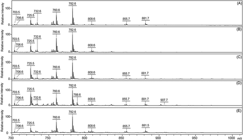
Figure 5. Three-dimensional PLS-DA score plot of samples from FBS-treated denuded oocytes (FBS-DO) and FBS-treated cumulus-oocyte complexes (FBS-COC). A good separation between two clusters was achieved. See Supplemental Figure S4 for related variable importance in projection (VIP) plot of the ions. FBS: fetal bovine serum; SSS: synthetic serum substitute.
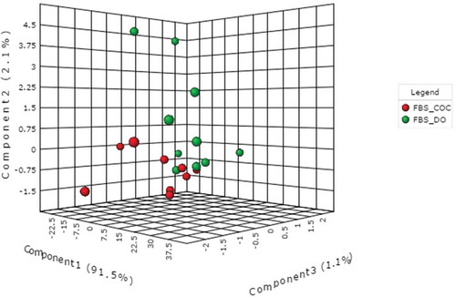
Figure 6. Three-dimensional PLS-DA score plot of samples from SSS-treated denuded oocytes (SSS-DO) and SSS-treated cumulus-oocyte complexes (SSS-COC). A clear separation between two clusters was achieved. See Supplemental Figure S6 for related variable importance in projection (VIP) plots reporting the ions responsible by clusters separation. FBS: fetal bovine serum; SSS: synthetic serum substitute.
In conclusion, the difference in lipid profile observed for DO and COC seems to indicate a CC modulation of the lipid profile of the oocytes according to the supplement used for the maturation.
Embryonic development of FCS and SSS-supplemented DO or COC during IVM
To investigate the modulatory role of cumulus cells on the oocyte lipid profile and developmental competence, oocytes were exposed to FBS or SSS in the presence or absence of cumulus cells. The resulting DO as well as intact COCs were then exposed to either a lipid-poor (SSS) or a lipid-rich (FBS) culture media during maturation. The developmental competence of the oocyte, as determined by the cleavage and blastocyst rates after fertilization, was altered under these two conditions (). Supplementation with FBS significantly increased blastocyst rates compared to SSS (p<0.05). The presence (COC) or absence (DO) of cumulus cells during maturation had no effect on either cleavage or blastocyst rates (p>0.10). However, within the FBS supplemented groups, the absence of cumulus cells (DO) reduced cleavage rates (57.9±1.9% vs 71.4±3.5% for FBS-COC; p<0.05). Blastocysts rates were greatest in the FBS-COC group (32.4±7.1%), with rates for FBS-DO (25.9±0.8%) and SSS-COC (19.8±5.8%) reduced, but not significantly (p=0.09 and 0.68, respectively). Developmental competence of SSS-DO (15.8±0.5%) was significantly reduced in the absence of both FBS and cumulus cells (p<0.05; ).
Table 2. Embryonic development of bovine denuded oocytes (DO) or cumulus-oocyte-complexes (COC) matured in vitro in TCM-199 medium supplemented with fetal bovine serum (FBS) or synthetic serum substitute (SSS).
Discussion
In the current study, MALDI-MS analysis of IVM with either COC or DO oocytes that were exposed to different lipid sources via FBS or SSS was used to investigate how the CC layer modulates the lipid profile of the oocyte. The lipid profiles of the TCM medium, FBS and SSS supplements, and immature oocytes and their respective CC were evaluated in order to define if the changes in lipid profiles of the CC and oocyte after IVM could reflect the composition of these sera and may be due to lipid uptake. The lipid profile of bovine oocytes matured in the presence or absence of cumulus cells, using both FBS and SSS supplements, was therefore determined.
MALDI-MS analysis in the positive ion mode and in the m/z 700–1200 range provided characteristic lipid profiles for the samples, in which the lipids were detected mainly as protonated or cationized molecules. The MALDI-MS profiles in both FBS and SSS supplements were comprised of numerous SM, PC, and TAG ions ( and , respectively). provides a summary for the tentative assignments of these lipid ions. The MALDI-MS lipid compositions of FBS and SSS were distinct as shown mainly by the greater relative abundance in FBS of ions of m/z 810.6, 836.6, and 856.6 corresponding to PC containing long-chain fatty acyl residues as C20:4 and C22:6, respectively [Berdeaux et al. Citation2010; Haysaka et al. Citation2008; Lessig et al. Citation2004]. In fact, FBS composition is characterized by elevated concentrations of arachidonic (AA, 20:4n6) and docosahexaenoic (DHA, 22:6n3) acids [Stoll and Spector Citation1984]. Particularly, the C20:4 content in FBS is of special interest since AA may be relevant to oocyte competence [Kim et al. Citation2001]. Indeed, we found a higher relative abundance of the ion of m/z 725.5 [SM (16:0) + Na]+ in FBS compared to SSS. Accordingly, SM constitutes a significantly high percentage of the total PL in fetal plasma [Crowley et al. Citation1963]. In SSS however, MALDI-MS detected predominantly unsaturated PC and a monounsaturated TAG containing palmitic and oleic acids. SSS is a protein source and its formulation contains no plasma lipids, except a human serum albumin (HSA) as an additive [Weathersbee et al. Citation1995; Meintjes et al. Citation2009]. We can therefore infer that HSA provided the lipid species detected by MALDI-MS in SSS.
Significant differences in SM, PC, and TAG profiles in iCC compared with CC harvested from oocytes matured in FBS or SSS have been detected by MALDI-MS using PLS-DA as a statistical approach, indicating that serum affects the lipid profile of CC during IVM. As recently reported, the lipid composition of CC analyzed by MALDI-MS in the range of m/z 300–1000 was significantly distinct before and after 18 hours of IVM in the absence of FA and serum [Sanchez-Lazo et al. Citation2014]. Interestingly, as reported the abundance of most of the species in the range between m/z 381.1 and 557.2 increased in CC after IVM, whereas the abundance of ions at m/z 578.7 up to 858.9 were decreased [Sanchez-Lazo et al. Citation2014] suggesting the occurrence of lipogenic and lipolytic activities in CC during IVM. In contrast, we observed an enrichment of matured CC with higher molecular weight species (>m/z 700) comprising some SM, PL, and TAG, which probably resulted from the uptake of lipids supplied by FBS or SSS in IVM medium. In support of this finding, the accumulation of lipid droplets that mainly contain TAG and the increase of various lipid species in CC after 18 hours of IVM have been described and also underlie de novo lipogenesis in CC [Sanchez-Lazo et al. Citation2014]. According to the PLS-DA analyses in our work, FBS-CC were enriched with two saturated SM species and a polyunsaturated PC whereas SSS-CC retained two polyunsaturated PC and a monounsaturated TAG. Remarkably, we observed that the TAG ion of m/z, 881.7 assigned to [POO(52:2) + Na]+, as well as the PC ion of m/z 760.6 assigned to [PC (34:1) + H]+ and 806.6 [PC (38:6) + H]+ and/or [PC(36:3)+ Na]+ were more abundant in mature CC harvested from the SSS-COC group. This finding may reflect a preferential uptake of these unsaturated PC and TAG species from SSS by CC. In fact, the uptake of PL and TAG involves the intact molecule and does not require hydrolysis [Gadella et al. Citation1999; Bailey et al. Citation1973]. Inversely, CC exposed to FBS presents a higher abundance of the ions of m/z 809.6, assigned as [SM (22:0) + Na] +, 725.5 [SM (16:0) + Na]+, and 728.6 assigned as [PC (32:3) + H]+. Noteworthy, TAG species were not detected in higher abundance in FBS-CC which could be linked to an increased hydrolysis of TAG in these cells and subsequent FA transfer to the oocyte. Consistent with this observation, we found that oocytes harvested from FBS-COC were enriched with various TAG after IVM. Even though we present data indicating that the lipid profile of CC varies in parallel with two different lipid sources, the precise way in which FBS and SSS differentially interact with CC remains unclear. These findings were confirmed by the PLS-DA analyses and demonstrate that the lipid profile varies when CC are cultured in media supplemented with each of the two types of serum.
The influence of CC on the lipid profile of the oocyte was yet observed by PLS-DA data comparing both COC and DO exposed to FBS or SSS during IVM in our study. Note that the relative abundance of TAG ion of m/z 935.7 [AOL (56:3) + Na]+, 955.7 [TAG (58:7) + Na]+, and/or [TAG (60:10) + H]+, 957.7 [TAG (59:13) + Na]+, 971.7 [PPO (50:1) + Na]+ was significantly higher in FBS-COC samples compared to FBS-DO (Supplemental ). The increase in TAG varied, therefore as a function of the presence of CC in the intact COC highlighting the role of CC on energy metabolism of the oocyte as previously described [Auclair et al. Citation2013, Cetica et al. Citation2002; Sanchez-Lazo et al. Citation2014]. These TAG species may be synthesized from FA supplied by CC to the oocyte in the intact COC during maturation, since these lipids were not detected in TCM medium, FBS, immature CC, and IO, in our experimental conditions. This finding is in agreement with previous studies that reported lower total lipid content in DO compared to COC [Auclair et al. Citation2013]. Parallel to this, an increased mRNA level for genes involved in lipid synthesis as ACLY and APOO and a downregulation of the gene ACADSB involved in FAO was observed in DO [Auclair et al. Citation2013] probably associated with a lack of FA provision from CC and decreased lipogenesis in the absence of CC. Cumulus cells are involved in providing energy substrates from surrounding fluids to the oocyte and could catabolize lipids by lipase activity, supplying FA to the oocyte as oxidative substrates [Cetica et al. Citation2002; Auclair et al. Citation2013]. The very low TAG abundance in the FBS-DO samples detected by MALDI-MS may therefore reflect a loss of FA intake/synthesis oriented by CC and/or a decreased uptake by DO of lipids from the culture medium. As previously reported, analysis of the proteins involved in lipid metabolism also revealed that the level of FA was significantly lower in DO than in COC [Auclair et al. Citation2013]. In all, these observations highlight the role of CC in orienting the synthesis and/or the consumption of TAG by the oocyte to provide energy for maturation [Auclair et al. Citation2013]. There are few available studies that investigate the influence of lipolytic, lipogenic, and oxidative pathways on the lipid profile of CC and oocytes during maturation [Auclair et al. Citation2013; Sanchez-Lazo et al. Citation2014; Uzbekova et al. Citation2015]. Recent evidence has indicated that the lipid profile can reflect subtle changes in lipogenesis and lipolysis in the COC during maturation once the pharmacological inhibition of mitochondrial FA β-oxidation (FAO) in CC during IVM induced an overabundance of PL species of m/z 804.8 and 832.8 in these cells [Sanchez-Lazo et al. Citation2014]. In line with this, MALDI-MS analysis has also detected increased and decreased abundances of various lipid species in CC after IVM [Sanchez-Lazo et al. Citation2014] probably associated to anabolism (lipid storage and/or a de novo lipogenesis) and catabolism (TAG hydrolysis and FA oxidation) events during maturation.
Further support for CC involvement in the lipid profile of the oocyte during IVM was achieved when another type of serum was tested in our study. Remarkably, when MALDI-MS profiles of SSS-COC and SSS-DO were compared (, , ) we noted an increased PL ion abundance in COC. A consistent separation of the two groups, indicating clear differences in the lipid composition as shown in . Interestingly, the PC and SM ions of m/z 700.6 [PC (30:3) + H]+, 703.5 [SM (16:0) + H]+, 706.6 [PC (30:0) + H]+, 725.5 [SM (16:0) + Na]+, and 720.6 [PCe (32:0) + H]+ showed an increased abundance in the SSS-COC samples (Supplemental ). This finding reinforces the already observed higher lipid ion abundance in FBS-COC and may reflect a loss of key factors involved in FA synthesis and/or FA entry in the DO. Although the lipid profiles were not similar in the two types of sera tested, cumulus-enclosed oocytes exposed to both FBS or SSS had a significantly higher abundance of lipid ions compared with DO. It is therefore possible to suggest that regardless of the exogenous lipid source, CC stimulated lipid biosynthesis and an enrichment of TAG or PL species in oocytes, according to lipids supplied by sera.
In our experiments, a tendency to lower cleavage and blastocyst rates of fertilized FBS-DO (57.89% and 25.9%, respectively) than that for the FBS-COC (71.37% and 32.4%, respectively) was observed and could be, in part, related to deficient neutral lipid storage by the oocyte for subsequent use as an oxidative substrate during embryogenesis. In line with this, lipid metabolism in CC has been reported to maintain metabolic homeostasis and may influence meiosis progression and survival of enclosed oocytes [Sanchez-Lazo et al. Citation2014]. In addition, accumulation of TAG in the oocytes has been related to improved developmental competence [Jeong et al. Citation2009]. Although changes in the TAG profile probably may be responsible to some extent for the slight differences in the embryo development rates between DO and COC in this study, other factors must have contributed to this effect, including cAMP and glutathione content in the oocyte [Luciano et al. Citation2005; Wang et al. Citation1997]. The oocyte metabolism is supported by related metabolic pathways as glycolysis, the pentose phosphate pathway (PPP), and lipolysis [Cetica et al. Citation2002]. However, the nature of COC metabolism is complex as it contains two different cell types with distinct metabolic profiles and requirements. Of note, the oocyte mainly undergoes oxidative phosphorylation and CCs have a high rate of glycolytic activity [Cetica et al. Citation2002; Thompson et al. Citation2007]. In CC, glucose oxidation is performed through phosphofructokinase and glucose-6-phosphate-dehydrogenase (G6PDH) pathways. G6PDH in CC could provide nicotinamide adenine dinucleotide phosphate-oxidase (NADPH) to the oocyte for its own synthesis of lipids and the regeneration of reduced glutathione [Cetica et al. Citation2002], supporting both energetic metabolism and potential redox of oocyte.
In our experiments, DO were not co-cultured with COC during IVF but reached slightly higher cleavage and blastocyst rates than that found when DO were co-cultured with COC during IVF [Auclair et al. Citation2013]. This could reflect differences in sample size and/or IVM media composition as the absence of FA in 199EM IVM medium used in this study. Moreover, oocyte metabolism during culture was reported to be affected by components of basal IVM media [Wang et al. Citation1997]. It should be noted that our IVM medium was supplemented with metabolites such as pyruvate and amino acids, which can be used as oxidative substrates by DO in the absence of CC, as well as FA supplied by FBS, which might be partially compensated by the lack of CC by DO utilizing, to some extent, its own FA uptake. However, oocytes are more susceptible to impaired embryonic development when IVM takes place under suboptimal conditions [Zhou et al. Citation2012], notably in the absence of CC. The presence of CC in the intact COC and/or supplementary CC used as a feeder layer during IVM was found to be effective in regulating multiple parameters of oocyte homeostasis as cell cycle kinases in the human oocyte [Combelles et al. Citation2005], gene expression levels of TGFβ ligands and its receptors in sheep COC [Kyasari et al. Citation2012], both involved in glycolysis and de novo cholesterol biosynthesis of the oocyte [Su et al. Citation2008], and to direct a set of metabolic pathways operating in the oocyte to maintain the energy supply and the intracellular redox potential as reported in mouse [Li et al. Citation2011], porcine [Hao et al. Citation2009; Yoon et al. Citation2015], and rat [Jiao et al. Citation2016].
Oocyte competence can be modulated and improved by stimulating in parallel the glycolytic activity of CC and oxidative phosphorylation and tricarboxylic acid (TCA) within the oocyte [Sutton-McDowall et al. Citation2012; Sutton-McDowall et al. Citation2015]. Besides glycolysis, PPP in CC improves oocyte maturation by both reducing oxidative stress and supplying energy through providing fructose-6-phosphate for glycolysis and NADPH for the lipid synthesis in the oocyte [Cetica et al. Citation2002]. Taking this into account, these reports highlight the importance of the metabolic cooperativity between the oocyte and CC during maturation on oocyte health.
Noteworthy, our study clearly demonstrates that DO exposed to SSS during IVM had a significant diminished ability to attain the blastocyst stage in vitro. This suggests that factors other than CC absence contributed to the low developmental potential of SSS-DO as a scarce source of lipid substrates in SSS supplement, since pyruvate was readily available in the IVM medium. In line with this, bovine DO mainly utilize lipolysis as a metabolic route during IVM [Cetica et al. Citation2002]. The composition of FBS and SSS differentially affected the embryo development of DO, underlying the importance of the exogenous nutrients and probably specific lipids present in FBS to oocyte metabolism. Importantly, there is evidence that in vitro matured bovine oocytes showed significant decreases in the proportion of AA compared with immature ones [Kim et al. Citation2001], indicating consumption by the oocyte of this FA present in high proportions in the FBS. Additionally, it has been described that polyunsaturated FA as AA are important to modulating the uptake of lipids and further regulate the development of the oocyte [Chen et al. Citation2016]. This indicates that the lipid uptake by DO could be differentially affected by FBS and SSS.
Cumulus-enclosed oocytes exposed to SSS, a poor-lipid serum, failed however to experience the same changes in blastocyst rates seen in FBS-DO suggesting that CC compensated, at least in part, the differences in composition between the two sera during IVM. Additional work is required to clarify the significance of this finding, but from the present results it seems that lipid fraction of the FBS was responsible for the better embryo development of the FBS-DO compared to SSS-DO. CC affecting the oocyte lipid profile was supported by this MALDI-MS lipidomics analysis, providing further evidence for the importance of CC in modulating the lipid dynamics of the oocyte during IVM through to different lipid’s source.
In conclusion, MALDI-MS measurements provided the first data on the changes in SM, PC, and TAG ions in bovine oocytes exposed to FCS or SSS during in vitro maturation in the presence or absence of CC layer. It was found that cumulus-enclosed and denuded oocytes showed quite unique MALDI-MS lipid profiles. Additionally, differences in lipid composition were observed between two types of sera and distinctly influenced the lipid profile of CC. The enrichment of some specific lipids in the oocyte and CC needs to be studied in more detail as well as the influence of membrane lipids on oocyte maturation. The present results and further MALDI-MS investigations of lipid composition of sera added to IVM media in parallel with COC lipid profile seems invaluable to guide rational design of IVM media for human oocyte, an unresolved potential approach in ART.
Material and methods
Reagents and culture media for in vitro embryo production
All culture media and reagents were purchased from Sigma Chemical Company (St. Louis, MO, USA), except where otherwise indicated. Phosphate buffer saline (PBS) solution was supplied by Gibco/BRL (Grand Island, NY, USA). Methanol (ACS/HPLC) grade was purchased from Burdik & Jackson (Muskegon, MI, USA) and 2,5-dihydroxybenzoic acid (DHB) was purchased from ICN Biomedicals (Aurora, OH, USA). Ultrapure water, purified by Direct-Q water system (Millipore, Bedford, MA, USA), was used for the preparation of solvents.
Culture media for in vitro maturation
The maturation medium was HEPES-buffered tissue culture medium-199 (TCM-199, Gibco/BRL, Grand Island, NY, USA) supplemented with 0.2 mM sodium pyruvate, 100 IU/mL penicillin G, 100 µg/mL streptomycin, 0.5 µg/mL follicle stimulating hormone (FSH, Folltropin-Bioniche, Canada), 5 µg/mL luteinizing hormone (LH, Lutropin-Bioniche, Canada) and 1 μg/mL 17β-estradiol. Fetal bovine serum (FBS, Invitrogen Gibco/BRL) or serum substitute supplement (SSS, Irvine Scientific, Santa Ana, CA, USA) were added to the standard TCM medium at 10% (v/v) according to the experimental design.
Collection of oocytes
The investigations on bovine oocytes have been approved by the ethics committee of the University of São Paulo (number 089/2010). Bovine ovaries were collected from a slaughterhouse and transported to the laboratory in 0.9% physiological saline supplemented with 0.05 g/L streptomycin at 35ºC. The ovaries were washed in 0.9% physiological saline at 37ºC and 3- to 8-mm antral follicles from 40–50 ovaries/experiment were aspirated with 18 G needles adapted to 20 mL syringes and pooled. COC with at least three compact layers of CC were selected and washed several times in TCM/HEPES medium supplemented with 50 mg/L gentamycin and 0.1% BSA and were morphologically evaluated under a stereomicroscope, as previously described [Vireque et al. Citation2009]. One-half of the oocytes were stripped of CC by gentle pipetting using a P100 pipette (Eppendorf, Hamburg, Germany) with a tip in several successive 100 µL drops of 0.5% hyaluronidase under observation with a stereomicroscope. Completely naked and morphologically undamaged oocytes were removed from the microdrops and washed from residual CC three times in a Petri dish containing 3 mL of TCM medium. Subsequently, COC or DO were randomly exposed to maturation medium supplemented with FBS or SSS.
In vitro maturation protocol
Pools of 20 COCs or DO were cultured in microdrops of 100µL in 35 mm Petri dishes covered with 3 mL of M8410 mineral oil at a density of 1/10 μL culture medium at 38.8°C for 22 h in a humidified atmosphere containing 5% CO2. Each experiment was replicated at least three times.
Experimental design
Lipid profile of cumulus cells collected from germinal vesicle (GV) and metaphase II (MII) oocytes
First, samples of FBS and SSS supplements and media batches prepared on the same day of the experiment were collected in order to analyze the lipid profile of the bovine-derived serum and serum substitute supplement (a human-derived serum) before solubilization to culture medium and COCs culture, and to determine what changes occurred as a result of an uptake of lipids from serum.
The CC were collect by pipetting from morphologically healthy selected immature COC and from matured FBS or SSS-supplemented COC. Briefly, the pooled CC from 75 COC of each experimental group were collected in 15 mL sterile polypropylene Falcon tubes (Corning, Corning, NY, USA) containing phosphate-buffered saline (PBS) at 4°C and centrifuged at 300 x g for 10 min. The resulting cell pellets were washed twice with 2 mL of PBS at 4°C and centrifuged for 10 min at 4°C. The supernatant was discarded, and the pellets were frozen at -80°C overnight until lipid analysis.
Lipid profile of immature oocytes and oocytes matured in the presence or absence of cumulus cells
Immature oocytes (IO) with several compact layers of CC were denuded and assigned to MALDI-MS analysis. Next, groups of either 20 COC or DO were subjected to IVM. After maturation, FBS-COC or SSS-COC were randomly submitted to in vitro fertilization (IVF) or stripped of CC. Cumulus-free oocytes from both COC and DO groups with a visible polar body were collected and then stored until lipid analysis. At least 75 immature or mature oocytes were analyzed for their lipid profile status in each experimental situation (COC and DO). In total, five groups of naked oocytes were analyzed as follows: i) IO, immature oocytes denuded from the compact COCs before IVM (n=75); ii) FBS-COC, oocytes matured enclosed in intact COCs at 10% FBS and denuded after IVM (n=75); iii) SSS-COC, oocytes matured enclosed in intact COCs at 10% SSS and denuded after IVM (n=75); iv) FBS-DO, oocytes denuded from the compact COCs before IVM and matured in 10% FBS (n=75); and v) SSS-DO, oocytes denuded from the compact COCs before IVM and matured in 10% SSS (n=75).
In vitro fertilization and embryonic development of FBS and SSS-supplemented oocytes during IVM
COC and DO from FBS or SSS-supplemented oocytes during IVM were washed in Fert TALP medium and inseminated with thawed spermatozoa obtained by the swim-up method [Parrish et al. Citation1988] using the Sperm TALP medium supplemented with 6 mg/mL BSA fraction V as previously described [Vireque et al. Citation2009]. Fertilization of COC and DO from FBS or SSS groups was performed in microdrops of Fert TALP medium (20 COCs or 20 DOs/100µL) supplemented with 20 µg/mL heparin and 6 mg/mL fatty acid-free BSA under mineral oil and the sperm was co-incubated with the COCs for 18 h in a humidified atmosphere containing 5% CO₂ at 38.5ºC. The concentration of sperm during fertilization was approximately 1 x 10⁶/mL. At the end of the fertilization period, presumptive zygotes were partially denuded by pipetting, washed three times in TALP medium and then cultured in a microdrop of CR2aa medium [Rosenkrans and First Citation1994] with 5% FBS and 1 mg/mL BSA (20-25 embryos/50 µL) under mineral oil at 38.8°C for 8 d in a water-saturated atmosphere of 5% CO2. The culture medium was changed every 48 h. Embryonic cleavage rates (ratio of the number of cleaved zygotes to the total number of the oocytes subjected to IVF) and blastocyst rates (ratio of the number of blastocysts to the number of fertilized oocytes cultured) were evaluated at day 3 (D3) and 7-8 (D7-D8), respectively. In total, developmental rates of 524 COC and 518 DO were checked in three independent IVF experiments.
Lipid profile analysis by MALDI-MS
Sample preparation
Each sample of FBS, SSS, or IVM medium (1 µL) was placed on a spot on the MALDI target plate, allowed to dry at room temperature and then covered with 1 μL of 1.0 mol L(-1) of DHB diluted in pure methanol just before analysis. Pools of oocytes from each group (IO, IVM COC, and IVM) completely denuded from their CC were washed five times in drops of PBS with no macromolecular supplement and stored in microtubes containing 2-4 μL of PBS at -80°C until analysis. Trying to avoid biased results, IO and iCC samples were immediately washed in PBS and were not exposed to culture media or serum supplements. The samples were thawed by pipetting 100 μL of (1:1, v/v) methanol/H2O solution into the microtube, and then washed five times in the same solution.
Pools of five oocytes were placed on a spot on the MALDI plate under the stereomicroscope. Samples were allowed to dry at room temperature, and their location was recorded. Prior to analysis, 1 μL of mol L(-1) DHB diluted in pure methanol was deposited on each target spot to cover the oocytes and the spots were allowed to dry at room temperature. The mass spectra were acquired from 15 spots, totaling 75 oocytes in each group.
MALDI-MS data acquisition
MALDI-MS data were acquired using an Autoflex III MALDI time-of-flight mass spectrometer operated in the reflectron mode (Bruker Daltonics, Bremen, Germany) and equipped with a 334 nm smart beam laser. The MS data were acquired averaging 1500 consecutive laser shots at a frequency of 200 Hz until signals in the region of interest were observed and then disappeared due to the consumption of the sample. The spectra were recorded in the positive ion mode and in m/z 700-1200 range. FlexAnalysis 3.0 software (BrukerDaltonics) was used to process the mass spectra. The most abundant ions from each spectrum, considering all ions that were clearly distinct from noise after the exclusion of isotopopologue ion peaks, were considered a starting point in the search for ions of m/z values corresponding to lipids. After attribution, only the m/z values for ions which were clearly distinct from noise level in the spectra were included in the partial least squares – discriminant analysis (PLS-DA). Ions corresponding to TAG and PL species were attributed () based on previous lipid profile studies of animal and human oocytes and embryos performed by MALDI-MS. The lipid ions are described by class abbreviation followed by the total number of carbons and double bounds in the acyl residues attached to the glycerol backbone (in parenthesis). Note that isomers cannot be resolved by MS and hence more than one isomer can occur for a single lipid ion.
Statistical analysis
Cleavage and blastocyst rates were analyzed using the general linear model/ANOVA procedures of SAS with supplement, group, and replicate as fixed effects. Mean comparisons were performed using Duncan´s multiple range test. A probability level of p < 0.05 was considered significant.
Univariate t-test and ANOVA and multivariate analysis was performed with m/z and relative intensity data from lipid profiles of CC and oocytes obtained from the analysis by MALDI-MS. Data lists of m/z values with relative ion abundances were exported from the Flex Analysis software, saved as .xls files in .csv format and uploaded into the MetaboAnalyst 3.0 software.
For multivariate analysis the data were aligned, preprocessed by autoscaling, and submitted to PLS-DA which is a supervised method that uses multivariate regression techniques to extract the information that can predict the class membership via linear combination of original variables. Based on the variable importance in projection (VIP) from PLS loading, the most important ions that explained the higher variance of the data were selected. VIP is a weighted sum of squares of the PLS loadings which considers the amount of explained variation in each dimension, calculated for each component, for prediction of feature importance.
Declaration of interest
The authors report no conflicts of interest.
IAAN_1289279_Supplementary_Files.zip
Download Zip (2 MB)Supplemental data
Supplemental data for this article can be accessed on the publisher’s website.
Additional information
Notes on contributors
Alessandra A. Vireque
Designed the study, data collection, performed the experiments, analyzed and interpreted the data, and wrote the manuscript: AAV; Responsible for the MALDI-MS analyses, interpretation of MALDI-MS data, helped in the critical discussion of results, and revised the manuscript: AT, KRAB, FNS, MNE; Performed data collection, in vitro embryo production experiments, analyzed and interpreted the data, and wrote the manuscript: JGVG, DRA, ACB; Interpreted the data and critically revised the manuscript: MFSS, RAF; Conceived and designed the study, analyzed the data, and wrote the manuscript: ACJSRS.
References
- Aardema, H., Lolicato, F., van de Lest, C.H., Brouwers, J.F., Vaandrager, A.B., van Tol, H.T., et al. (2013) Bovine cumulus cells protect maturing oocytes from increased fatty acid levels by massive intracellular lipid storage. Biol Reprod 88: 1–15. doi: 10.1095/biolreprod.112.106062
- Auclair, S., Uzbekov, R., Elis, S., Sanchez, L., Kireev, I., Lardic, L., et al. (2013) Absence of cumulus cells during in vitro maturation affects lipid metabolism in bovine oocytes. Am J Physiol Endocrinol Metab 15: 304, 599–613.
- Bailey, J.M., Howard, B.V., Tillman, S.F. (1973) Lipid metabolism in cultured cells. XI. Utilization of serum triglycerides. J Biol Chem 248: 1240–1247.
- Berdeaux, O., Juaneda, P., Martine, L., Cabaret, S., Bretillon, L., Niyazi, A. (2010) Identification and quantification of phosphatidylcholines containing very-long-chain polyunsaturated fatty acid in bovine and human retina using liquid chromatography/tandem mass spectrometry. J Chromatography 1217: 7738–7748.
- Brugger, B., Erben, G., Sandhoff, R., Wieland, F.T., Lehmann, W.D. (1997) Quantitative analysis of biological membrane lipids at the low picomole level by nano-electrospray ionization tandem mass spectrometry. Proc Natl Acad Sci USA 94: 2339–2344.
- Cetica, P.D., Pinto, L.N., Dalvit, G.C., Beconi, M.T. (2002) Activity of key enzymes involved in glucose and triglyceride catabolism during bovine oocyte maturation in vitro. Reproduction 124: 675–681.
- Chen, W.W., Yi, Y.H., Chien, C.H., Hsiung, K.C., Ma, T.H., Lin, Y.C., et al. (2016) Specific polyunsaturated fatty acids modulate lipid delivery and oocyte development in C. elegans revealed by molecular-selective label-free imaging. Scientific Reports 6: 32021. doi: 10.1038/srep32021
- Combelles, C.M.H., Fissore, R.A., Albertini, D.F., Racowsky, C. (2005) In vitro maturation of human oocytes and cumulus cells using a co-culture three-dimensional collagen gel system. Hum Reprod 20: 1349–1358. doi:10.1093/humrep/deh750
- Crowley, J., Ways, P., Jones, J.W. (1963) Human fetal erythrocyte and plasma lipids. J Clin Inv 44: 989–998.
- Ferguson, E.M., Leese, H.J. (1999) Triglyceride content of bovine oocytes and early embryos. J Reprod Fertil 116: 373–387.
- Ferreira, C.R., Saraiva, S.A., Catharino, R.R., Garcia, J.S., Gozzo, F.C., Sanvido, G.B., et al. (2010) Single embryo and oocyte lipid fingerprinting by mass spectrometry. J Lipid Res 51: 1218–1227.
- Ferreira, C.R., Jarmusch, A.K., Pirro, V., Alfaro, C.M., González-Serrano, A.F., Niemann, H. et al. (2015) Ambient ionisation mass spectrometry for lipid profiling and structural analysis of mammalian oocytes, preimplantation embryos and stem cells. Reprod Fertil Dev 27: 621–637. doi: 10.1071/RD14310.
- Ford, J.H., Tavendale, R. (2010) Analysis of fatty acids in early midlife in fertile women: implications for reproductive decline and other chronic health problems. Am J Hum Biol 22: 134–136.
- Fuchs, B., Jakop, U., Goritz, F., Hermes, R., Hildebrandt, T., Schiller, J. et al. (2009) MALDI-TOF “fingerprint” phospholipid mass spectra allow the differentiation between ruminantia and feloideae spermatozoa. Theriogenology 71:568–575.
- Gadella, B.M., Miller, N.G.A., Colenbrander, B., Van Golde, L.M.C., Harrison, R.A.P. (1999) Flow cytometric detection of transbilayer movement of fluorescent phospholipid analogues across the boar sperm plasma membrane: elimination of labeling artifacts. Mol Reprod Dev 53:108-125.
- Gilchrist, R.B., Thompson, J.G. (2007) Oocyte maturation: emerging concepts and technologies to improve developmental potential in vitro. Theriogenology 67: 6–15.
- Gilchrist, R.B. (2011) Recent insights into oocyte-follicle cell interactions provide opportunities for the development of new approaches to in vitro maturation. Reprod Fertil Dev 23: 23–31.
- Haggarty, P., Wood, M., Ferguson. E., Hoad, G., Srikantharajah, A., Milne, E., et al. (2006) Fatty acid metabolism in human preimplantation embryos. Hum Reprod 21: 766–773.
- Hannun, Y.A., Obeid, L.M. (2008) Principles of bioactive lipid signaling: lessons from sphingolipids. Nat Rev Mol Cell Biol 9: 139–150.
- Hao, Z.D., Liu, S., Wu, Y., Wan, P.C., Cui, M.S., Chen, H., et al. (2009) Abnormal changes in mitochondria, lipid droplets, ATP and glutathione content, and Ca2+ release after electro-activation contribute to poor developmental competence of porcine oocyte during in vitro ageing. Reprod Fertil Dev 21: 323–332. doi.org/10.1071/RD08157
- Hayasaka, T., Goto-Inoue, N., Sugiura, Y., Zaima, N., Nakanishi, H., Ohishi, K., et al. (2008) Matrix-assisted laser desorption/ionization quadrupole ion trap time-of-flight (MALSI-QIT-TOF)-based imaging mass spectrometry reveals a layered distribution of phospholipid molecular species in the mouse retina. Rapid Commun Mass Spectrum 22: 3415.
- Homa, S.T., Racowsky, C., McGaughey, R.W. (1986) Lipid analysis of immature pig oocytes. J Reprod Fertil 77: 425–434.
- Jeong, W.J., Cho, S.J., Lee, H.S., Deb, G.K., Lee, Y.S., Kwon, T.H. et al. (2009) Effect of cytoplasmic lipid content on in vitro developmental efficiency of bovine IVP embryos. Theriogenology 72: 584–589.
- Jiao G.Z., Cui, W., Yang, R., Lin, J., Gong, S., Lian H.Y., et al. (2016) Optimized protocols for in vitro maturation of rat oocytes dramatically improve their developmental competence to a level similar to that of ovulated oocytes. Cell Reprogram 18: 17–29. doi: 10.1089/cell.2015.0055.
- Jungheim, E.S.; Macones, G.A.; Odem, R.R.; Patterson, B.W.; Lanzendorf, S.E.; Ratts, V.S., et al. (2011) Associations between free fatty acids, cumulus oocyte complex morphology and ovarian function during in vitro fertilization. Fertil Steril 95: 1970–1974.
- Kim, J.Y., Kinoshita, M., Ohnishi, M., Fukui, Y. (2001) Lipid and fatty acid analysis of fresh and frozen-thawed immature and in vitro matured bovine oocytes. Reproduction 122: 131–138.
- Kyasari, O.R., Valojerdia, M.R., Farrokhid, A., Ebrahimi, B. (2012) Expression of maturation genes and their receptors during in vitro maturation of sheep COCs in the presence and absence of somatic cells of cumulus origin. Theriogenology 77: 12–20. doi:10.1016/j.theriogenology.2011.07.007
- Lapa, M., Marques, C.C., Alves, S.P., Vasques, M.I., Baptista, M.C., Carvalhais, I., et al. (2011) Effect of trans-10 cis-12 conjugated linoleic acid on bovine oocyte competence and fatty acid composition. Reprod Domest Anim 46: 904–910.
- Lepage, N., Miron, P., Hemmings, R., Roberts, K.D., Langlais, J. (1993) Distribution of lysophospholipids and metabolism of platelet-activating factor in human follicular fluid and peritoneal fluids. J Reprod Fertil 98: 349–356.
- Lessig, J., Gey, G., Süss, R., Schiller, J., Glander, H.J., Arnhold, J. (2004) Analysis of the lipid composition of human and boar spermatozoa by MALDI-TOF mass spectrometry, thin layer chromatography and 31 P NMR spectroscopy. Comp Biochem Physiol B Biochem Mol Biol 137: 265–277.
- Li, Q., Miao, D.Q., Zhou, P., Wu, Y.G., Gao, D., Wei, D.L. (2011) Glucose metabolism in mouse cumulus cells prevents oocyte aging by maintaining both energy supply and the intracellular redox potential. Biol Reprod 84: 1111–1118. doi: 10.1095/biolreprod.110.089557
- Lolicato, F., Brouwers, J.F., de Lest, C.H., Wubbolts, R., Aardema, H., Priore, P., et al. (2015) The cumulus cell layer protects the bovine maturing oocyte against fatty acid-induced lipotoxicity. Biol Reprod. 92: 16. doi 10.1095/biolreprod.114.120634
- Loutradis, D., Kiapekou, E., Zapanti, E., Antsaklis, A. (2006) Oocyte maturation in assisted reproductive techniques. Ann NY Acad Sci 1092: 235–246.
- Luciano, A.M., Lodde, V., Beretta, M.S., Colleoni, S., Lauria, A., Modina, S. (2005) Developmental capability of denuded bovine oocyte in a co-culture system with intact cumulus-oocyte complexes: role of cumulus cells, cyclic adenosine 3,5-monophosphate, and glutathione. Mol Reprod Dev 71: 389–397.
- Marei, W.F., Wathes, D.C., Fouladi-Nashta, A.A. (2010) Impact of linoleic acid on bovine oocyte maturation and embryo development. Reproduction 39: 979–988.
- Matorras, R., Ruiz, J.I., Mendoza, R., Ruiz, N., Sanjurjo, P., Rodriguez-Escudero, F.J. (1988) Fatty acid composition of fertilization-failed human oocytes. Hum Reprod 13: 2227–2230.
- Meintjes, M., Chantilis, S.J., Ward, D.C., Douglas, J.D., Rodriguez, A.J., Guerami, A.R. et al., (2009) A randomized controlled study of human serum albumin and serum substitute supplement as protein supplements for IVF culture and the effect on live birth rates. Hum Reprod 24: 782–789. doi:10.1093/humrep/den396
- Milne, S., Ivanova, P., Forrester, J., Alex Brown, H. (2006) Lipidomics: an analysis of cellular lipids by ESI-MS. Methods 39: 92–103.
- Montani, D.A., Cordeiro, F.B., Regiani, T., Victorino, A.B., Pilau, E.j., Gozzo, F.C., et al. (2012) The follicular microenviroment as a predictor of pregnancy: MALDI-TOF MS lipid profile in cumulus cells. J Assist Reprod Genet 29: 1289–1297.
- Ohvo-Rekila, H., Ramstedt, B., Leppima, K., Slotte, J.P. (2002) Cholesterol interactions with phospholipids in membranes. Prog Lipid Res 41: 66–97.
- Ouandaogo, Z.G., Haouzi, D., Assou, S., Dechaud, H., Kadoch, I.J., De Vos, J., et al. (2011) Human Cumulus Cells Molecular Signature in Relation to Oocyte Nuclear Maturity Stage. PLoS One 6: e27179. doi:10.1371/journal.pone.0027179
- Parrish, J.J., Susko-Parrish, J., Winer, M.A., First, N.L. (1988) Capacitation of bovine sperm by heparin. Biol Reprod 38: 1171–1180.
- Petkovic, M., Schiller, J., Müller, M., Bernard, S., Reichl, S., Arnold, K., et al. (2001) Detection of individual phospholipids in lipid mixtures by matrix-assisted laser desorption/ionization time-of-flight mass spectrometry: phosphatidylcholine prevents the detection of further species. Anal Biochem 289: 202–206.
- Rosenkrans, C.F. Jr., First, N.L. (1994) Effect of free amino acids and vitamins on cleavage and developmental rate of bovine zygotes in vitro. J Anim Sci 72: 434–437.
- Sanchez-Lazo, L., Brisard, D., Ellis, S., Maillard, V., Usbekov, R., Labas, V., et al. (2014) Fatty acid synthesis and oxidation in cumulus cells support oocyte maturation in bovine. Mol Endocrinol 28: 1502–1521. doi: 10.1210/me.2014-1049
- Shrestha, B., Sripadi, P., Reschke, B.R., Henderson, H.D., Powell, M.J., Moody, S.A., et al. (2014) Subcellular Metabolite and Lipid Analysis of Xenopus laevis Eggs by LAESI Mass Spectrometry. PLoS ONE 9(12): e115173.
- Silva-Santos, K.C., Ferreira C.R., Santos G.M.G., Eberlin M.N, Siloto L.S., Rosa C.O., et al. (2014) MALDI-MS lipid profiles of oocytes recovered by ovum pickup from bos indicus and 1/2 indicus 3 taurus with high vs low oocyte yields. Reprod Dom Anim 49: 711–718.
- Stoll, L.L., Spector, A.A. (1984) Changes in serum influence the fatty acid composition of established cell lines. In vitro 20: 732–738.
- Su, Y.Q., Sugiura, K., Wigglesworth, K., O’Brien, M.J., Affourtit, J.P., Pangas, S.A., et al. (2008) Oocyte regulation of metabolic cooperativity between mouse cumulus cells and ocytes: BMP15 and GDF9 control cholesterol biosynthesis in cumulus cells. Development 135: 111–121.
- Sudano, M.J., Santos, V.G., Tata, A., Ferreira, C.R., Paschoal, D.M., Machado, R. et al., (2012) Phosphatidylcholine and sphingomyelin profiles vary in Bos taurus indicus and Bos taurus taurus in vitro- and in vivo-produced blastocysts. Biol Reprod 87: 130.
- Sutton, M.L., Gilchrist, R.B., Thompson, J.G. (2003) Effects of in-vivo and in-vitro environments on the metabolism of the cumulus-oocyte complex and its influence on oocyte developmental capacity. Hum Reprod Update 9: 35–48.
- Sutton-McDowall, M.L., Muttershead, D.G., Gardner, D.K., Gilchrist R.B., Thompson J.G. (2012) Metabolic differences in bovine cumulus-oocyte complexes matured in vitro in the presence or absence of follicle-stimulating hormone and bone morphogenetic protein. Biol Reprod 87: 81–88.
- Sutton-McDowall, M.L., Purdey, M., Brown, H.M., Abell, A.D., Muttershead, D.G., Cetica, P.D., et al. (2015) Mol Reprod Dev 82: 281–294.
- Tata, A., Sudano, M.J., Santos, V.G., Landim-Alvarenga, F.D., Ferreira, C.R., Eberlin, M.N. (2013) Optimal single-embryo mass spectrometry fingerprinting. J Mass Spectrom 48: 844–849.
- Thompson J.G., Lane, M., Gilchrist R.B. (2007) Metabolism of the bovine cumulus-oocyte complex and influence on subsequent developmental competence. Soc Reprod Fertil Suppl 64: 179–190.
- Uzbekova, S., Elis, S., Teixeira-Gomes, A.P., Desmarchais, A., Maillard, V., Labas, V. (2015) MALDI mass spectrometry imaging of lipids and gene expression reveals differences in fatty acid metabolism between follicular compartments in porcine ovaries. Biology 4: 216–236. doi:10.3390/biology4010216
- Van Hoeck, V., Sturmey, R.G., Bermejo-Alvarez, P., Rizos, D., Gutierrez-Adan, A., Leese, H.J., et al. (2011) Elevated non-esterified fatty acid concentrations during bovine oocyte maturation compromise early embryo physiology. PLoS One 6: e23183. doi:10.1371/journal.pone.0023183
- Valckx, S.D., Arias-Alvarez, M., de Pauw, I., Fievez, V., Vlaeminck, B., Fransen, E., et al. (2014) Fatty acid composition of the follicular fluid of normal weight, overweight and obese women undergoing assisted reproductive treatment: A descriptive cross-sectional study. Reprod Biol Endocrinol 12: 13. doi:10.1186/1477-7827-12-13
- Vireque, A.A., Camargo, L.S., Serapiao, R.V., Rosa-e-Silva, A.A.M., Watanabe, Y.F., Ferreira, E.M., et al. (2009) Preimplantation development and expression of Hsp-70 and Bax genes in bovine blastocysts derived from oocytes matured in alpha-MEM supplemented with growth factors and synthetic macromolecules. Theriogenology 71: 620–627.
- Wang, W.H., Abeydeera, L.R., Cantley, T.C., Day, B.N. (1997) Effects of oocyte maturation media on development of pig embryos produced by in vitro fertilization. J Reprod Fertil 111: 101–108.
- Weathersbee, P.S., Pool T.B., Ord, T. (1995) Synthetic serum substitute (SSS): a globulin-enriched protein supplement for human embryo culture. J Assist Reprod Genet 12: 354–360.
- Yoon, J.D., Jeon, Y., Cai, L., Hwang, S.U., Kim, E., Lee, E. (2015) Effects of coculture with cumulus-derived somatic cells on in vitro maturation of porcine oocytes. Theriogenology 83: 294–305.
- Zhou, P., Lian, H.U., Cui, W., Wei, D.L., Li, Q. Liu, Y.X. (2012) Maternal-restraint stress increases oocyte aneuploidy by impairing metaphase I spindle assembly and reducing spindle assembly checkpoint proteins in mice. Biol Reprod 86: 83.