
ABSTRACT
Monosodium glutamate (MSG) is widely used in food preparation industry and has been consumed regularly. Previous studies had reported on effects of MSG when given at extremely high dosages, the results are not applicable to human equivalent intake. Therefore, the present study aimed to evaluate the effect of MSG on sperm quality and changes in reproductive organs of adult male rats when taken at average human daily intake (ADI). Twenty-four adult male rats were randomly assigned into three groups; NC (Normal control), MSG60 and MSG120 where MSG was given orally at 60 mg/kg and 120 mg/kg to each respective group. All treatments were conducted for 28 consecutive days. MSG at estimated ADI of 120 mg/kg body weight resulted in a significant drop in sperm quality (p < 0.05) when compared to both control and MSG60 groups. A significant decrease in the weight of reproductive organs was also apparent (p < 0.05). Moreover, oxidative status evaluation showed that treatment of MSG induces oxidative stress in the testis, more severely at a dose of 120 mg/kg body weight. These findings are supported by alterations in the observed histology of reproductive organs. This study shows that an intake dose of 120 mg/kg body weight MSG could cause significant damage to the reproductive system.
Abbreviations: MSG: Monosodium glutamate; ADI: average daily intake; PUFA: polyunsaturated fatty acid; FSH: follicle stimulating hormone; LH: luteinizing hormone; TCA: tricarbocylic acid; PF: prostatic fluid
Introduction
Monosodium glutamate (MSG), a glutamic acid salt derivative, is widely consumed as a flavor enhancer all over the world (Eweka Citation2007). It is commonly marketed and acknowledged as a safe food additive that requires no specific daily intake limit. Excessive intake of MSG, however, happens rather commonly because of the high MSG content in processed foods that are sold without labeling.
Since its first introduction for communal consumption in 1909 by Kikunae Ikeda, the estimated average daily intake (ADI) of this substance has been rising due to its popularity and efficacy in inciting palatability of food (Sand Citation2005). Brosnan et al. (Citation2014) estimated in their report that MSG taken by Asian (10 to 20 fold that of European and American ADI) ranges from 1200 to 3000 mg per day. Most of the previous studies mentioned were mainly reported on the MSG toxicity on rats given at extremely high dose ranging from 2000 to 8000 mg/kg body weight, which is very unlikely to be taken by human at any equivalent dose. According to allometric conversion by Shin et al. (Citation2010), this dose is equivalent to 120 mg/kg body weight in rats.
In general, the reproductive system is an easy target for glutamate-induced injury due to the abundance of glutamate receptors found in the reproductive organs and sperm itself rendering them susceptible to excitatory damage by excessive glutamate in the body. Moreover, glutamate toxicity is also known to cause direct toxic effect to the hypothalamic-pituitary-gonadal axis, consequently causing reproductive homeostatic imbalance (Igwebuike et al. Citation2010). Previous studies have shown that oral supplementation of MSG could cause reproductive toxicity; however, due to the high doses used in the studies the toxic effects to reproductive system due to MSG consumption could hardly be correlated to human consumption (Igwebuike et al. Citation2010; Mondal et al. Citation2018; Ochiogu et al. Citation2015). Therefore, we investigated the effects of oral consumption of MSG on rats when taken at moderate dose, which has been directly extrapolated from human daily intake.
Results and discussion
In this study, the dose of 120 mg/kg body weight of MSG was chosen based on allometric extrapolation from human average daily intake of this substance as described by Shin et al. (Citation2010). MSG is regularly taken by the consumer and in human the ADI is between 1200 and 3000 mg/kg (Brosnan et al. Citation2014). Very limited studies have been done to examine the effect of continuous intake of monosodium glutamate on reproductive system. The effect of continuous and regular intake of this substance is becoming a concern to reproductive health.
Body weight and organ weights
shows the effect of MSG on the body weight of the rats in the experimental groups. In comparison with the normal control group, no marked differences were found between the body weights of all group. However, significant decrease in weight was observed in all organs of rats treated with 120 mg/kg body weight of MSG when compared to the normal control (p < 0.05).
Table 1. Body and organ weights. Values are expressed as mean ± SD, n = 8 in each group.
The male reproductive system is very susceptible to many factors including chemicals, environmental and industrial pollutants as well as dietary (Nordkap et al. Citation2012). MSG is the most frequent food additives as preservatives and flavor enhancer. Previous studies suggest that MSG could increase body weight of rodents and could cause obesity (França et al. Citation2014; Miranda et al. Citation2017). In contrast, our result demonstrated that MSG intake in adult rats did not result in significant weight increase at both doses. This finding is most probably due to both the dose given being lower compared to the dose of previous studies (Igwebuike et al. Citation2010; Iamsaard et al. Citation2014) and in line with study of Kondoh and Torii (Citation2008) and Yahya et al. (Citation2018). Moreover, previous studies that correlate MSG intake with the development of obesity were done in immature rats, making the findings irrelevant in regard to adult consumption (Tordoff et al. Citation2012).
Reduction in reproductive organs weights in MSG120 was significant. Decreased reproductive organ mass is an index of reproductive toxicity and could indicate atrophic and degeneration of tissue (Michael et al. Citation2007). Testis weight is dependent on the mass of the differentiated spermatogenic cells; therefore, the reduction in the testis weight may be due to the decreased density of germ cells and mature spermatids. Similarly, reduction in sperm production would ultimately cause decrease in epididymal weight due to reduced sperm storage (Mesbah et al. Citation2007). Degeneration of germinal cells in the tubule lining could be due to the disturbance of reproductive hormone, especially testosterone. Our results support the previous findings by Fernandes et al. (Citation2012) and Iamsaard et al. (Citation2014) where the reduction of reproductive organs weights was significantly seen in rats treated with MSG at 120 mg/kg body weight.
Sperm characteristics
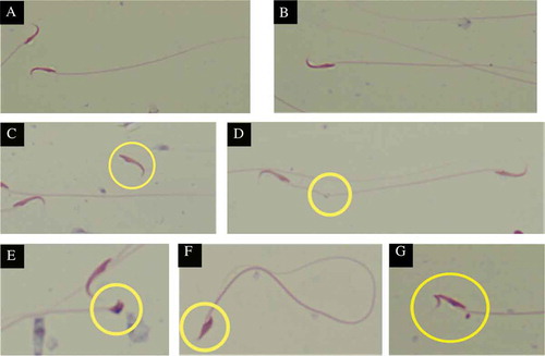
The sperm characteristics of the experimental groups are shown in . Treatment of MSG at 120 mg/kg body weight significantly lowered the epididymal sperm count, motility and viability as well increased the percentage of sperm with abnormal morphology in rats (p < 0.05) when compared to normal rats. Treatment of MSG at 60 mg/kg body weight did show detrimental effect to the sperm characteristics measured, however did not differ significantly from the normal control. depicts the differences in normal and abnormal morphology of sperm.
Figure 1. Effects of MSG on sperm (A) count, percentages of (B) motility, (C) abnormal morphology, and (D) viability. ap < 0.05, as compared to normal control group, bp < 0.05, as compared to 60 mg/kg group.
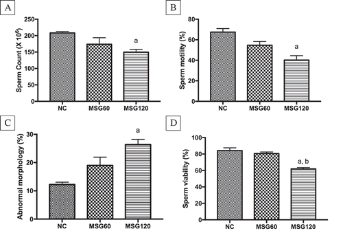
Figure 2. Comparison of normal and abnormal sperm morphology, X400. (A and B) Show normal sperm morphology; hook head and long tail. (C) Shows abnormal tailless sperm. (D) Depicts a bend at a point on the sperm tail. (E–G) Show abnormally developed sperm head; knobbed, short and no hook, and bent sperm head, respectively. Sperm were stained with Diff-Quik staining kit.
Sperm are highly vulnerable to oxidative damage attributable to the high polyunsaturated fatty acid (PUFA) content and low antioxidant protection and very susceptible to ROS attack (Agarwal et al. Citation2014) besides having numerous glutamate receptors. This reflects the finding of this study which showed that treatment of MSG at 120 mg/kg caused a significant decline in sperm quality which is reflected in the reduction of sperm count, sperm motility and viability as well as increased abnormally morphed sperm. Reduction of sperm count is supported by the decrease in sperm mass in epididymis as seen in histological observation. This finding agrees with previous study which revealed reduction in sperm count in epididymis cauda in animals treated with MSG (Nayanatara et al. Citation2008). However, these alterations were not obviously apparent in sperm of rats treated with MSG at 60 mg/kg body weight indicating that the dose is not significant to cause oxidative damage, contradicting to previous report by Hamza and Al-Harbi (Citation2014).
Reproductive hormonal assays
The hormonal assays for levels of testosterone, follicle-stimulating hormone (FSH) and luteinizing hormone (LH) of the experimental groups are shown in . Treatment of MSG at 120 mg/kg body weight caused a significant decrement in the LH level while there are no significant differences in the FSH and testosterone levels of these rats when compared to the normal control group. However, disturbance in these hormone levels were observable by the change in trends where the level of FSH increased in MSG120 while that of testosterone decreased when compared to the normal control group.
Figure 3. Effects of MSG on the levels of (A) luteinizing hormone (LH), (B) follicle stimulating hormone (FSH), and (C) testosterone levels. ap < 0.05, as compared to normal control group.

Previous reports have suggested that MSG could cause toxicity through neurotoxic effect (Onaolapo et al. Citation2016; Yonden et al. Citation2016). In regard to reproductive system, MSG alters the neural control of reproductive hormone due to its ability to damage nerve cells of the hypothalamus, which leads to defect in synthesis and secretion of FSH and LH (Ismail Citation2012). However, the reduction in the weight of all reproductive organs were not seen in the MSG60 group suggesting that the aforementioned damage that promotes a reduction of organ weights in MSG120 rats might not occur in the MSG60 rats. Although this study found that there is a decrease in the testosterone level after MSG supplementation, the difference is not significant when compared to the normal control group. This shows that MSG did not cause extensive damage to the Leydig cells in the testis that are responsible for its production. The significant decrease in LH supports this finding where low LH causes reduction in testosterone level. In contrast, increased FSH level shows that there is a disturbance in the spermatogenesis which cause excessive signaling to the brain to secret FSH. Both unstable levels of FSH and LH after MSG supplementation show that there might be a damage or disturbance to the hormonal secretion pathways in the hypothalamus caused by this substance (Belluardo et al. Citation1990).
Oxidative status evaluation
The effect of MSG administration to rats’ testicular oxidative status is illustrated in . Administration of MSG to rats showed to aggravate oxidative stress in the testis as unveiled in the significantly decreased levels of SOD activity and GSH as well as increased levels of MDA and AOPP with respect to normal control group (p < 0.05).
Figure 4. Effects of MSG on the levels of (A) superoxide dismutase (SOD) activity, (B) reduced glutathione (GSH), (C) malondiadehyde (MDA), and (D) advanced oxidation protein products (AOPP) of the testis. ap < 0.05, as compared to normal control group, bp < 0.05, as compared to 60 mg/kg group.
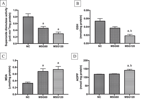
Our results demonstrated a significant increase in testicular MDA and AOPP, agreeing to the plausible oxidative damage effect of MSG on reproductive organs as stated by Ismail (Citation2012). In this study, the marked decrease of antioxidants in the testes might be due to their consumption during the breakdown of ROS generated from the hyperactivity of glutamate receptors in the testis.
Glutamate is the end product of MSG metabolism and its receptors and transporters are reported to be abundantly present in the sperm and reproductive organs of rats, including testis, epididymis, prostate and seminal vesicle (Takarada et al. Citation2004). The high level of circulatory glutamate will affect the tricarbocylic acid (TCA) cycle, which eventually increases the level of alpha-ketoglutarate dehydrogenase activity. This event will stimulate the production or ROS hence increasing the oxidative stress (Sharma Citation2015). In addition, attachment of glutamate to its receptors will cause instantaneous calcium ion influx into the cells, causing the cells to be hyperactive which eventually cause cell damage (Tan et al. Citation2002).
Histological observation
Testis
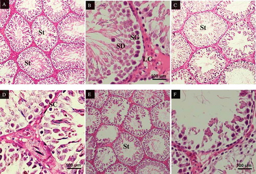
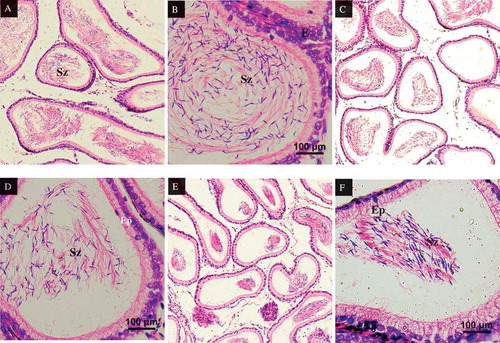
shows the testicular section of (5A) normal rat, (5B) rat treated with 60 mg/kg body weight of MSG and (5C) rat treated with 120 mg/kg body weight of MSG. There are no significant changes in the testicular morphology of MSG60 group when compared to normal control except for the lesser density of spermatids in the seminiferous tubules. In contrast, most of the seminiferous tubules in the testes of rats treated with 120 mg/kg of MSG were missing of spermatids, whereas the germinal epithelium linings are thinner than normal control. There is no obvious difference in the tubules size.
Figure 5. Testicular cross sections of rats, stained with H&E. (A) Testis section of control group (NC), showing normal histological structure of seminiferous tubules (St) containing spermatocytes and late spermatids in the lumen, X100. (B) At higher magnification, the seminiferous epithelium consisting of spermatogonia (SG), spermatocytes and spermatids (SD) can be observed clearly, X400. (C) The effect of MSG treatment at 60 mg/kg is not significant as the normal histostructure of the testis is not disturbed, X100. (D) The seminiferous epithelium is also well intact, X400. (E) However, at the dose of 120 mg/kg, MSG treatment caused deformation to the seminiferous tubules, where some of the seminiferous tubules showed atrophied epithelium layer, X100. (F) At higher magnification, the damage is even more obvious with the loss of spermatocytes and thinning of the epithelium layer, X400.
Epididymis
All rats in the control group showed normal histological structure (). A packed mass of spermatozoa (Sz) was seen in control group within the lumen of the epididymis (). The epididymis in rats in MSG60 group showed a reduction in spermatozoa mass within the epididymal lumen compared to control group; however, the epithelium lining the epididymis of MSG60 did not show any difference compared to control group (). Meanwhile, MSG120 groups revealed a degenerative alteration in epithelium cells lining the epididymis and reduced mass of spermatozoa compared to MSG60 and control group ().
Figure 6. Epididymis cross-sections of rats, stained in H&E. (A) Epididymis of rats in control group, X100s. Epididymis layered by pseudostratified epithelium (Ep). A packed mass of spermatozoa (Sz) were seen in control group within the lumen of the epididymis. (B) Epididymis of rats in control group, X400. Epididymis layered by pseudostratified epithelium (Ep). A packed mass of spermatozoa (Sz) were seen in control group within the lumen of the epididymis. (C) Epididymis of rats in MSG60 group, X100. The epididymis showed reduced in spermatozoa (Sz) mass within the lumen. (D) Epididymis of rats in MSG120 group, X400. The epididymis showed reduced in spermatozoa (Sz) mass within the lumen. (E) Epididymis of rats in MSG120 group, X100. MSG 120 groups revealed reduced mass of spermatozoa (Sz) compared to MSG60 and control group. (F) Epididymis of rats in MSG120 group, X400. Alteration in epithelium (Ep) cells lining the epididymis in which nucleus appear irregular. Mass of spermatozoa (Sz) greatly reduced compared to MSG60 and control group.
Prostate gland
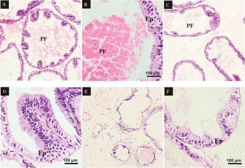
Prostate structure in control group shows normal lumens containing prostatic fluid (PF) and epithelium folding that form the glands (). The glandular epithelium (Ep) is lined with simple to pseudostratified to columnar epithelium surrounded by connective tissue and smooth muscle (). However, sections of prostate gland of rats in MSG60 group showed an atrophy of the prostate lumen compared to the control group (). The amount of prostatic fluid in the lumen of the prostate gland is greatly reduced in MSG120 group compared to control group and MSG60 group (). In addition, the epithelium of prostate in MSG120 rats appears thinner and less folded when compared with the MSG60 and control groups.
Figure 7. Prostate cross-sections of rats, stained in H&E. (A) Prostate gland of rats in control group, X100. The lumen contains prostatic fluid (PF). The glandular epithelium (Ep) are lined with simple to pseudostratified to columnar epithelium. (B) Prostate gland of rats in control group, X400. The lumen contains prostatic fluid (PF) and the glandular epithelium (Ep) are lined with simple to pseudostratified to columnar epithelium cells. (C) Prostate gland of rats in MSG60 group, X100. Atrophy of the prostate lumen compared to control group was observed. (D) Prostate gland of rats in MSG60 group, X400. No changes in the glandular epithelium cells (E) as the cells are still in intact. (E) Prostate gland of rats in MSG120 group, X100. Prostatic fluid in the lumen of the prostate gland is greatly reduced in MSG120 group compared to MSG 60 and control groups. (F) Prostate gland of rats in MSG120 group, X400. Epithelium of prostate in MSG120 rats appear thinner and less folded when compared with the MSG60 and control groups.
Seminal vesicle
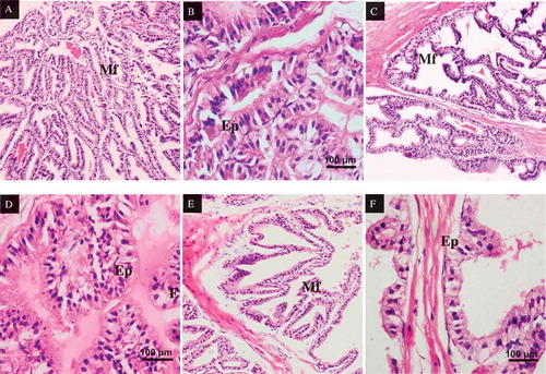
All rats in the control group showed a normal histological pattern (). Cross sections of seminal vesicle in control group shows a mucous layer, which are thin in structure, branching out and folded to form an uneven anastomose branches (F). The epithelium of seminal vesicle also consists of closely arranged columnar cells. However, the prostate gland of rats in MSG60 group showed decrease in number of mucosal folding (Mf) compared to the control group (). Rats treated with 120 mg/kg dose of MSG showed an alteration in the nucleus of seminal vesicle epithelium which appears smaller under microscope. The cell structure arrangements also observed to be irregular and uneven in the epithelium lining of MSG120 group compared to MSG60 control groups ().
Figure 8. Seminal vesicle cross-sections of rats, stained in H&E. (A) Seminal vesicle of rats in control group, X100. Mucous layer are thin in structure, branching out and folded to form an uneven anastomos branches (Mf). (B) Seminal vesicle of rats in control group, X400. The epithelium of seminal vesicle also consists of closely arranged columnar cells. (C) Seminal vesicle of rats in MSG60 group, X100. The mucosal folding (Mf) decreases compared to the control group. (D) Seminal vesicle of rats in MSG60 group, X400. The cell structure arrangements observed to be irregular and uneven in the epithelium lining. (E) Seminal vesicle of rats in MSG120 group, X100. Decrease in number of mucosal folding (Mf) compared to the control MS 60 groups. (F) Seminal vesicle of rats in MSG120 group, X400. The cell structure arrangements observed to be irregular and uneven in the epithelium (Ep) lining of MSF120 group compared to MSG60 and control groups.
Our findings show marked restructuring of reproductive organs in the rats treated with MSG at 120 mg/kg body weight. In regard to the testis damage, it is possible that high glutamate levels may hyper-activate the glutamate receptors in spermatogenic cells, which may induce testicular pathological changes such as sloughing of spermatogenic cells into the tubular lumen as seen in this study. The damage of the accessory reproductive organs, which are also androgenic dependent organs such as epididymis, seminal vesicle and prostate gland, is obviously seen in this study. Androgens are important in ensuring the secretory function of accessory gland (Lina et al. Citation2018). Decrease in the prostatic fluid as seen in this study could possibly be due to the reduction in androgen, which is suggested by Seo et al. (Citation2010). Furthermore, the reproductive organs are also rich with glutamate receptors (Takarada et al. Citation2004). The irregular and uneven of the epithelium lining and reduction in the mucosal folding of the seminal vesicle are obviously seen in this study. This is possibly due to the oxidative damage caused by ROS activity and reduction in the antioxidant status (Taib et al. Citation2015). In contrast, the histological changes in rats treated with 60 mg/kg body weight of MSG are not apparent when compared to MSG120 group. Contradicting to previous studies, MSG at dose 60 mg/kg body weight does not give prominent negative effects on the reproductive systems; however, the observable changes and alterations in this system when MSG is taken at this dose is still somehow should be a concern (Alalwani Citation2014; Hamza and Al-Harbi Citation2014).
The results of the present study have shown that MSG at the dose 120 mg/kg could inflict damage on the reproductive system when taken at estimated ADI of human, which is extrapolated to be 120 mg/kg of body weight. MSG treatment may contribute to the causes that led to infertility problem in rats, which correlates to an increased fear to this substance effect to human health in the recent years. Thus, it is important to reconsider the usage of MSG as a flavor enhancer in a long-term usage.
Materials and methods
Chemical
Commercial brand of 95% food-grade package of MSG was used in the study.
Animals and treatments
A total of 24 male Sprague-Dawley rats weight ranging between 230 and 250 g, age between 9 and 10 weeks old. The animals were obtained from Animal Unit, Faculty of Medicine, Universiti Kebangsaan Malaysia. All rats were maintained in well-ventilated room under standardized conditions of 12/12 light and dark cycle. They were given free access to standard feed pellet and drinking water ad libitum.
Experimental design
All rats were randomized into three groups (n:8); Normal Control (NC), MSG at 60 mg/kg body (MSG60) and MSG at 120 mg/kg body weight (MSG120). The MSG groups were given MSG at 60 mg/kg body weight (Hamza & Al-Harbi Citation2014) and 120 mg/kg body weight (Brosnan et al. Citation2014). Normal control rats were given vehicle (distilled water). On the 28th day, the rats were put under anesthesia before being culled to harvest the reproductive organs for analysis. This experimental design has been approved by Universiti Kebangsaan Malaysia Animal Ethic Committee (UKMAEC) (Ethic code: FSK/2016/BALKIS/23-MAR/741-APR.-2016-DEC.-2018).
Sperm characteristics analysis
The sperm was collected immediately after dissection. 10 μl of sperm suspensed in Hank’s balances salt solution (HBSS) was placed on ‘Makler Counting Chamber’ (Sefi-Medical Instruments, USA). By using the counting chamber, the epididymal sperm count and motility were determined under a light microscope at 100× magnification. Sperm count was expressed as million sperm cells per ml of suspension while sperm motility in percentage of motile sperm.
For sperm viability assessment, 10 μl of sperm suspension was added with 10 μl eosin-nigrosin stain and thick smear were done on the slides. Normal live sperm will not take up the eosin stain and appear white in color, whereas ‘dead’ sperm will take up the eosin stain and appear pinkish. The percentage of abnormal sperm morphology was calculated by using a thin smear of sperm suspension which was Diff-Quik staining kit to assist observation. Two hundred sperm were examined per slide to measure the morphological abnormalities under oil immersion. The data are presented as percentage of abnormal sperm morphology. All sperm characteristics analysis was in accordance to guidelines lined by WHO (Citation2010) whereby the rat sperm abnormal morphology analysis was performed according to guidelines by IRDG (2000).
Reproductive hormonal assays
Testosterone, FSH and LH hormones were analyzed by using enzyme-linked immunoassay (ELISA) kit purchased from Caymen Chemical (USA). All procedures adhered to the standard protocol supplied.
Oxidative status of testis
The oxidative status of the testis is analyzed by measuring the levels of antioxidants and peroxidation products in the testis homogenate. The testis homogenate was obtained by homogenizing the testis in buffer solution which was then centrifuged before collecting the supernatant for biochemical analysis. Superoxide dismutase (SOD) activity level was evaluated according to Beyer and Fridovich (Citation1987) whereas reduced glutathione (GSH) level was measured according to the method by Ellman (Citation1959). Level of malondialdehyde (MDA) in the sample was estimated by using method by Stocks and Dormandy (Citation1971). Advanced oxidation of protein products level (AOPP) was measured by using the method described by Witko-Sarsat et al. (Citation1996).
Histological study of reproductive organs
The reproductive organs (testis, epididymis, prostate and seminal vesicle) were rinsed in buffer saline before being fixed in freshly prepared 10% formalin. They were dehydrated in graded concentrations of ethyl alcohol, cleared in xylene and embedded in paraffin wax. The sections were then cut in 5 μm thick, mounted on glass slides before being stained with hematoxylin and eosin and these sections were examined by light microscope.
Statistical analysis
Data analysis was conducted by using GraphPad Prism (Version 7.0, San Diego). One-way analysis of variance (ANOVA) and post hoc Tukey’s test were performed to determine the significant difference between groups. Results are presented as mean ± standard deviation (SD). Values were considered statistically significant if p < 0.05.
Authors' contributions
Experiment carried out: FFJ, RDM. Wrote the manuscript: FJJ, SBB. Supported in writing manuscript: MMN. Helped supervise the project: MMN, IST. Conceived the original idea: FFJ, SBB. Supervised the project: SBB.
Acknowledgments
The authors are thankful to the Faculty of Health Sciences, Universiti Kebangsaan Malaysia for innumerable support throughout the study.
Disclosure statement
No potential conflict of interest was reported by the authors.
Additional information
Funding
References
- Agarwal A, Virk G, Ong C, du Plessis SS. 2014. Effect of oxidative stress on male reproduction. World J Mens Health. 32(1):1–17.
- Alalwani A. 2014. Monosodium glutamate induced testicular lesions in rats (histological study). Middle East Fertil Soc J. 19:274–280.
- Belluardo N, Mudo G, Bindoni M. 1990. Effects of early destruction of the mouse arcuate nucleus by monosodium glutamate on age-dependent natural killer activity. Brain Res. 534(1–2):225–253.
- Beyer W, Fridovich I. 1987. Assaying for superoxide dismutase activity: some large consequences of minor changes in conditions. Anal Biochem. 161:559–566.
- Brosnan JT, Drewnowski A, Friedman MI. 2014. Is there a relationship between dietary MSG and obesity in animals or humans? Amino Acids. 46(9):2075–2078.
- Ellman G. 1959. Tissue sulfyhdryl groups. Arch Biochem Biophys. 82(1):70–77.
- Eweka O. 2007. Histological studies of the effects of monosodium glutamate on the kidney of adult Wistar rats. Internet J Health. 6:2.
- Fernandes GSA, Arena AC, Campos KE, Volpato GT, Anselmo-Franci JA, Damasceno DC, Kempinas WG. 2012. Glutamate-induced obesity leads to decreased sperm reserves and acceleration of transit time in the epididymis of adult male rats. Reprod Biol Endocrinol. 10:105.
- França LM, Freitas LNC, Chagas VT, Coêlho CFF, Barroso WA, Costa GC, de Andrade Paes AM. 2014. Mechanisms underlying hypertriglyceridemia in rats with monosodium L-glutamate-induced obesity: evidence of XBP-1/PDI/MTP axis activation. Biochem Biophys Res Com. 443:725–730.
- Hamza RZ, Al-Harbi MS. 2014. Monosodium glutamate induced testicular toxicity and the possible ameliorative role of vitamin E or selenium in male rats. Toxicol Rep. 1:1037–1045.
- Iamsaard S, Sukhorum W, Samrid R, Yimdee J, Kanla P, Chaisiwamongkol K, Hipkaeo W, Fongmoon D, Kondo H. 2014. The sensitivity of male rat reproductive organs to monosodium glutamate. Acta Med Acad. 43(1):3–9.
- Igwebuike UM, Nwankwo IA, Ochiogu IS. 2010. Effects of oral administration of monosodium glutamate (MSG) on serum testosteron levels and muscle mass development in male rats. Anim Res Int. 7(2):1212–1217.
- Ismail NH. 2012. Assessment of DNA damage in testes from young Wistar male rat treated with monosodium glutamate. Life Sci J. 9(1):930–939.
- Kondoh T, Torii K. 2008. MSG intake suppresses weight gain, fat deposition, and plasma leptin levels in male Sprague-Dawley rats. Physiol Behav. 95(1–2):135–144.
- Lina S, Eliza H, Hashida NH, Ibrahim SF, Osman K. 2018. Androgen receptor and ultrastructural features of Nigella sativa oil and nicotine-treated male rat reproductive glands. Sains Malays. 47(8):1827–1833.
- Mesbah SF, Shokri S, Karbalay-Doust S, Mirkhani H. 2007. The effect of nandrolone decanoate on the body, testis and epididymis weight and semen parameters in adult male rats. Iran J Med Sci. 32(2):93–99.
- Michael B, Yano B, Sellers RS, Perry R, Morton D, Roome N, Johnson JK, Schafer K. 2007. Evaluation of organ weights for rodent and non-rodent toxicity studies: a review of regulatory guidelines and a survey of current practices. Toxicol Pathol. 35:742–750.
- Miranda RA, da Silva Franco CC, de Oliveira JC, Barella LP, Tófolo LP, Ribeiro TA, Pavanello A, da Conceição EP, Torrezan R, Armitage J, et al. 2017. Cross-fostering reduces obesity induced by early exposure to monosodium glutamate in male rats. Endocrine. 55(1):101–112.
- Mondal M, Sarkar K, Nath PP, Paul G. 2018. Monosodium glutamate suppresses the female reproductive function by impairing the functions of ovary and uterus in rat. Environ Toxicol. 33(2):198–208.
- Nayanatara A, Vinodini N, Damadar G, Ahamed B, Ramaswamy C, Shabarinath M, Bhat M. 2008. Role of ascorbic acid in monosodium glutamate mediated effect on testicular weight sperm morphology and sperm count in rat testis. J Chin Clin Med. 3:1–5.
- Nordkap L, Joensen UN, Blomberg Jensen M, Jørgensen N. 2012. Regional differences and temporal trends in male reproductive health disorders: semen quality may be a sensitive marker of environmental exposures. Mol Cell Endocrinol. 355(2):221–230.
- Ochiogu IS, Ogwu D, Uchendu CN, Okoye CN, Ihedioha JI, Mbegbu EC. 2015. Effects of monosodium-L-glutamate administration on serum levels of reproductive hormones and cholesterol, epididymal sperm reserves and testicular histomorphology of male albino rats. Acta Vet Hung. 63(1):125–139.
- Onaolapo OJ, Onaolapo AY, Akanmu MA, Gbola O. 2016. Evidence of alterations in brain structure and antioxidant status following ‘low-dose’ monosodium glutamate ingestion. Pathophysiology. 23(3):147–156.
- Sand J. 2005. A short history of MSG: good science, bad science and taste cultures. Gastronomica. 5(4):38–49.
- Seo HJ, Ham HD, Jin HY, Lee WH, Hwang HS, Park SA. 2010. Chronic administration of monosodium glutamate under chronic variable stress impaired hypothalamic-pituitary-adrenal axis function in rats. Korean J Physiol Pharm. 14(2):13–21.
- Sharma A. 2015. Monosodium glutamate-induced oxidative kidney damage and possible mechanisms: a mini-review. J Biomed Sci. 22:93.
- Shin JW, Seol IC, Son CG. 2010. Interpretation of animal dose and human equivalent dose for drug development. J Korean Oriental Med. 31(3):1–7.
- Stocks J, Dormandy TL. 1971. The autooxidation of human red cell lipids induced by hydrogen peroxide. J Hematol. 20(1):95–111.
- Taib IS, Budin SB, Ghazali AR, Jayusman PA, Louis SR, Mohamed J. 2015. Palm oil tocotrienol-rich fraction attenuates testicular toxicity induced by fenitrothion via an oxidative stress mechanism. Toxicol Res. 4:132–142.
- Takarada T, Hinoi E, Balcar VJ, Taniura H, Yoneda Y. 2004. Possible expression of functional glutamate transporters in the rat testis. J Endocrinol. 181:233–244.
- Tan S, Schubert D, Maher P. 2002. Oxytosis, a novel form of programmed cell death. Curr Top Med Chem. 1(6):497–506.
- Tordoff MG, Aleman TR, Murphy MC. 2012. No effects of monosodium glutamate consumption on the body weight or composition of adult rats and mice. Physiol Behav. 107:338–345.
- WHO. 2010. WHO laboratory manual for the examination and processing of human semen. Geneva: World Health Organization.
- Witko-Sarsat V, Friedlander M, Capeillére-Blandin C, Nguyen-Khoa T, Nguyen AT, Zingraff J, Jungers P, Descamps-Latscha B. 1996. Advanced oxidation protein products as a novel marker of oxidative stress in Uremia. Kidney Int. 49(5):1304–1313.
- Yahya NJ, Abd Hamid Z, Abu Hanipah EN, Ajik EM, Yusoff NA, Taib IS. 2018. Oxidative stress and morphological assessment of bone marrow in monosodium glutamate-treated rat. Jurnal Teknologi. 80(2):105–111.
- Yonden Z, Ozcan O, Cimen AY, Delibas N. 2016. The effects of monosodium glutamate and aspartame on rat hippocampal N-methyl-D-aspartate receptor subunits and oxidative stress biomarkers. Int J Clin Exp Med. 9(2):1864–1870.