
ABSTRACT
Helicobacter pylori (H. pylori) infection is the strongest known risk factor for the development of gastric cancer. DNA damage response (DDR) and autophagy play key roles in tumorigenic transformation. However, it remains unclear how H. pylori modulate DDR and autophagy in gastric carcinogenesis. Here we report that H. pylori infection promotes DNA damage via suppression of Rad51 expression through inhibition of autophagy and accumulation of p62 in gastric carcinogenesis. We find that H. pylori activated DNA damage pathway in concert with downregulation of repair protein Rad51 in gastric cells, C57BL/6 mice and Mongolian gerbils. In addition, autophagy was increased early and then decreased gradually during the duration of H. pylori infection in vitro in a CagA-dependent manner. Moreover, loss of autophagy led to promotion of DNA damage in H. pylori-infected cells. Furthermore, knockdown of autophagic substrate p62 upregulated Rad51 expression, and p62 promoted Rad51 ubiquitination via the direct interaction of its UBA domain. Finally, H. pylori infection was associated with elevated levels of p62 in gastric intestinal metaplasia and decreased levels of Rad51 in dysplasia compared to their H. pylori- counterparts. Our findings provide a novel mechanism into the linkage of H. pylori infection, autophagy, DNA damage and gastric tumorigenesis.
Introduction
Helicobacter pylori (H. pylori), a human pathogen, colonizes approximately half of the world’s population, and its colonization can last for life without antibiotic intervention.Citation1,Citation2 H. pylori infection typically causes chronic inflammation and cellular injury, which leads to gastric carcinogenesis through the histopathological Correa cascade from chronic gastritis (CG), intestinal metaplasia (IM) and dysplasia (Dys) culminating in gastric cancer.Citation3,Citation4 Cytotoxin-associated gene A (CagA), encoded by the cag pathogenicity island, is a well-characterized virulence factor of H. pylori. CagA is delivered into gastric epithelial cells through the type IV secretion system, and dysregulates a series of intracellular signaling pathways, resulting in severe tissue damage and inflammation.Citation5 It has been reported that CagA was associated with a higher risk of gastric cancer in experimental animals and human samples.Citation6
The maintenance of human genome integrity depends on DNA damage response. Although single-strand breaks (SSB) are the most common types of DNA damage, double-strand breaks (DSB) are lethal to cells and need to be rapidly repaired for ensuring cell survival. In response to DSB, DNA damage sensor Ataxia-telangiectasia mutated serine/threonine kinase (ATM) is rapidly translocated to the sites of DNA damage where it is activated.Citation7,Citation8 The activation of ATM phosphorylates histone H2AX (γH2AX) and induces damage-pathway downstream effectors, such as Chk2, p53 and BRCA1, leading to DNA repair, cell cycle arrest and apoptosis.Citation9 The highly conserved protein Rad51 plays a central role in homologous recombination of DNA during DSB repair. It has been shown that accumulation of DNA damage by elevated mutation rate, a potential cause of cancer, is associated with altered Rad51 expression.Citation10,Citation11 Accumulating evidence has indicated that H. pylori infection increases production of reactive oxygen species (ROS) and DNA damage levels, which can contribute genome instability and gastric carcinogenesis.Citation12 Also, our previous studies indicated a progressively elevated γH2AX levels in gastric pathological cascade from CG, IM to Dys, but slightly declined in GC, suggesting that DSBs appear to be an early event in H. pylori-mediated genetic instability and gastric tumorigenesis.Citation13 Although DNA damage can be efficiently repaired, it has been reported that long-term bacterial infection leads to accumulation of unrepaired breaks due to saturation of repair capabilities.Citation14 However, the molecular mechanism underlying H. pylori-induced DSBs and the DNA damage repair has not been fully elucidated.
Recently, DNA damage has been linked to autophagy, an intracellular degradation process by which cytoplasmic materials are delivered to the lysosome for digestion.Citation15 In response to various cellular stress, activation of autophagy is crucial for maintaining cellular energy levels through synthesis, degradation and recycle of constituents. Induction of autophagy serves as a cell defense mechanism against intracellular pathogenic microorganisms.Citation16 Several microbes such as Listeria monocytogenes have been shown to stimulate formation of autophagosomes, which in turn initiates bacterial degradation.Citation17 Emerging evidence over the past decade has indicated that H. pylori infection was linked to autophagy.Citation18 The virulence factor cytotoxin (VacA) is sufficient to induce autophagy and cell death in H. pylori-infected gastric epithelial cells.Citation19 Furthermore, another important bacterial oncogenic protein CagA has been found to be degraded by autophagy, whereas CAPZA1 overexpression promoted CagA to escape from autophagic degradation.Citation20 In addition, H. pylori CagA protein was reported to negatively regulate autophagy and promote inflammation.Citation21
Activation of autophagy by various cellular stress has been implicated in DNA repair mechanisms, such as homologous recombination (HR) and non-homologous end-joining (NHEJ).Citation22 Degradation of p62/SQSTM1 (p62), the best-known autophagic substrate, has been widely identified as an indicator of autophagy. p62 interacts with ubiquitinated proteins via its C-terminal ubiquitinated-associated domain (UBA) and results in their autophagic degradation.Citation23 Wang et al. have found that p62 accumulation due to deficiency of autophagy can bind to and inhibit the activity of RNF168, an E3 ligase required for histone H2A ubiquitination. As a result, DSB repair pathway was significantly impaired.Citation24 In addition, overexpression of p62 has been shown to induce proteasomal degradation of homologous recombination marker Rad51.Citation25 Currently, the precise mechanisms by which autophagy mediates DDR and genome stability remain unknown in H. pylori infection-associated gastric tumorigenesis.
In this study, we show that prolonged exposure to H. pylori infection inhibited autophagosome formation, which contributed to excessive DNA damage and eventual gastric tumorigenesis. Mechanistically, accumulation of p62 due to autophagy inhibition caused DNA repair marker Rad51 ubiquitination and degradation. Our findings indicated a critical role for autophagy and DDR in H. pylori infection-induced gastric carcinogenesis.
Results
H. pylori infection promotes ATM-dependent DNA damage response
We have previously shown that γH2AX, a reliable marker of DSB, was progressively increased in tissues in gastric lesions from CG, IM to Dys.Citation13Given an important role of H. pylori infection in gastric carcinogenesis, we investigated whether H. pylori infection induces DNA damage in gastric epithelial cells. Human normal gastric epithelial GES-1 cells were co-cultured with CagA+VacA+H. pylori strain NCTC11637 (ATCC43504) at various time-points and at different MOIs. γH2AX was significantly increased in a time- and MOI-dependent manner following infection ( and S3A). In addition, a percentage of tail DNA in the comet assay was used to represent the abundance of fragmented DNA due to DNA damage. In GES-1 cells, H. pylori infection led to significant increases of DNA DSB levels over the infection duration (). To further evaluate these in vitro findings, Mongolian gerbil, a widely used model for investigating H. pylori-induced gastric tumorigenesis,Citation26 was challenged with Brucella broth (negative control) or with carcinogenic H. pylori 43503 strain. Immunohistochemistry staining was performed to detect H. pylori colonization (Figure S1D). Consistent with our in vitro data, H. pylori infection significantly increased gastric γH2AX expression levels compared to uninfected control and elevated γH2AX expression at 12 months post-infection (MPI) compared to 6 MPI using immunohistochemistry (). It has been documented that ATM is recruited and activated at sites of DSB where it phosphorylates serine/threonine kinase Chk2 at priming site Thr68, leading to cell cycle arrest through phosphorylation of p53.Citation27 We next examined whether H. pylori-induced DNA damage is associated with the activation of ATM. Following H. pylori infection on gastric GES-1 cells, levels of phosphorylated ATM were increased in a time-dependent manner. In addition, its downstream substrates Chk2 and p53 were also activated via phosphorylation ( and S3B). Furthermore, the percentage of S phase was significantly increased in gastric epithelial cells at 12 h or 24 h of infection, suggesting that H. pylori could induce S-phase cell cycle arrest ().
Figure 1. H. pylori infection affects DNA damage response signaling pathway. (a) Representative images of Western blot analysis for γH2AX in GES-1 cells with H. pylori NCTC11637 infection at different MOI or at different time points. (b) The comet assay for assessing DNA damage in GES-1 cells with or without H. pylori. Scale bar, 10 μm. (c) Immunochemistry for γH2AX in gastric tissues collected from Mongolian gerbils that were challenged with H. pylori NCTC11637 strain or Brucella broth for 6 months (H. pylori, n = 30; control, n = 15) or 12 months (H. pylori, n = 30; control, n = 10). (d) Representative images (200× magnification) of Western blot analysis for p-p53 (Ser20), p-p53 (Ser15), p-Chk2 (Thr168), Chk1, p-ATM (Ser1981), ATM in GES-1 cells infected with H. pylori NCTC11637 at different time points. (e) Flow cytometry analysis for cell cycle distribution after H. pylori infection. (f) Representative immunohistochemical staining for p-ATM (S1981), p-Chk2 (T68) and p-p53 (S15) in a series of human gastric tissues with Correa cascade from CNAG, IM, Dys and GC. Quantitative analysis of the immunohistochemical staining results in gastric tissues. *P < .05, **P < .01. (g) Representative images of Western blot analysis for Rad51 in GES-1 cells with H. pylori NCTC11637 infection. (h) Representative images of Western blot analysis for γH2AX, p-p53 (Ser20) and p-p53 (Ser15) in GES-1 cells, which were treated with H. pylori in combination with plasmid expressing Rad51 or Rad51 siRNAs. A representative blot of three independent experiments was shown. (i) After transfected with Rad51 expression plasmid or Rad51 siRNAs, the comet assay was performed to detect DNA damage and the length of tail DNA was evaluated in gastric cells with H. pylori infection. (j) Immunohistochemistry staining images (200× magnification) for Rad51 in gastric tissues of Mongolian gerbils infected with H. pylori. Scale bar: 50 μm. All in vitro experiments were independently repeated three or more times. *P < .05, **P < .01.
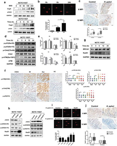
To characterize the role of the DNA damage response pathways in H. pylori-associated gastric carcinogenesis, we examined and compared the protein levels of phosphorylated ATM, Chk2 and p53 in human gastric tissues with CG, IM to Dys and between H. pylori-positive and -negative tissues. Levels of p-ATM (S1981), p-Chk2 (T68) and p-p53 (S15) were gradually increased during neoplastic progression from CG, IM to Dys, suggesting the suppressive effect of ATM/CHK2 in early gastric carcinogenesis. H. pylori infection induced the significant increases of p-ATM at CG/IM/Dys, p-Chk2 at IM and p-p53 at CG/M compared to the H. pylori-negative subjects. Interestingly, the activity of these protein targets was decreased in GC stage ( and Figure S1 C). These data suggest that reduced DDR activity could promote cell cycle progression and facilitate the generation of gene mutations, finally enhancing GC development. In addition, there were no significant differences on total ATM, Chk2 levels in human gastric tumorigenesis cascade (Figure S1A and S1B). Taken together, our findings clearly indicate that H. pylori infection promoted the DNA damage response pathway in the early stages of gastric tumorigenesis.
Persistent H. pylori infection promoted DNA damage via suppression of Rad51
Accurate DNA damage repair is required for maintaining genome stability. Our previous studies have shown that H. pylori infection was correlated with the expression of NHEJ marker Ku70/80 in human GC tissues.Citation28 To explore how H. pylori infection modulates DSB HR repair, GES-1 cells were co-cultured with H. pylori NCTC11637 strain at different time-points. H. pylori infection resulted in the reduction of Rad51 protein, a crucial protein in HR DNA repair process, but did not significantly affect another DNA repair marker BRCA2, which can facilitate the loading of the recombination protein Rad51 at DNA breaks ( and S3 C).Citation29 In addition, DSB marker γH2AX and HR repair marker Rad51 focus formation were further detected by immunofluorescent microscopy. Intracellular H. pylori colonization in cells was firstly confirmed by Immunofluorescence staining (Figure S1E). We found that H. pylori infection led to statistically significant increase in the ratio of γH2AX and Rad51 (Figure S1 F). These findings suggested that H. pylori-induced elevation of DSBs may be due to downregulation of Rad51. To further elucidate the role of Rad51 in DSB repair, gastric GES-1 cells were treated with Rad51-expressing plasmid or Rad51 siRNA and then co-cultured with H. pylori NCTC11637 strain. H. pylori-induced elevation of γH2AX and downstream phosphorylated p53 levels was suppressed in cells transfected with the Rad51 plasmid. Conversely, siRNAs-mediated Rad51 knockdown led to an increase of levels of γH2AX and phosphorylated p53 (, S3D and S3E). Moreover, overexpression of Rad51 significantly suppressed H. pylori-induced DNA damage, whereas inhibition of Rad51 upregulated DNA damage as demonstrated in the comet assay, further indicating the importance of Rad51 in H. pylori-induced DNA damage response (). To evaluate if these in vitro mechanisms are operable in vivo, Rad51 levels in the gastric mucosa of Mongolian gerbils at 6 and 12 MPI were examined and quantified using immunohistochemistry analysis. At 6 MPI, elevated Rad51 levels were found between infected and control groups, but there was no statistic difference. By 12 MPI, Rad51 expression was significantly decreased in the H. pylori-infected gerbils versus the control animals (). Collectively, these data indicate that persistent H. pylori infection suppressed expression of homologous recombination marker Rad51, which resulted in the accumulation of DNA damage.
Autophagy is dynamically changed in response to H. pylori infection
Autophagy, a cellular self-degradation process, plays a pivotal role in host–pathogen interaction and gastric carcinogenesis. LC3B is a well-established autophagy marker. Conversion of soluble LC3B-I to lipid bound LC3B-II has been considered as a hallmark of autophagosome formation.Citation30 To investigate how H. pylori infection affects autophagy, gastric cells were co-cultured with H. pylori at different time-points. Western blot showed that levels of LC3B-I and II were significantly increased up to 6 hours post-infection (HPI) and then were gradually decreased ( and S4A). Consistently, transmission electron microscopy (TEM) revealed that there was an increase of autophagosomes or autolysosomes-like structures at 6 HPI, which were significantly reduced by 24 HPI (). P62, a well-known substrate for autophagic degradation, can be used as an indicator of autophagic flux.Citation31 As shown in ,, p62 levels in the gastric mucosa of H. pylori-infected gerbils were significantly increased at 12 MPI.
Figure 2. Autophagy is dynamically changed in response to H. pylori infection. (a) Representative images of Western blot analysis for LC3 in GES-1 cells co-cultured with H. pylori NCTC11637. (b) Transmission electron microscopy showing autophagosomes in gastric cells with or without H. pylori infection. Scale bar: 500 μm. (c) The Mongolian gerbils were challenged with H. pylori NCTC11637 strain or Brucella broth 6 months (H. pylori, n = 30; control, n = 15) or 12 months (H. pylori, n = 30; control, n = 10). Representative p62 immunohistochemical staining images in gastric tissues were shown. Scale bar: 50 μm. (d) Quantification of staining scores for p62 in gastric epithelium. (e) After transfection with mRFP-GFP-LC3 adenovirus, cells were incubated with H. pylori and Confocal microscopy was used to monitor the autophagic flux. Scale bar, 10 μm. (f) The numbers of autophagosome (mRFP+GFP+) and autolysosome (mRFP+GFP−) were quantified. All in vitro experiments were independently repeated three or more times. *P < .05, **P < .01, ***P < .001.
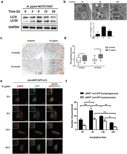
To directly visualize and quantitatively characterize autophagy in situ, adenovirus expressing LC3B fused to fluorescent proteins mRFP and GFP (Ad-mRFP-GFP-LC3) was transfected into GES-1 cells in which, autophagosome formation with the mRFP-positive/GFP-positive (yellow) puncta can be distinguished from autolysosome with the mRFP-positive/GFP-negative puncta (red). The number of autophagosomes and autolysosomes in the cells were increased at 6 HPI. However, by 24 HPI, both the abundance of autophagosomes and autolysosomes were rapidly decreased (,). Taken together, these findings indicate that H. pylori infection induces autophagy at 6 HPI, which was followed by a rapid decrease of autophagy by 24 HPI.
Inhibition of autophagy increased DNA damage in response to H. pylori infection
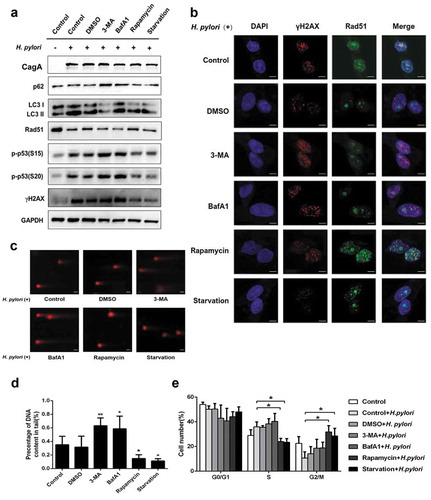
Recent studies have documented that cell autophagy contributes to the maintenance of the genomic stability and loss of autophagy impairs DNA repair.Citation15 We next investigated whether autophagy plays a role in DNA damage response to infection in gastric GES-1 cells. GES-1 cells were treated with the autophagy inhibitor 3-MA, or BafA1, the autophagy inducer rapamycin, or starvation, and then co-cultured with H. pylori strain NCTC11637. Inhibition of autophagy by the pharmacological inhibitors significantly aggravated the H. pylori-induced DNA damage signaling pathway, which were reflected by the upregulation of DNA damage marker γH2AX, increased the phosphorylation of p53 and the downregulation of DNA repair protein Rad51, and opposite is true with the treatment of rapamycin or starvation that activates autophagy ( and S4B). These results were further supported using immunofluorescence ( and S1 G). In addition, the tail lengths of H. pylori-induced-DNA fragmentation were significantly increased in response to the treatment of 3-MA or BafA1, but decreased after treatment with rapamycin or starvation (,). Furthermore, H. pylori infection concurrently with the treatment of the autophagy activator Rapamycin or nutrient starvation accelerated cell cycle progression from S to G2/M phase compared to H. pylori infection alone (). Taken together, these data suggested that persistent H. pylori infection led to the accumulation of DNA damage and cell cycle arrest via the suppression of autophagy.
Figure 3. Inhibition of autophagy increased DNA damage in response to H. pylori infection. (a) Representative images of Western blot analysis for p62, LC3, Rad51, p-p53 (S15), p-p53 (S20), γH2AX protein in GES-1 cells infection with H. pylori (MOI 200) for 12 hand treatment with autophagy inhibitors (3-MA, BafA1) or autophagy activators (Rapamycin, Starvation). (b) Immunofluorescence assay showing γH2AX (red) and Rad51 (green) expressions in GES-1 cells treated with autophagy inhibitors or autophagy activators and then infected with H. pylori (MOI 200) for 12 h. Scale bar, 10 μm. (c) After treatment with autophagy-related drugs and H. pylori, DNA damage was determined by the Comet assay in GES-1 cells. Scale bar, 10 μm. (d) Comet tail lengths showing DNA damage. (e) Flow cytometry analysis showing cell cycle progression in cells treated with autophagy inhibitor or activators and then infected with H. pylori (MOI 200) for 12 h. All experiments were independently repeated three or more times. *P < .5, **P < .01.
Rad51 is integral for autophagy-mediated DNA damage repair response to H. pylori infection
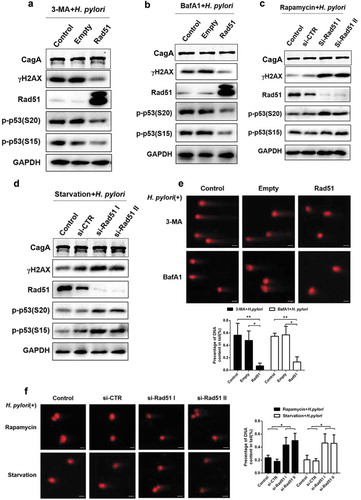
Our aforementioned results indicated that H. pylori infection suppressed autophagy and DNA repair protein Rad51. Additionally, inhibition of autophagy led to decreased expression of Rad51 in the setting of H. pylori infection. We hypothesize that Rad51 could serve as a crucial adaptor in autophagy-regulated DNA damage in response to H. pylori infection. To test this hypothesis, GES-1 cells were transfected with Rad51-expressing plasmid, in combination with H. pylori NCTC11637 infection and treatment of autophagy inhibitors 3-MA or BafA1. Overexpression of Rad51 suppressed elevation of H. pylori-induced DNA damage signals such as γH2AX and its downstream factors due to impairment of autophagy (,, S5A and S5B). Conversely, knocking-down endogenous Rad51 by siRNA significantly increased H. pylori-induced DNA damage signal suppressed by the activation (rapamycin or starvation) of autophagy (,, S5C and S5D). Consistent with the Western blot data, transient overexpression of Rad51 decreased the H. pylori-induced DNA fragmentation, despite inhibition of autophagy, whereas, the knockdown of Rad51 elevated autophagy-inhibited DNA damage (,). Collectively, these data suggest that autophagy modulates H. pylori-induced DNA damage via Rad51.
Figure 4. Rad51 is a crucial adaptor in autophagy-regulated DNA damage in response to H. pylori infection. (a, b) Representative images of Western blot analysis for γH2AX, Rad51, p-p53 (S20) and p-p53 (S15) protein levels in GES-1 cells following H. pylori infection (MOI 200) for 12 h, and transfection with Rad51 expression plasmids in the presence of autophagy inhibitor treatment (a) 3-MA or (b) BafA1. (c, d) Representative images of Western blot showing expression of γH2AX, Rad51, p-p53 (S20) and p-p53 (S15) in GES-1 cells transfected with Rad51 siRNAs, infected with H. pylori (MOI 200) for 12 h and treated with autophagy inducer (c) rapamycin or (d) starvation. (e) Comet assay showing DNA damage in GES-1 cells transfected with Rad51 expression plasmid, and then infected with H. pylori for 12 h in the presence of autophagy inhibitors 3-MA or BafA1. Scale bar, 10 μm. (f) Comet assay showing DNA damage in GES-1 cells transfected with Rad51 siRNAs, and then infected with H. pylori for 12 h in the presence of autophagy inducers rapamycin or starvation. Scale bar, 10 μm. All in vitro experiments were independently repeated three or more times. *P < .5, **P < .01.
H. pylori-induced DNA damage response and suppression of autophagy are cytopathic events common to different CagA+H. pylori strains, cell types and animal models
To examine whether H. pylori-induced DNA damage response and alteration of autophagy are strain-specific. CagA+H. pylori strain 7.13 was co-cultured with GES-1 cells with different MOI for 24 h. Consistent with the results with NCTC11637 (,), the strain 7.13 infection in GES-1 cells increased levels of the DSBs marker γH2AX in concert with the decrease of the DNA repair protein Rad51 in a MOI-dependent manner. Also, accumulation of autophagy substrate p62 ascended in line with an increase of MOIs following 7.13 infection ( and S6A). TEM assay also showed that 7.13 infection increased the number of autophagosomes at 6 HPI, and then led to the inhibition of autophagy at 24 HPI. These findings with 7.13 strain were consistent with the results with H. pylori NCTC11637 strain (). To exclude a possibility that H. pylori-induced cytopathic effects were cell type-dependent, gastric adenocarcinoma AGS cells were co-cultured with 7.13 strain. The responses of the DNA damage marker γH2AX, DNA repair protein Rad51 and the autophagy substrate p62 to 7.13 infection in AGS cells were similar to those in GES-1 cells ( and S6B). CagA protein is one of the most important virulence factors in H. pylori-induced gastric carcinogenesis. We therefore investigated the role of CagA in DNA damage and autophagy regulation. AGS and GES-1 cells were co-cultured with H. pylori strain 7.13 or its isogenic CagA-mutant. Unlike wild-type H. pylori 7.13, the loss of CagA failed to promote DNA damage and accumulation of autophagic receptor p62, and to suppress expression of Rad51 ( and S6 C).
Figure 5. H. pylori-Susceptible DNA damage response and autophagy are shared between different bacterial strains, cell types and animal models. (a) Representative images of Western blot analysis for p62, Rad51 and γH2AX in GES-1 cells infected with H. pylori 7.13 at different MOI or at different time points. (b) Transmission electron microscopy for autophagic vacuoles in gastric cells infected with H. pylori 7.13. Scale bar: 500 μm. (c, d) Representative images of Western blot analysis for CagA, p62, Rad51 and γH2AX in gastric cells (c) GES-1, or (d) AGS infected with H. pylori strain 7.13 or its CagA-knockout mutant. (e) H&E staining for gastric pathology in gastric tissues of H. pylori-infected C57BL/6 mice. (f) Immunohistochemistry staining for Rad51 and p62 levels in C57BL/6 mice infected with H. pylori. Scale bar: 50 μm. (g) Western blot analysis for Rad51 and p62 protein levels in gastric tissues of H. pylori-infected mouse model. Images are representative of n = 3 mouse stomachs. All in vitro experiments were independently repeated three or more times. **P < .01, ***P < .001.
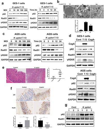
To explore whether H. pylori infection-regulated DNA damage and autophagy responses are operable in different animal models, C57BL/6 male mice were infected with H. pylori NCTC11637 strain for 3 months. H. pylori colonization of mouse gastric mucosa was visualized using immunohistochemistry and FISH (Figure S2A). Real-time PCR analysis showed that infection with H. pylori led to transcriptional upregulation of inflammatory factors including IL-1β, IL-6, IL-10 and IL-17, suggesting inflammation response induced by H. pylori (Figure S2B). H&E staining showed that H. pylori infection resulted in significantly gastric inflammation compared to control group at 3MPI (). In addition, levels of Rad51were significantly decreased in concert with increased p62 levels in H. pylori-infected gastric mucosa, compared to uninfected controls (). This result was supported by Western blot analysis showing the reduction of Rad51 level and the elevation of p62 level in H. pylori-infected mouse gastric mucosa ( and S6D). These results demonstrate that accumulation of DNA damage and loss of autophagy due to H. pylori infection was operable by other CagA+H. pylori strains, in different cell lines or different animal models.
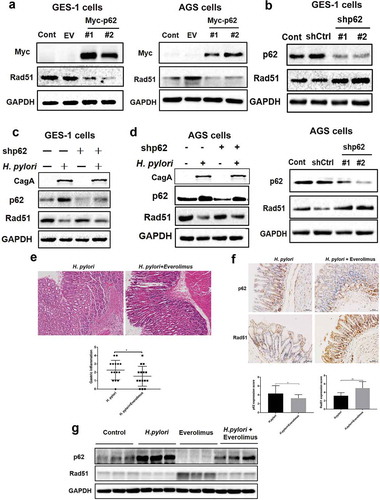
Accumulation of p62 promoted H. pylori-induced DNA damage via suppression of the DNA repair protein Rad51
Given that H. pylori-induced suppression of autophagy was linked to persistent DNA damage and genome instability via inhibition of DNA repair protein Rad51, we next investigated the interaction between autophagy and Rad51. P62 is the best-known autophagic substrate. We hypothesize that p62 is a crucial factor in modulating expression of Rad51 since levels of p62 were inversely related to levels of Rad51 levels in H. pylori-infected gastric cells and gastric mucosa of animal models. Overexpression of p62 in AGS or GES-1 cells significantly reduced the expression of Rad51 ( and S7A). Whereas knockdown of p62 led to upregulation of Rad51 protein ( and S7B). To further assess how H. pylori-induced accumulation of p62 regulates expression of Rad51. Normal gastric epithelial GES-1 cells were transfected with p62 shRNA plasmid and cocultured with or without H. pylori 7.13 strain. The knockdown of p62 with the p62 shRNA plasmid increased the expression of Rad51 in both H. pylori 7.13-infected GES-1 and AGS cells (,, S7C and S7D). Furthermore, we assessed whether p62 also suppressed Rad51 expression in H. pylori NCTC11637-infected C57BL/6 mice in combination with treatment of everolimus, a well-known autophagy inducer of inhibiting mTOR. As shown in , everolimus treatment significantly attenuated H. pylori-induced inflammatory response at 3 MPI (). Immunohistochemistry staining revealed that the gastric mucosa of C57BL/6 mice treated with both H. pylori infection and everolimus contained lower p62 levels and higher Rad51 levels, compared to the mice infected with H. pylori alone (). The loss of p62 caused by everolimus was significantly correlated with the elevation of Rad51 levels following H. pylori infection ( and S7E). These results indicate that accumulation of the autophagic substrate p62 in H. pylori-infected gastric epithelial cells promoted H. pylori-induced DNA damage via suppression of the DNA repair marker Rad51.
Figure 6. Accumulation of p62 suppressed DNA repair protein Rad51 in response to H. pylori infection. (a) Representative images of Western blot analysis for p62 and Rad51 proteins in GES-1 and AGS cells transfected with plasmid expressing p62. (b) Representative images of Western blot analysis for p62 and Rad51 proteins in GES-1 and AGS cells transfected with p62 shRNAs. (c, d) After transfected with p62 shRNAs, (c) GES-1 cells or (d) AGS cells showing p62 and Rad51 levels by immunoblot assay following H. pylori infection. (e) H&E staining showing the extent of gastric inflammatory response in C57BL/6 mice infected with H. pylori alone or combined with everolimus treatment. (f) Immunohistochemistry staining showing p62 and Rad51 levels in C57BL/6 mice treated with H. pylori and autophagy inducer everolimus by gastric gavage. Scale bar: 50 μm. (g) Western blot analysis for p62 and Rad51 in gastric tissues of mouse models. Images are representative of n = 3 mouse stomachs. All in vitro experiments were independently repeated three or more times. *P < .05, **P < .01.
The ubiquitination associated (UBA) domain of p62 was essential for H. pylori infection-mediated Rad51 ubiquitination
To test whether H. pylori infection induced proteasomal degradation of Rad51, AGS cells were pre-treated with a proteasome inhibitor MG132 prior to H. pylori 7.13 infection. Inhibition of proteasomal function by MG132 led to an increase of Rad51 levels ( and S7 F). In addition, AGS or GES-1 cells were co-transfected with plasmids expressing Rad51 and ubiquitin, then treated with proteasomal inhibitor MG132 for 6 hours prior to H. pylori 7.13 or its CagA− mutant infection. CagA+ H. pylori 7.13 but not its strain its CagA− mutant significantly promoted Rad51 ubiquitination (). These results indicate that CagA+H. pylori infection decreased Rad51 through the ubiquitination and degradation of Rad51. Next, we dissected the mechanism underlying H. pylori-induced accumulation of p62 and suppression of Rad51. Our data showed that H. pylori infection suppressed Rad51 protein levels, but not mRNA, suggesting that post-transcriptional process affected Rad51 protein levels (Figure S2C). Given that p62, an ubiquitin and LC3 binding protein directly binds to an ubiquitinated protein via its UBA domain and sequesters them into inclusion for degradation by autophagy,Citation32 we examined the possible interaction between p62 and Rad51. Immunofluorescence staining showed that p62 was mostly localized in the cytoplasm and Rad51 was in the nucleus of uninfected cells, respectively. H. pylori infection could enhance p62 levels and decreased Rad51 expression. Furthermore, H. pylori infection resulted in the nuclear translocation of p62 where p62 was colocalized with Rad51, suggesting that H. pylori infection promoted p62 nuclear entry and interaction with Rad51 (). To demonstrate the physical interaction between p62 and Rad51, Plasmids Flag-Rad51 or Myc-p62 were transfected into gastric AGS or GES-1 cells followed by the immunoprecipitation assay. As shown in , p62 was coimmunoprecipitated with Rad51, indicating that p62 interacted with Rad51 ().
Figure 7. P62 induced H. pylori infection-mediated Rad51 ubiquitination. (a) Gastric epithelial cells were co-cultured with H. pylori strain and incubated with proteasome inhibitor MG132. P62 (green) and Rad51 (red) protein levels were measured by Western blot assay. A representative blot of three experiments was shown. (b) AGS or GES-1 cells were co-transfected with plasmids expressing Rad51 and ubiquitin, then infected with H. pylori strain 7.13 or its CagA-knockout mutant and incubated with proteasome inhibitor MG132. Lysates were detected for polyubiquitinated Rad51 by immunoprecipitation assay. (c) Immunofluorescence assay for the intracellular localization of Rad51 and p62 in gastric epithelial cells following H. pylori infection. Scale bar, 10 μm. (d) The interaction between Myc-p62 and Flag-Rad51 was examined using immunoprecipitation assay. (e) AGS or GES-1 cells were co-transfected with plasmids of Flag-Rad51, Myc-p62, P62-△UBA and HA-ubiquitin, and then incubated with MG132. Immunoprecipitation assay was used to determine polyubiquitinated Rad51. (f) Stability of Rad51 protein was analyzed in gastric epithelial cells transfected with p62 shRNA using the cycloheximide chase method. (g) AGS or GES-1 cells were co-transfected with plasmids of Flag-Rad51, Myc-p62, P62-△UBA and HA-ubiquitin, and then incubated with MG132. Immunoprecipitation assay was used to determine polyubiquitinated Rad51. All experiments were independently repeated three times.
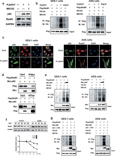
To determine the role of p62 in modulating Rad51 ubiquitination, AGS or GES-1 cells were co-transfected with plasmids expressing p62, Rad51 and ubiquitin, and then treated with a proteasomal inhibitor MG132. We found that overexpression of p62 resulted in an increase in the polyubiquitination of Rad51, compared with transfection controls (). Moreover, Cycloheximide chase assay showed that the knockdown of p62 by shRNA significantly extended the half-life of Rad51 protein (). This effect is likely attributable to the interaction of p62, ubiquitin and Rad51 as predicted in the STRING database (Figure S2D). Because the UBA domain of p62 is required for its binding to an ubiquitinated substrate, the plasmid expressing p62-ΔUBA was generated by deleting its C-terminal UBA domain. Rad51 was ubiquitinated on by intact p62, but not p62ΔUBA, indicating that p62 UBA domain is essential for p62-mediated Rad51 ubiquitination (). Taken together, these findings indicate that H. pylori infection induces the accumulation of p62, which promoted Rad51 ubiquitination and degradation via through the direct interaction between its UBA domain and Rad51.
Expression profiles of p62 and Rad51 over the histopathologic cascade of human gastric cancer
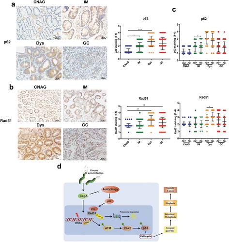
To examine relevance of our in vitro and in vivo findings to clinical manifestations, CNAG, IM to Dys, GC from human subjects were collected and evaluated for the expression profiles of p62 and Rad51. Levels p62 were progressively elevated in the gastric tissues from CNAG, IM to Dys. However, p62 level was slightly lower in GC tissues than in Dys, although it was still significantly higher compared to CNAG group (). Interestingly, we found that the levels of Rad51 were significantly increased in Dys and GC (the severe precancerous and cancerous lesions) than in CNAG and IM (the mild and moderate lesions) (). These findings suggested that multiple factors, not just H. pylori infection, were involved in Rad51 regulation and genome integrity in gastric tumorigenesis. To further characterize the effect of H. pylori infection on their expression, gastric tissues were divided into H. pylori+ and H. pylori- groups. There were significantly decreased levels of Rad51 in H. pylori-infected Dys tissues, as compared to H. pylori non-infected Dys tissues. In addition, levels of p62 were significantly higher in H. pylori+ IM tissues compared to H. pylori- IM tissues (). These data indicate that H. pylori infection could promote elevation of p62 and reduction of Rad51 in the select histopathologic stages of human gastric cancer.
Figure 8. The expression of p62 and Rad51 in the human gastric carcinogenesis cascade (a, b) Immunohistochemical staining for p62 and Rad51 in serial tissue sections from human gastric mucosa with different stages (CNAG, IM, Dys and GC) of the Correa histopathological cascade. (c) Expression levels of p62 and Rad51 were compared between H. pylori+ and H. pylori- gastric tissues with different stages (CNAG, IM, Dys and GC) of the Correa histopathological cascade. (d) Models of the regulation of autophagy-DNA damage axis in response to H. pylori infection. **P < .01, ***P < .001. All experiments were independently repeated three times.
Discussion
H. pylori has been recognized to be one of the most common human pathogens. Epidemiological data suggest that approximately 90% of gastric cancer are attributable to H. pylori infection.Citation33 Genomic instability is one of the cancer hallmarks.Citation34 Recently, DNA damage has been linked to autophagy, an intracellular degradation process by which cytoplasmic materials are delivered to the lysosome for digestion.Citation15 Despite that autophagy has been implicated in H. pylori pathogenesis,Citation35 the mechanism is still lacking. In this study, in vivo and in vitro data showed that H. pylori infection induced DSBs, triggered activation of DDR and impaired DNA repair via reduction of the DNA repair proteinRad51; this process is apparently CagA-dependent in vitro. Such reduction of Rad51 was mediated by H. pylori-induced suppression of autophagy, which led to accumulation of the autophagic substrate p62. Subsequently, the accumulation of p62 resulted in ubiquitination and degradation of Rad51 through the interaction of the UBA domain of p62 with Rad51. Based on these findings, a working model is proposed to depict the molecular mechanism underlying elevation of H. pylori CagA-induced DSBs ().
Cellular DNA damage can be caused by exposure to exogenous genotoxic agents. DSBs are a major cytotoxic lesion and must be repaired properly to maintain chromosomal integrity. Our data demonstrated that carcinogenic H. pylori can activate DNA damage response signaling, including induction of DSBs assessed by γH2AX, activation of checkpoint response by phosphorylation of ATM, Chk2 and p53. As a result, infection with H. pylori induced S cell cycle arrest, suggesting the rate of DNA synthesis has been decreased. HR and NHEJ are two main pathways to repair DSBs. Our previous studies have shown that H. pylori infection is correlated with the levels of NHEJ marker Ku70/80.Citation28 In this study, we found that H. pylori stimulation induces the expression of Rad51 that is crucial for HR repair. However, upon exposure to prolonged infection of H. pylori (at 12 h or 24 h), DNA repair was impaired due to the loss of repair protein Rad51 that could result in genome instability. Such effect could lead to an increase of mutation rates and is associated with tumorigenic potential.Citation36 The H. pylori-induced loss of Rad51 supports previous reports showing that the effects of genomic instability were induced by H. pylori infection.Citation37 It is worth nothing that Rad51 was overexpressed in human GC tissues compared with CNAG tissues, which is apparently inconsistent with H. pylori-modulated expression patterns of Rad51 noted in gastric epithelial cells, gerbils or C57BL/6 mice. Several factors could contribute to this discrepancy. Firstly, most of human GC tissues in this study were obtained from the patients with advanced gastric tumor. Consistently, it has been reported that elevated Rad51 was observed in a wide range of human tumors.Citation10 On one hand, upregulation of Rad51 could compensate for the homologous recombination defects in another repair genes-deficient (e. g. BRCA1/2) tumors. On the other hand, overexpression of Rad51 in tumor cells is responsible for radio- and chemotherapeutic resistance through excessive DNA repair mechanism.Citation38 Secondly, due to limited tissue samples, it may result in no significant downregulation of Rad51 expression in H. pylori+ tissues compared to H. pylori- tissues at CNAG, and IM and GC stages, except for Dys stage. Finally, H. pylori infection initiates host precancerous lesions and numerous factors contribute to gastric carcinogenesis. Therefore, the expression of Rad51 in human stomach tissues is regulated by multiple factors, not only the effect of H. pylori infection.
It has been documented that that initial H. pylori infection transiently induced autophagy, which may be a host defense mechanism to mitigate toxin-induced damage.Citation39 And VacA was found to be sufficient to induce autophagosome formation.Citation40 Consistent with these reports, our in vitro studies showed that autophagy was induced following H. pylori infection at 6 HPI, whereas prolonged exposure to H. pylori infection resulted in loss of autophagy in both gastric epithelial cells at 24 HPI and gerbils at 6 MPI. Furthermore, the virulence factor CagA played an important role in H. pylori-suppressed autophagy. To rule out the effect of VacA on autophagy, we used H. pylori 7.13 that contains a type s1/m2 vacA allele and does not express a functional VacA toxic in this study.Citation41 As shown in , CagA+7.13 strain infection resulted in p62 accumulation, compared to CagA−7.13 strain infection. These findings are supported by several previous studies reporting that persistent H. pylori CagA negatively regulated autophagy and promoted chronic inflammation.Citation21,Citation42 It is plausible that prolonged H. pylori-disrupted autophagy may facilitate accumulation of genotoxic products, which thereby contributing to gastric tumorigenesis.Citation35
A growing evidence has linked autophagy to DNA damage response. Autophagy can be activated by diverse DNA damage factors such as radiation, oxidative stress and chemicals. It has been reported that various DNA damage regulators including ATM, PARP1, and p53 are associated with rapid induction of autophagy, which in turn modulate DNA repair pathways. However, when DNA damage is beyond repair, autophagy is typical for the initiation of cell apoptosis and cell death.Citation22,Citation27 In our study, our data indicate that inhibition of autophagy aggravated DNA damage via suppression of Rad51 in H. pylori-infected gastric epithelial cells. Treatment with autophagy inducer rapamycin or starvation induced G2/M cell cycle arrest, which prevented G2/M cells from entering mitosis. These findings suggest that dysregulated DNA damage response to H. pylori infection is attributed to altered cell cycle progression. This premise is supported by previous data the interaction between autophagy and regulation of cell cycle.Citation43 Taken together, we hypothesize that early infection with H. pylori induced DSBs, which can be sufficiently repaired by concurrently induced autophagy. However, long-term H. pylori infection can lead to loss of autophagy, which led to accumulation of unrepair DSBs through inhibition of the DNA repair protein Rad51. In the present study, we showed the effect of autophagy on DNA repair in H. pylori-infected animal models. Everolimus, activating autophagy by inhibiting mTOR complex, could decrease p62 levels and increase Rad51 levels in H. pylori-infected animal models. As a result, everolimus treatment reduced H. pylori-induced inflammation. Consistently, recent studies have shown that everolimus suppresses inflammation by inducing autophagy.Citation44 Taking together, these findings elucidated the mechanism by which autophagy mediates DNA repair in H. pylori infection pathogenesis.
It has been reported that the selective autophagy adaptor p62 could connect two biological processes: autophagy and DNA repair. P62 accumulation can result in inhibition of DNA-damage-induced histone H2A ubiquitination through the regulation of RNF168.Citation45 Also, it has been found that p62 suppressed Rad51-mediated repair mechanism by proteasomal degradation of FLNA.Citation25 Our data indicate: (1) H. pylori infection upregulated p62 and downregulated Rad51 protein levels in vitro and in vivo models; (2) Transient overexpression of p62 apparently suppressed Rad51 expression, whereas knockdown of p62 increased Rad51 levels; (3) we found that p62 directly interacts with Rad51; (4) overexpression of p62 was associated with increased Rad51 ubiquitination, knockdown of p62 significantly prolonged the half-life of Rad51, and p62 induced Rad51 ubiquitination via the direct interaction of its UBA domain with Rad51. Based on our findings, we proposed that accumulation of p62 caused by H. pylori induced Rad51 ubiquitination and proteasomal degradation, which resulted in impairment of DSBs repair and genome instability.
In summary, our findings presented in this study provide new mechanistic insights into the relationship between autophagy and DNA damage response in H. pylori-associated gastric carcinogenesis. Chronic H. pylori infection induces the loss of autophagy and subsequent accumulation of autophagic substrate p62. The accumulated p62 results in ubiquitination of Rad51 via the direct interaction of its UBA with Rad51, thereby suppressing capability of the DNA damage repair. These cellular events allow H. pylori to elevate DSBs and genomic instability which potentially promotes gastric carcinogenesis. Further elucidation of this molecular mechanism will shed new insight into the development of more effective therapeutic strategies for preventing and curing H. pylori infection-associated gastric diseases.
Materials and methods
Cell culture and H. pylori strains
Human gastric cancer AGS cells were cultured with DMEM/F12 (Gibco, CA, USA) containing 10% fetal bovine serum (FBS) and 1% penicillin/streptomycin (Gibco). The immortalized human gastric GES-1 epithelial cells were cultured in RPMI 1640 (Gibco) with 10% FBS and 1% penicillin/streptomycin. Cells were incubated at 37°C under a 5% CO2 atmosphere.
Previously characterized H. pylori strain CagA+ATCC43504 (NCTC11637) was used in this study.Citation46 Additionally, H. pylori strain 7.13 and its isogenic cagA mutant were kindly provided by Dr. Richard Peek at Vanderbilt University Medical Center, Nashville, TN, USA. The vacA+ and vacA− H. pylori strains 26695 were kindly provided by Dr. Chunhui Lan from Daping Hospital of the Army Medical University (Chongqing, China). All H. pylori strains were cultured on Campylobacter agar plates containing 10% sheep serum and incubated at 37°C under microaerophilic conditions. The bacteria were suspended in DMEM/F12 or RPMI 1640 and the concentration was estimated by spectrophotometry (OD600 nm). Subsequently, cells were supplemented with fresh medium without antibiotic, followed by incubation with H. pylori at specified time and different multiplicities of infection (MOIs).
Animals and infections
All experiments and procedures carried out on animals were approved by the Ethics Committee of The First Affiliated Hospital of Nanchang University. Specific pathogen free (SPF) male Mongolian gerbils were challenged with H. pylori by previously described methods. Briefly, Mongolian gerbils were infected with H. pylori for 6 months (30 of H. pylori group; 15 of control group) or 12 months (24 of H. pylori group; 10 of control group).Citation47 In addition, five to six-week-old male SPF C57BL/6 mice (Hunan SJA Laboratory Animal Co Ltd, Hunan, China) were divided into four groups: control group (n = 6), H. pylori group (n = 9), everolimus group (n = 6), H. pylori in combination with group (n = 9). H. pylori-infected mice were administrated with 2 × 10⁸ colony forming units (CFU)/ml bacterial suspension of H. pylori NCTC11637 once every other day for 5 times. After infection with H. pylori for 3 months, mice were given with autophagy inducer everolimus (1.5 mg/kg, every other day) for a total of 6 times. Mice were euthanized and gastric tissues were harvested for immunohistochemistry staining, Western blot, and qRT-PCR analyses.
Histopathology
Then, mice were euthanized and linear strips of gastric tissues extending from the squamocolumnar junction through proximal duodenum were collected. Colonization of H. pylori of animals was examined by immunohistochemistry or fluorescence in situ hybridization (FISH) staining. H&E staining was used to evaluate gastric pathology. Gastric lesions were graded on a scale from 0 to 4 as the extent of inflammatory cells infiltration in the mucosa and submucosa.Citation48
Gastric specimens and immunohistochemistry
Human gastric tissue samples were obtained from patients undergoing gastroduodenoscopy or gastrectomy at the First Affiliated Hospital of Nanchang University. Embedded-paraffin tissues were collected as described previously,Citation13 which included 56 of CG, 53 of IM, 47 of Dys, 146 of GC. The patients who received previous H. pylori eradication treatment were excluded. The distribution of gender and age among patients was shown in . Immunohistochemistry staining was used to detect H. pylori infection. The study was approved by the Ethics Committee of The First Affiliated Hospital of Nanchang University. Immunohistochemistry (IHC) was performed as previously described.Citation49 Briefly, the slides were deparaffinized, rehydrated. Endogenous peroxidase activity was blocked in 3% H2O2, and antigens retrieval utilizing microwave oven heating. Tissue sections were incubated with primary antibodies overnight at 4°C, and followed by incubation with secondary antibody. Finally, slides were developed in diaminobenzidine (DAB) solution, and counterstained with hematoxylin and mounted with coverslips. The immunoreactivity of the human and animal samples was evaluated and scored for intensity (scale 0–3) and frequency (scaled 0–4) by two pathologists blinded to sample identity. For the purpose of statistical analysis for animal samples, the intensity and frequency of targets were transformed into a composite expression score using the following formula intensity × frequency. The score ranges from 0 to 12. For the statistical analysis of human samples, a score of 0–2 was considered negative (grade 1), 3–4 weakly positive (grade 2), 5–8 positive (grade 3), 9–12 strongly positive (grade 4)..Citation49,Citation50
Table 1. The distribution of gender and age between different groups.
Antibodies, siRNA, plasmids and reagents
Antibodies and their sources were as follows: CagA (sc-28368) from Santa Cruz Biotechnology (Santa Cruz, CA, USA); Anti-γH2AX (05–636), Anti-Rad51 from Millipore; Anti-H. pylori (ab7788), Anti-ATM (ab78), p-ATM (S1981) (ab36810), Chk2 (ab47433), p-Chk2 (ab85743), BRCA2 (ab27976), p-P53 (S15) (ab1431), p-P53 (S20) (ab59206) from Abcam (Cambridge, MA, USA); LC3B (3868 S), and GAPDH (2118 S) from Cell signaling Technology (Beverly, MA, USA). Goat anti-Rabbit and anti-mouse secondary antibodies were purchased from Invitrogen (Thermo Fisher Scientific, Suwanee, GA, USA). For immunofluorescence assay, 4ʹ,6-diamidino-2-phenylindole (DAPI), anti-rabbit or anti-mouse conjugated to Alexa Fluor 488, anti-rabbit or anti-mouse conjugated to Alexa Fluor 594 were from Invitrogen.
Pharmacological agents: rapamycin (R8781), everolimus (SML2282), 3-methyladenine (3-MA) (M9281), Bafilomycin A1 (BafA1) (B1793), Cycloheximide (C7698) were obtained from Sigma (St. Louis, MO, USA). Proteasomal inhibitor MG132 was purchased from Selleck Chemicals.
The recombinant plasmid of pcDNA3.0 HA-Rad51 was kindly provided by Dr. Xingzhi Xu at Capital Normal University. Myc-p62, Flag-Rad51, HA-Ubiquitin plasmids and Rad51 siRNA, p62 shRNA were purchased from Gene-Chem, Shanghai, China.
Western blotting
Tissue or cells were lysed in lysis buffer with protease inhibitor cocktail (Roche, Amherst, CA, USA). Protein concentrations were determined by the BCA assay kit (Thermo Fisher Scientific). Protein samples (25 μg) were separated by SDS-PAGE and transferred to nitrocellulose membranes. PageRuler Prestained Protein Ladder (cat. #26616) was obtained from Thermo Fisher Scientific (Waltham, MA, USA). The membranes were blocked in 5% blocking buffer (5% nonfat dry milk in TBST buffer) at room temperature for 1 h and then incubated with primary antibodies overnight at 4°C. The membranes were then incubated with secondary antibodies at room temperature for 1 h. The protein bands were imaged and quantified by using the BioRad-ChemiDoc XR+ system. The signal intensity of each target protein band was normalized to GAPDH.
Immunofluorescence
For immunofluorescence, human gastric epithelial cells were fixed with 4% formaldehyde in PBS for 15 min at room temperature. The cells were permeabilized with 0.25% Triton X-100 for 15 min and then blocked in PBS with 3% BSA for 1 h. Subsequently, the cells were incubated with primary antibody and then with a secondary fluorescent antibody. Cell nuclei were counter-stained with DAPI. All slides were evaluated using a fluorescent microscope (Nikon C2).
Flow cytometry
Cell pellets were resuspended in Buffer A (1 mg/ml Sodium citrate, 0.1%Triton-X-100, 20 μg/ml RNase A and 100 μg/ml propidium iodide) at 4°C for 30 min in the dark, and then were analyzed by a flow cytometer (BD Accuri C6).
Comet assay
The comet assay was performed as previously described. Briefly, cells were suspended in low-melting-point agarose and spread on glass slide. The slides were incubated in lysis buffer at 4°C for 1 h and then were electrophoresed at 30 V for 30 min. Comet tails were stained with propidium iodide (PI). More than 70 cells were analyzed under the Nikon C2 confocal microscope.
Transmission electron microscopy
The gastric cells were cocultured with H. pylori for indicated time. Cells were fixed in glutaraldehyde for 1.5 h at 4°C, then washed and fixed again in 1% OsO4, and dehydrated in graded ethanol and embedded in Epon-Araldite resin. Ultrathin sections were cut and stained with uranyl acetate and lead citrate, and visualized under a HT7700-SS electron microscope (HITACHI, Japan).
Real-time quantitative PCR analysis
Transcript levels of IL-1β, IL-6, IL-8, IL-10, IL-17 and Rad51 were assayed using qPCR as previously methods.Citation49 The following primers were used: mouse IL-1β, forward primer 5ʹ-GACCTTCCAGGATGAGGACA-3ʹ, reverse primer 5ʹ-AGGCCACAGGTATTTTGTCG; mouse IL-6, forward primer 5ʹ-TCTCCAGCAACGAGGAGAAT-3ʹ, reverse primer 5ʹ-TGTGATCTGAAACCTGCTGC-3ʹ; mouse IL-10, forward primer 5ʹ- AAGCTGAGAACCAAGACCCAGAC-3ʹ, reverse primer 5ʹ- AGCTATCCCAGAGCCCCAGATCCGA-3ʹ; mouse IL-17, forward primer 5ʹ- ACCTCAACCGTTCCACGTCA-3ʹ, reverse primer 5ʹ-CAGGGTCTTCATTGCGGTG-3ʹ; mouse GAPDH, forward primer 5ʹ-GTAGCAAAGGGAATGGGTCT-3ʹ, reverse primer 5ʹ-AGATGGTGAAGGGCTAATGG-3ʹ; human Rad51, forward primer 5ʹ-CTATGTAGCAAAGGGAATGGG-3ʹ, reverse primer 5ʹ-AAGCAGGTAGATGGTGAAGG-3ʹ; human GAPDH, forward primer 5ʹ-GTAGCAAAGGGAATGGGTCT-3ʹ, reverse primer 5ʹ-AGATGGTGAAGGGCTAATGG-3ʹ.
Cycloheximide chase assay
Cycloheximide chase assay was performed to characterize Rad51 protein stability. Briefly, AGS cells were transfected with p62 shRNA plasmid and then treated with cycloheximide (50 ng/ml). Rad51 protein levels were examined at specified times.
Immunoprecipitation assay
After transfected with expressing p62 or Rad51 plasmid, cells were collected and lysed with RIPA lysis buffer. All samples were processed using the Immunoprecipitation Kit (Abcam, ab206996) according to the manufacture recommendations. In brief, lysed cells were incubated with a primary antibody overnight at 4°C. Prior to immunoprecipitation, protein A/G beads were washed with the wash buffer. After antibody binding, p62 or Rad51 was immunoprecipitated with agarose beads for 2 h at 4°C. Western blotting was performed to detect the levels of immunoprecipitated proteins.
In vitro ubiquitination assay
AGS cells were transfected with plasmids (Flag-Rad51 or Myc-p62) and ubiquitin for 48 h, then co-cultured with H. pylori strain and treated with MG132 for 6 h. Immunoprecipitation assay was performed as described.Citation51
FISH assay
FISH assay was performed to detect H. pylori colonization in animal paraffin-embedded animal gastric biopsy specimens using a H. pylori-specific fluorescent probe-Hpy (5ʹ-CACACCTGACTGACTATCCCG-3ʹ). The probe labeled with fluorochrome Cy3 at 5ʹ-end were synthesized by Genscript (shanghai, China). The specimens were overlaid with a hybridization buffer (0.9 M NaCl, 20 mM Tris-HCl [pH 7.2], 0.01% sodium dodecyl sulfate) containing 30% formamide and an Hpy oligonucleotide probe (10ng/μl). Hybridization was performed at 48°C overnight in a humid chamber. Then, the slides were washed in the pre-warmed washing buffer I (0.9 M NaCl, 20 mM Tris-HCl [pH 7.2], and 0.01% sodium dodecyl sulfate) and the washing buffer II (0.9 M NaCl, 20 mM Tris-HCl [pH 7.2]) for 15 mins, respectively. Subsequently, the slides were stained with DAPI and examined under the fluorescent microscope (Nikon C2).
Statistical assay
All data were presented as mean ± standard error of mean (SEM). All statistical analyses were performed using SPSS 20.0 software. Mann–Whitney, Student’s t-test and one-way analysis of variance (ANOVA) were applied depending on the data set. P < .05 (***, P < .001, **, P < .01, *, P < .05) were considered significant.
Author contributions
CX, HW, NSL, NHL conceived, designed the study and analyzed data. NSL and DJW drafting the manuscript. YH, CP and JC collected human specimens and analyzed immunohistochemical data.CH, DJW, SX, YZ and YX provided technical support. CX and NHL Obtained funding support. NHL supervised and oversaw the study. All the authors critically revised the manuscript and provided intellectual content.
Ethics approval
The biopsy specimens were obtained under protocols and all animal experiments were approved by the ethics committees of The First Affiliated Hospital of Nanchang University.
Disclosure of potential conflicts of interest
No potential conflicts of interest were disclosed.
Supplemental Material
Download Zip (26.4 MB)Acknowledgments
We thank Prof. Zhongming Ge (Massachusetts Institute of Technology, Cambridge, MA, USA), Prof. William K.K. and Lin Zhang (The Chinese University of Hong Kong, Hong Kong, China) for editing of the manuscript. We also thank Prof. Richard Peek (Vanderbilt University Medical Centre, Nashville, TN, USA) for kindly providing the H. pylori strain 7.13 and its CagA-mutant. We also thank Dr. Xingzhi Xu (Capital Normal University, Beijing, China) for pcDNA3.0 HA-Rad51 plasmid. We also thank Prof. Chunhui Lan for providing vacA+ and vacA− H. pylori strains 26695.
Supplementary material
Supplemental data for this article can be accessed on the publisher’s website
Additional information
Funding
References
- Hooi JKY, Lai WY, Ng WK, Suen MMY, Underwood FE, Tanyingoh D, Malfertheiner P, Graham DY, Wong VWS, Wu JCY, et al. Global prevalence of helicobacter pylori infection: systematic review and meta-analysis. Gastroenterology. 2017;153:420–429. doi:10.1053/j.gastro.2017.04.022.
- Zamani M, Ebrahimtabar F, Zamani V, Miller WH, Alizadeh-Navaei R, Shokri-Shirvani J, Derakhshan MH. Systematic review with meta-analysis: the worldwide prevalence of Helicobacter pylori infection. Aliment Pharmacol Ther. 2018;47:868–876. doi:10.1111/apt.14561.
- Correa P, Houghton J. Carcinogenesis of helicobacter pylori. Gastroenterology. 2007;133:659–672. doi:10.1053/j.gastro.2007.06.026.
- Kodaman N, Pazos A, Schneider BG, Piazuelo MB, Mera R, Sobota RS, Sicinschi LA, Shaffer CL, Romero-Gallo J, de Sablet T, et al. Human and Helicobacter pylori coevolution shapes the risk of gastric disease. Proc Natl Acad Sci USA. 2014;111:1455–1460. doi:10.1073/pnas.1318093111.
- Hatakeyama M. Helicobacter pylori CagA and gastric cancer: a paradigm for hit-and-run carcinogenesis. Cell Host Microbe. 2014;15:306–316. doi:10.1016/j.chom.2014.02.008.
- Amieva M, Peek RM Jr. Pathobiology of helicobacter pylori-induced gastric cancer. Gastroenterology. 2016;150:64–78. doi:10.1053/j.gastro.2015.09.004.
- Blackford AN, Jackson SP. ATM, ATR, and DNA-PK: the Trinity at the heart of the DNA damage response. Mol Cell. 2017;66:801–817. doi:10.1016/j.molcel.2017.05.015.
- Scully R, Panday A, Elango R, Willis NA. DNA double-strand break repair-pathway choice in somatic mammalian cells. Nat Rev Mol Cell Biol. 2019;20(11):698–714. doi:10.1038/s41580-019-0152-0.
- Huen MS, Chen J. The DNA damage response pathways: at the crossroad of protein modifications. Cell Res. 2008;18:8–16. doi:10.1038/cr.2007.109.
- Gachechiladze M, Skarda J, Soltermann A, Joerger M. RAD51 as a potential surrogate marker for DNA repair capacity in solid malignancies. Int J Cancer. 2017;141:1286–1294. doi:10.1002/ijc.30764.
- Lose F, Lovelock P, Chenevix-Trench G, Mann GJ, Pupo GM, Spurdle AB. Kathleen cuningham foundation consortium for research into familial breast C. variation in the RAD51 gene and familial breast cancer. Breast Cancer Res. 2006;8:R26. doi:10.1186/bcr1415.
- Xie C, Yi J, Lu J, Nie M, Huang M, Rong J, Zhu Z, Chen J, Zhou X, Li B, et al. N-acetylcysteine reduces ROS-mediated oxidative DNA damage and PI3K/Akt pathway activation induced by helicobacter pylori infection. Oxid Med Cell Longev. 2018;2018:1874985. doi:10.1155/2018/1874985.
- Xie C, Xu LY, Yang Z, Cao XM, Li W, Lu NH. Expression of gammaH2AX in various gastric pathologies and its association with Helicobacter pylori infection. Oncol Lett. 2014;7:159–163. doi:10.3892/ol.2013.1693.
- Toller IM, Neelsen KJ, Steger M, Hartung ML, Hottiger MO, Stucki M, Kalali B, Gerhard M, Sartori AA, Lopes M, et al. Carcinogenic bacterial pathogen Helicobacter pylori triggers DNA double-strand breaks and a DNA damage response in its host cells. Proc Natl Acad Sci USA. 2011;108:14944–14949. doi:10.1073/pnas.1100959108.
- Eliopoulos AG, Havaki S, Gorgoulis VG. DNA damage response and autophagy: A meaningful partnership. Front Genet. 2016;7:204. doi:10.3389/fgene.2016.00204.
- Sudhakar P, Jacomin AC, Hautefort I, Samavedam S, Fatemian K, Ari E, Gul L, Demeter A, Jones E, Korcsmaros T, et al. Targeted interplay between bacterial pathogens and host autophagy. Autophagy. 2019;1–14. doi:10.1080/15548627.2019.1590519.
- Huang J, Brumell JH. Bacteria-autophagy interplay: a battle for survival. Nat Rev Microbiol. 2014;12:101–114. doi:10.1038/nrmicro3160.
- Horvat A, Noto JM, Ramatchandirin B, Zaika E, Palrasu M, Wei J, Schneider BG, El-Rifai W, Peek RM Jr., Zaika AI. Helicobacter pylori pathogen regulates p14ARF tumor suppressor and autophagy in gastric epithelial cells. Oncogene. 2018;37:5054–5065. doi:10.1038/s41388-018-0343-8.
- Zhu P, Xue J, Zhang ZJ, Jia YP, Tong YN, Han D, Li Q, Xiang Y, Mao XH, Tang B. Helicobacter pylori VacA induces autophagic cell death in gastric epithelial cells via the endoplasmic reticulum stress pathway. Cell Death Dis. 2017;8:3207. doi:10.1038/s41419-017-0011-x.
- Tsugawa H, Mori H, Matsuzaki J, Sato A, Saito Y, Imoto M, Suematsu M, Suzuki H. CAPZA1 determines the risk of gastric carcinogenesis by inhibiting helicobacter pylori CagA-degraded autophagy. Autophagy. 2018:1–17. doi:10.1080/15548627.2018.1515530.
- Li N, Tang B, Jia YP, Zhu P, Zhuang Y, Fang Y, Li Q, Wang K, Zhang WJ, Guo G, et al. Helicobacter pylori CagA protein negatively regulates autophagy and promotes inflammatory response via c-Met-PI3K/Akt-mTOR signaling pathway. Front Cell Infect Microbiol. 2017;7:417. doi:10.3389/fcimb.2017.00417.
- Hewitt G, Korolchuk VI. Repair, reuse, recycle: the expanding role of autophagy in genome maintenance. Trends Cell Biol. 2017;27:340–351. doi:10.1016/j.tcb.2016.11.011.
- Peng H, Yang J, Li G, You Q, Han W, Li T, Gao D, Xie X, Lee BH, Du J, et al. Ubiquitylation of p62/sequestosome1 activates its autophagy receptor function and controls selective autophagy upon ubiquitin stress. Cell Res. 2017;27:657–674. doi:10.1038/cr.2017.40.
- Wang Y, Zhu WG, Zhao Y. Autophagy substrate SQSTM1/p62 regulates chromatin ubiquitination during the DNA damage response. Autophagy. 2017;13:212–213. doi:10.1080/15548627.2016.1245262.
- Hewitt G, Carroll B, Sarallah R, Correia-Melo C, Ogrodnik M, Nelson G, Otten EG, Manni D, Antrobus R, Morgan BA, et al. SQSTM1/p62 mediates crosstalk between autophagy and the UPS in DNA repair. Autophagy. 2016;12:1917–1930. doi:10.1080/15548627.2016.1210368.
- Peng C, Li NS, Hu Y, Lu NH. Impact factors that modulate gastric cancer risk in Helicobacter pylori-infected rodent models. Helicobacter. 2019:e12580. doi:10.1111/hel.12580.
- Roos WP, Thomas AD, Kaina B. DNA damage and the balance between survival and death in cancer biology. Nat Rev Cancer. 2016;16:20–33. doi:10.1038/nrc.2015.2.
- Li W, Xie C, Yang Z, Chen J, Lu NH. Abnormal DNA-PKcs and Ku 70/80 expression may promote malignant pathological processes in gastric carcinoma. World J Gastroenterol. 2013;19:6894–6901. doi:10.3748/wjg.v19.i40.6894.
- Jensen RB, Carreira A, Kowalczykowski SC. Purified human BRCA2 stimulates RAD51-mediated recombination. Nature. 2010;467:678–683. doi:10.1038/nature09399.
- Pugsley HR. Assessing autophagic flux by measuring LC3, p62, and LAMP1 Co-localization using multispectral imaging flow cytometry. J Vis Exp. 2017. doi:10.3791/55637.
- Moscat J, Karin M, Diaz-Meco MT. p62 in cancer: signaling adaptor beyond autophagy. Cell. 2016;167:606–609. doi:10.1016/j.cell.2016.09.030.
- Lee Y, Weihl CC. Regulation of SQSTM1/p62 via UBA domain ubiquitination and its role in disease. Autophagy. 2017;13:1615–1616. doi:10.1080/15548627.2017.1339845.
- Moss SF. The clinical evidence linking helicobacter pylori to gastric cancer. Cell Mol Gastroenterol Hepatol. 2017;3:183–191. doi:10.1016/j.jcmgh.2016.12.001.
- Hanahan D, Weinberg RA. Hallmarks of cancer: the next generation. Cell. 2011;144:646–674. doi:10.1016/j.cell.2011.02.013.
- Greenfield LK, Jones NL. Modulation of autophagy by Helicobacter pylori and its role in gastric carcinogenesis. Trends Microbiol. 2013;21:602–612. doi:10.1016/j.tim.2013.09.004.
- Tubbs A, Nussenzweig A. Endogenous DNA damage as a source of genomic instability in cancer. Cell. 2017;168:644–656. doi:10.1016/j.cell.2017.01.002.
- Koeppel M, Garcia-Alcalde F, Glowinski F, Schlaermann P, Meyer TF. Helicobacter pylori infection causes characteristic DNA damage patterns in human cells. Cell Rep. 2015;11:1703–1713. doi:10.1016/j.celrep.2015.05.030.
- Ward A, Khanna KK, Wiegmans AP. Targeting homologous recombination, new pre-clinical and clinical therapeutic combinations inhibiting RAD51. Cancer Treat Rev. 2015;41:35–45. doi:10.1016/j.ctrv.2014.10.006.
- Hu W, Zhang L, Li MX, Shen J, Liu XD, Xiao ZG, Wu DL, Ho IHT, Wu JCY, Cheung CKY, et al. Vitamin D3 activates the autolysosomal degradation function against Helicobacter pylori through the PDIA3 receptor in gastric epithelial cells. Autophagy. 2019;1–19. doi:10.1080/15548627.2018.1557835.
- Terebiznik MR, Raju D, Vazquez CL, Torbricki K, Kulkarni R, Blanke SR, Yoshimori T, Colombo MI, Jones NL. Effect of Helicobacter pylori’s vacuolating cytotoxin on the autophagy pathway in gastric epithelial cells. Autophagy. 2009;5:370–379. doi:10.4161/auto.5.3.7663.
- McClain MS, Shaffer CL, Israel DA, Peek RM, Cover TL. Genome sequence analysis of Helicobacter pylori strains associated with gastric ulceration and gastric cancer. BMC Genomics. 2009;10:3. doi:10.1186/1471-2164-10-3.
- Muhammad JS, Nanjo S, Ando T, Yamashita S, Maekita T, Ushijima T, Tabuchi Y, Sugiyama T. Autophagy impairment by Helicobacter pylori-induced methylation silencing of MAP1LC3Av1 promotes gastric carcinogenesis. Int J Cancer. 2017;140:2272–2283. doi:10.1002/ijc.30657.
- Zheng K, He Z, Kitazato K, Wang Y. Selective autophagy regulates cell cycle in cancer therapy. Theranostics. 2019;9:104–125. doi:10.7150/thno.30308.
- Lee SC, Kim KH, Kim OH, Lee SK, Kim SJ. Activation of autophagy by everolimus confers hepatoprotection against ischemia-reperfusion injury. Am J Transplant. 2016;16:2042–2054. doi:10.1111/ajt.13729.
- Wang Y, Zhang N, Zhang L, Li R, Fu W, Ma K, Li X, Wang L, Wang J, Zhang H, et al. Autophagy regulates chromatin ubiquitination in DNA damage response through elimination of SQSTM1/p62. Mol Cell. 2016;63:34–48. doi:10.1016/j.molcel.2016.05.027.
- Crabtree JE, Farmery SM, Lindley IJ, Figura N, Peichl P, Tompkins DS. CagA/cytotoxic strains of Helicobacter pylori and interleukin-8 in gastric epithelial cell lines. J Clin Pathol. 1994;47:945–950. doi:10.1136/jcp.47.10.945.
- Yang Z, Xie C, Xu W, Liu G, Cao X, Li W, Chen J, Zhu Y, Luo S, Luo Z, et al. Phosphorylation and inactivation of PTEN at residues Ser380/Thr382/383 induced by Helicobacter pylori promotes gastric epithelial cell survival through PI3K/Akt pathway. Oncotarget. 2015;6:31916–31926. doi:10.18632/oncotarget.5577.
- Ge Z, Sheh A, Feng Y, Muthupalani S, Ge L, Wang C, Kurnick S, Mannion A, Whary MT, Fox JG. Helicobacter pylori-infected C57BL/6 mice with different gastrointestinal microbiota have contrasting gastric pathology, microbial and host immune responses. Sci Rep. 2018;8:8014. doi:10.1038/s41598-018-25927-2.
- Li N, Feng Y, Hu Y, He C, Xie C, Ouyang Y, Artim SC, Huang D, Zhu Y, Luo Z, et al. Helicobacter pylori CagA promotes epithelial mesenchymal transition in gastric carcinogenesis via triggering oncogenic YAP pathway. J Exp Clin Cancer Res. 2018;37:280. doi:10.1186/s13046-018-0962-5.
- Zhu S, Soutto M, Chen Z, Peng D, Romero-Gallo J, Krishna US, Belkhiri A, Washington MK, Peek R, El-Rifai W. Helicobacter pylori-induced cell death is counteracted by NF-kappaB-mediated transcription of DARPP-32. Gut. 2017;66:761–762. doi:10.1136/gutjnl-2016-312141.
- Hu Y, Liu JP, Li XY, Cai Y, He C, Li NS, Xie C, Xiong ZJ, Ge ZM, Lu NH, et al. Downregulation of tumor suppressor RACK1 by Helicobacter pylori infection promotes gastric carcinogenesis through the integrin beta-1/NF-kappaB signaling pathway. Cancer Lett. 2019;450:144–154. doi:10.1016/j.canlet.2019.02.039.