
ABSTRACT
Candida albicans is a pathobiont that inflicts serious bloodstream fungal infections in individuals with compromised immunity and gut dysbiosis. Genomic diversity in the form of copy number alteration, ploidy variation, and loss of heterozygosity as an adaptive mechanism to adverse environments is frequently observed in C. albicans. Such genomic variations also confer a varied degree of fungal virulence and drug resistance, yet the factors propelling these are not completely understood. DNA polymerase delta (Polδ) is an essential replicative DNA polymerase in the eukaryotic cell and is yet to be characterized in C. albicans. Therefore, this study was designed to gain insights into the role of Polδ, especially its non-essential subunit Pol32, in the genome plasticity and life cycle of C. albicans. PCNA, the DNA clamp, recruits Polδ to the replication fork for processive DNA replication. Unlike in Saccharomyces cerevisiae, the PCNA interaction protein (PIP) motif of CaPol32 is critical for Polδ’s activity during DNA replication. Our comparative genetic analyses and whole-genome sequencing of POL32 proficient and deficient C. albicans cells revealed a critical role of Pol32 in DNA replication, cell cycle progression, and genome stability as SNPs, indels, and repeat variations were largely accumulated in pol32 null strain. The loss of pol32 in C. albicans conferred cell wall deformity; Hsp90 mediated azoles resistance, biofilm development, and a complete attenuation of virulence in an animal model of systemic candidiasis. Thus, although Pol32 is dispensable for cell survival, its function is essential for C. albicans pathogenesis; and we discuss its translational implications in antifungal drugs and whole-cell vaccine development.
Introduction
In eukaryotes, DNA replication is coordinated by three essential DNA polymerases Polα, Polδ, and Polε. Extensive genetic analyses in Saccharomyces cerevisiae suggest that Polε is involved in only leading strand DNA synthesis, whereas Polδ synthesizes both leading and lagging strands of DNA.Citation1,Citation2 Polα-primase provides the RNA-DNA primer to initiate DNA replication from the origin. Both Polδ and Polε interact with the proliferating cell nuclear antigen (PCNA) sliding clamp to carry out processive DNA synthesis.Citation3,Citation4 Polδ holoenzyme from S. cerevisiae consists of three subunits: the catalytic Pol3 and the accessory structural Pol31 and Pol32 proteins. In humans, the corresponding subunits are POLD1, POLD2, and POLD3.Citation5 In vertebrates and Schizosaccharomyces pombe, Polδ has one additional subunit p12/cdm1.Citation5,Citation6 Coherent with the central role of Polδ in genome replication, Pol3, and Pol31 subunits are indispensable for cell survival, whereas, in budding yeast, the Pol32 subunit is nonessential.Citation7 The catalytic subunit Pol3 has both DNA synthesizing and proofreading activities, however, despite Pol31 being essential, its exact function in Polδ holoenzyme has not been demonstrated except that it bridges Pol3 and Pol32 subunits. Interestingly, the Pol32 homologue Cdc27 in S. pombe is indispensable, suggesting its organism-specific importance in DNA replication.Citation8 The pol32 null strain of S. cerevisiae exhibits severe defects in DNA replication, repair, and mutagenesis.Citation7 Moreover, the combination of pol32Δ with conditional mutations in POL3, POL31, or POL30 (PCNA) causes synthetic lethality. Surprisingly, deletion analyses of ScPol32 suggest that the phenotypes exhibited by pol32Δ cells are due to the essential role of the N-terminal domain that is involved in binding to Pol31 or Pol1 subunit of Polα, but not due to the C-terminal tail that harbors the PCNA interaction protein motif (PIP). Thus, it was assumed that the role of Pol32 is to stabilize the interaction between Pol3 and Pol31 proteins. Recent reports also suggest the Pol31-Pol32 subcomplex is a part of Polζ holoenzyme, a mutagenic translesion DNA synthesis (TLS) polymerase.Citation9,Citation10 Therefore, the damage-induced mutagenesis gets reduced significantly in POL32 deficient cells.Citation11,Citation12 However, in DT40 cells, it was shown that the reduction in UV or MMS induced mutagenesis due to the lack of POLD3 is mostly Polζ independent.Citation13 Further, the study reported that Polδ promotes the bypass of an abasic site only when POLD3 is present. Thus, Pol32 could possess different species-specific functions. In our earlier study, we have shown that PIP motifs in all three subunits contribute to PCNA-stimulated DNA synthesis by ScPolδ, and mutational inactivation of all three PIP motifs abrogates its ability to synthesize DNA in the presence of PCNA.Citation3 Genetic analyses further revealed that in the absence of functional Pol32 PIP, PCNA binding by both Pol3 and Pol31 becomes essential for cell viability. Contrarily, the cryoEM structures of the Polδ-PCNA-DNA complex both from S. cerevisiae and humans suggest that only Pol3/POLD1 protein binds PCNA.Citation14,Citation15 Moreover, the orientation of the Pol31 and Pol32 subcomplex in the Polδ structure is such that it prevents the interaction of the subcomplex with the DNA template as well, despite Pol31 harboring a DNA binding oligonucleotide binding (OB) fold. Thus, the precise role of Pol31 and Pol32 subunits in an eukaryotic cell remains a mystery.
Polδ has not been characterized yet in the pathogenic yeast Candida albicans, a causative agent of candidiasis. Despite existing antifungal arsenals, the mortality rate of systemic candidiasis caused by C. albicans remains as high as 40–70%, especially in severely immune deficient patients.Citation16 No approved vaccine is available yet to mitigate any fungal infections.Citation17 Therefore, the identification of new drug targets and potential vaccine candidates are of paramount importance. Genomic diversity in the form of base substitutions, indels, ploidy variation, karyotypic changes, and loss of heterozygosity (LOH) is frequently observed in C. albicans.Citation18 While genomic alteration is deleterious to cells, in certain contexts, it facilitates rapid adaptation to unfavorable environments and is often considered a hallmark of pathogenicity, drug resistance, and cancer development.Citation19,Citation20 The opportunistic fungal pathogen C. albicans has emerged as an ideal model organism to study the association of genome dynamics with its pathogenesis. Since Polδ takes part in several DNA transaction processes, it might play a key role in the genome stability of C. albicans. Therefore, this study was designed to gain insights into the role of Polδ, especially its non-essential subunit Pol32 in genome stability and C. albicans biology. We find that CaPol32 plays a critical role in Polδ’s function, and its absence results in complete attenuation of fungal pathogenesis.
Results
Identification of the Pol32 subunit of CaPolδ and its interaction with PCNA
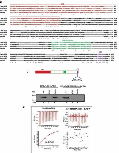
To identify the putative homologue of S. cerevisiae Pol32 in C. albicans, we performed a BLAST search in its genome database (http://www.candidagenome.org/) and found orf19.2465 as an uncharacterized protein that could encode for the smallest subunit of Polδ. The putative CaPOL32 gene (C1_05850W_A/B) is located in the largest chromosome Chr 1 and both the alleles display synonymous substitution suggesting the expression of a single protein with exactly similar amino acid composition (Length = 403 aa; MW = 46.0 kDa). Our primary sequence alignment of CaPol32 with already characterized Pol32 homologues showed a mere 17–25% identity (). Pol32 from S. cerevisiae is known to possess mutually exclusive-binding sites such as the N-terminal 103 aa (NTD), middle 269–310 aa, and C-terminal PIP for Pol31, Pol1, and PCNA, respectively. Except for the canonical PIP motif, the other two binding regions are relatively poorly conserved in CaPol32. Since CaPol32 showed poor conservation of amino acids, we sought to compare its structure with ScPol32. ScPol32 (PDB:6P1H) was taken as a template and the corresponding model structure of CaPol32 was predicted. Similarly, the model structures of the PIP motif of Pol32 from C. albicans, S. cerevisiae, and S. pombe were predicted using the PEP fold 3.0 online server. Despite the limited amino acid similarity, NTD from Sc- and Ca-Pol32 displayed exceptionally overlapping V-shaped structures and formed a winged helix-turn-helix (wHTH) domain (Supplementary Figure 1i and ii). Similarly, the PIP structures of ScPol32, p68, and CaPol32 were indistinguishable and they formed a 310 helix that stably bound to the hydrophobic pocket of PCNA (Supplementary Figure 1 iii and iv). To authenticate the involvement of the PIP motif of CaPol32 in PCNA binding, the highly conserved aromatic amino acids (F398 and F399) of the putative PIP motif were mutated to alanines as these residues in ScPol32 are involved in hydrophobic interaction with the inter-domain connecting loop of PCNA.Citation21 The wild-type and site-directed CaPol32 (F398A, F399A) mutant proteins were purified, and their interaction with CaPCNA was monitored by GST-pulldown and Isothermal calorimetry (ITC) analyses. An equimolar concentration of CaPCNA was incubated with wild-type or mutant GST-CaPol32 and a pull-down assay was performed using glutathione-Sepharose beads (). While the wild-type GST-Pol32 was able to pull down CaPCNA (compare lanes 1 and 3), GST-CaPol32 F398A, F399A protein failed to interact with CaPCNA and thus, no PCNA was precipitated in the elution fraction (compare lanes 4 and 6). For the ITC assay, CaPCNA was injected into the sample cell of the calorimeter either containing wild type or mutant of CaPol32, and heat exchange was monitored (). While the titration of CaPCNA against the wild-type protein resulted in an exothermic reaction with the estimated values of ΔH, ΔG, and KD as ~-27 kcal/mol, ~-29.5 kcal/mol, and ~6.75 μM, respectively, no perceivable heat change was detected when PCNA was titrated against the PIP mutant of CaPol32 or the buffer alone. The titration assay also revealed that one molecule of CaPol32 binds to one molecule of a trimeric PCNA. These results suggested that like in ScPol32 and human p68, CaPol32 also possesses a bonafide PIP motif (392-QSSLMSFF-399) at the extreme C-terminal end.
Figure 1. In silico analyses and identification of PIP motif of CaPol32. a. Multiple amino acid sequence alignment of CaPol32 with ScPol32, SpCdc27, and human p68 (Hsp68) using CLUSTAL W. Identical residues are designated by * and similar residues are designated with symbols: . N-terminal domain (NTD), Pol1 binding site, and PIP motif are shaded with brown color, green, and violet color, respectively. b. Glutathione sepharose beads bound with GST-CaPol32 (Lanes 1–3) and GST-CaPol32 (F398A, F399A) (Lanes 4–6) were mixed with CaPCNA in the equilibration buffer. After the incubation, beads were washed with equilibration buffer and bound PCNA was eluted by protein loading dye. Various fractions were resolved in 12.5% SDS-PAGE and binding of PCNA was detected by an anti-CaPCNA antibody. c. ITC analysis of binding of CaPCNA with CaPol32 and CaPol32 (F398A, F399A). In each figure, the upper panels show the measure of heat exchanges during each CaPCNA injection, while the lower panels show enthalpy changes as a function of the molar ratio of CaPol32 or CaPol32 (F398A, F399A) binding to a CaPCNA monomer.
PCNA interaction by Pol32 is indispensable for Polδ’s function in C. albicans
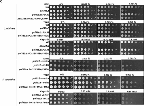
S. cerevisiae cells harboring a mutant Pol32 that is defective in PCNA binding did not show any noticeable growth defects in cold temperature and in the presence of hydroxyurea (HU).Citation3,Citation7 We examined the phenotypes of CaPol32 and its PIP motif by expressing them at the original genomic locus of a pol32 homozygous deletion strain (pol32ΔΔ) of C. albicans (Supplementary Figures 2 and ). Surprisingly, C. albicans cells possessing CaPol32 with F398A, F399A mutation exhibited almost similar sensitivity to HU, UV radiation, and high temperatures (42°C) as shown by the pol32 null strain (). At 42°C or 32–48 J/m2 of UV exposure or in the presence of 20–30 mM HU, both wild type and pol32ΔΔ::POL32 strains grew normally; however, almost no growth was observed in the cells expressing CaPol32 PIP mutant. Contrarily and as reported earlier also, S. cerevisiae pol32Δ cells possessing PIP mutant grew as good as the cells expressing the wild-type Pol32 at all temperatures tested and upon HU and UV radiation exposures. However, pol32Δ S. cerevisiae cells exhibited slow growth phenotypes at 16° and 37°C and sensitivity to HU and UV. To further strengthen this result, colony forming units (CFU) of each strain were determined in the presence of genotoxic agents and found that while in C. albicans, the PIP motif of Pol32 appears to be as critical as the full-length protein, in S. cerevisiae, Pol32 subunit possesses other essential functions that can be separated from the PCNA interaction (). To find out whether the essential role of the PIP motif of CaPol32 is also required during DNA repair synthesis by Polδ, C. albicans and S. cerevisiae cells expressing wild type or Pol32 PIP mutant were subjected to MMS, TBHP, and cisplatin DNA damaging agents. Surprisingly, both the wild type and PIP mutant of Pol32 conferred protection to genomic DNA from damage as only the pol32 defective strains of C. albicans and S. cerevisiae showed susceptibility to various DNA damaging agents (). This result suggested that in genomic DNA replication, PCNA interaction by Pol32 subunit of CaPolδ is indispensable, whereas for most of the DNA repair function PCNA binding to CaPol32 may not be essential, except when cells are exposed to UV radiation.
Figure 2. PIP motif of CaPol32 is essential for CaPolδ’s function. a. Serially diluted cells of WT, pol32ΔΔ, pol32ΔΔ::POL32, pol32∆∆::POL32 F398A, F399A C. albicans strains and pol32Δ deletion S. cerevisiae strain harboring vector alone or ScPol32 or ScPol32 F344A, F345A were spotted on YPD plates with the indicated concentration of HU. Similar YPD plates spotted with various strains were exposed to different doses of UV. All the plates were incubated at 30°C for 48 hr and then imaged. For the temperature sensitivity, YPD plates were incubated at 16°C, 30°C. 37°C, and 42°C for 48 hr and then photographed. b. CFU analysis of various strains of C. albicans and S. cerevisiae was performed by diluting logarithmically growing cells to 400 cells/ml and from which 250 µl cells were spreaded on YPD plates with HU (i) and UV treatments (ii). c. Equal number of cells of WT, pol32ΔΔ, pol32ΔΔ::POL32, pol32∆∆::POL32 F398A, F399A C. albicans and pol32Δ S. cerevisiae strain harboring a vector alone or ScPol32 or ScPol32 F344A, F345A plasmids were spotted on YPD plates without or with MMS, cisplatin, and TBHP. All the plates were incubated at 30°C for 48 hr and then imaged.
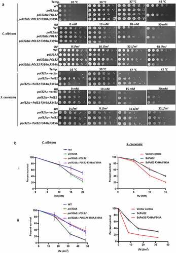
Figure 2. (Continued).
Loss of PCNA interaction by the Pol32 subunit of CaPolδ renders chromosomal instability
In order to investigate the occurrence of genomic alterations that could have happened due to the loss of the PCNA interaction motif of Pol32 or due to the complete deletion of the POL32 gene, we compared the karyotype of C. albicans strains (). Our pulse field gel electrophoresis (PFGE) analysis of total genomic DNA revealed no gross chromosomal rearrangement in any of the strains (WT, pol32ΔΔ, pol32ΔΔ::POL32, pol32ΔΔ::POL32 F398A, F399A), and DNA from all the eight chromosomes migrated according to their molecular sizes as of the parental C. albicans strain. Thus, despite the absence of Pol32, the overall structure and number of chromosomes in these cells were not greatly affected. To understand the role of Pol32 in processive DNA synthesis by CaPolδ, these strains were exposed to a sub-lethal dose of HU and further allowed to replicate DNA by growing them on fresh media. Since HU depletes cellular dNTPs level, a low processive DNA polymerase becomes inefficient in completing timely replication of the genome, therefore single-stranded DNA and breaks on DNA often get accumulated. Thus, the accumulation of fragmented smaller sized chromosomal DNA is indicative of compromised processive DNA synthesis. Alkali agarose gel electrophoresis of total genomic DNA isolated at various time points of recovery revealed that an equal amount of genomic DNA degradation was found in each cell type prior to recovery (, lanes 1, 5, and 9). With the increase in the duration of the recovery period, a comparatively higher accumulation of larger DNA fragments was observed in WT than that in pol32ΔΔ or the cells expressing Pol32 PIP mutant (lanes 2–4, 6–8, and 10–12). Since F398A, F399A mutations in Pol32 abrogated binding with PCNA, the accumulation of fragmented DNA in C. albicans strain expressing Pol32 PIP mutant even after 24 hr of recovery phase suggested a critical role of PCNA interaction of Pol32 in Polδ’s function in the cell. Next, we investigated the efficiency of spontaneous LOH events in C. albicans cells using a counter-selectable URA3 gene marker. Heterozygosity of URA3 was artificially created in different genetic backgrounds of C. albicans by knocking out one of the alleles and such strains were subjected to 5FOA treatment to measure LOH frequency. Cells grow in the presence of 5FOA, only when the other URA3 copy becomes nonfunctional. The appearance of 5FOAR colonies suggests an increased rate of LOH in that corresponding strain of C. albicans (). The efficiency of LOH was negligible in the wild type C. albicans (6–9 colonies per 5 × 10⁵ cells), whereas it increased by 8–10 folds and 2–4 folds in the POL32 deficient and strain possessing PIP mutant of Pol32, respectively. Interestingly, a single copy integration of the POL32 gene into the pol32ΔΔ strain exhibited a similar level of LOH frequency as by a diploid WT strain, thus the gene dosage of POL32 in regulating LOH frequency appears not to be critical in C. albicans.
Figure 3. Role of CaPol32 in chromosomal stability and cell cycle progression. a. DNA plugs were prepared for various strains of C. albicans and resolved by PFGE. b. Alkaline agarose gel electrophoresis of total DNA isolated from Pol32ΔΔ::POL32 (lanes 1–4), pol32ΔΔ (lanes 5–8), and pol32ΔΔ::POL32 F398A, F399A (lanes 9–12) strains of C. albicans during the recovery period after HU treatment was carried out. After neutralization, DNA fragments were visualized by ethidium bromide staining. c. The efficiency of LOH of various strains of C. albicans was measured by estimating the number of FOA resistant colonies. d. Synchronized cells were chased for 150 mins at an interval of 30 mins to check the cell cycle progression of C. albicans cells without or with HU treatment. Cells were stained with cytox green and a single cell gated population was analyzed by flow cytometry.
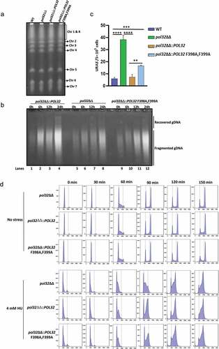
S. cerevisiae cells lacking Pol32 exhibit a delay at the G2/M phase of the cell cycle and accumulate non-segregated chromosomes.Citation22 To decipher a possible similar role of CaPol32, synchronized populations of pol32ΔΔ and its derivative strains C. albicans cells at the G1 phase of the cell cycle were treated with or without HU, and the cell cycle progression of the gated single nucleated cells was monitored at different time intervals by flow cytometry analysis as described previously.Citation23 Without HU, all three strains (pol32ΔΔ, pol32ΔΔ::POL32, and pol32ΔΔ::POL32 F398A, F399A strains followed a normal cell cycle and took ~90 min to complete a cycle from 2 N (G1) to 4 N (G2/M) (). Up to 150 mins, the progression profiles of all of the cells remained undistinguishable suggesting that the loss of the POL32 gene or PIP motif mutant of POL32 did not result in any replication stress to activate cell cycle arrest under optimal growth conditions. However, chronic exposure to a nontoxic dose of HU (4 mM) was sufficient enough to delay the cell cycle progression and more cells were accumulated at the S phase of pol32ΔΔ and pol32ΔΔ::POL32 F398A, F399A C. albicans cells. Altogether, these results suggested that although Pol32 is non-essential for growth, its binding to the processivity factor PCNA stimulates processive DNA synthesis by Polδ, and its absence leads to a higher rate of spontaneous and HU induced chromosomal instability, and early to late S phase arrest.
Genomics of pol32ΔΔ and its isogenic parental strain ofC. albicans
Genomic analysis of clinical isolates of C. albicans and isolates subjected to environmental stresses revealed that many of those contained loss of heterozygosity (LOH) and large-scale genomic changes such as whole or segmental chromosome aneuploidy.Citation24,Citation25 Although our PFGE analysis of pol32ΔΔ C. albicans chromosomal DNA did not show any abnormal chromosomal mobility, the POL32 defective strain exhibited an increased rate of LOH. To further strengthen our understanding of the role of Pol32 in genome stability, whole-genome sequencing of the pol32ΔΔ and its parental wild-type strain of C. albicans was performed concurrently and compared with the reference genome. Since mutations have been identified in C. albicans isolates passaged both in vitro and in vivo conditions during laboratory culture and in the mammalian host by earlier reports,Citation25,Citation26 it was important to re-sequence our laboratory parental wild-type strain. C. albicans genome consists of eight chromosomes, with the reference isolate SC5314 harboring ∼70,000 heterozygous positions representing ∼0.5% of the 14.3 Mb genome. After filtering, trimming and removal of redundans from the raw reads, a total of 259 and 234 contigs corresponding to 14528623 and 14626883 bp of total reads were obtained for wild type and pol32ΔΔ genome, respectively, and are equivalent to the reference genome size. First, we used a web-based tool YMAP (Yeast Mapping Analysis Pipeline) that generates a chromosomal map and analyzes the genome sequence to map copy number variations (CNV), SNPs, and LOH.Citation27 This pipeline uses the FASTQ files, makes stringent corrections, and compares with the reference sequence to result in a HapMap that illustrates each chromosome ( i and II, and Supplementary Figure 3). The constriction that divides each chromosomal arm denotes a centromere. The X-axis represents the chromosomal distance, with 0.2 representing 200 KB. A graph on the left of the chromosome represents the allelic ratio. The right-hand side number represents the ploidy of that specific chromosome, i.e. 2 for two alleles, 1 for a single allele, and any numbers between >1 and <2 represent cases of aneuploidy. The regions with copy number variations are depicted as prominent black histograms along the length of the chromosome. The dots on the X-axis denote the positions of the major repeat sequences (MRS). YMAP distinguishes between the haplotypes of each chromosome (A or B) by using a color code. Heterozygous (AB) single nucleotide polymorphisms (SNPs) are shown as vertical gray bars in the background of each chromosome, and increasing shades of dark gray indicate regions with higher numbers of SNPs. Homozygous SNPs are displayed in cyan for haplotype A and in magenta for haplotype B. Aneuploidy is determined by the weighted average of the colors assigned to the individual SNPs. As shown in , there are two copies of all chromosomes in both the C. albicans strains. A large region on the small arm of chromosome 5 (colored in cyan) and a small region close to the MRS on chromosome 2 (colored in magenta) have undergone loss of heterozygosity (LOH). Since these LOH events are commonly found in both strains, these events happened at an early stage and not during passaging in our laboratory ( i and II). Among these chromosomes, Chr 3 which is relatively smaller in size looks very stable as it has minimal LOH events. The appearance of several new cyan and magenta lines in the pol32ΔΔ strain is indicative of loss and gain of homozygosity accumulated due to faulty DNA replication that was more evident in Chr 7. Interestingly, Chr 7 also exhibited localized segmental aneuploidy in the larger arm (chromosome number is estimated to lie between 1.2 and 2). These genetic variations can cause the loss or gain of new phenotypes in the pol32ΔΔ strain.
Figure 4. WGS of pol32ΔΔ strain of C. albicans. a. The raw FASTQ genome sequence files of our laboratory WT (i) and pol32ΔΔ (II) strains of C. albicans were mapped with reference sequence using YMAP. YMAP provided the chromosomal map. The gray color lines indicate heterozygous regions in both haplotypes and the cyan and magenta lines indicate heterozygous regions in any of the haplotypes. The histogram at the left arm of the chromosome represents the proportion of each haplotype (assigned as a or b). The histogram at the right end of the chromosome indicates the copy number variation. Chromosome 7 of pol32ΔΔ strain showed segmental aneuploidy and brown circles indicate some of the unique variation regions. b. Numbers of Indels (i) and SNPs (ii) specifically accumulated in the genome of pol32ΔΔ strain of C. albicans. Blue color indicates deletion, brown color indicates insertion, gray color indicates homozygous Indels, yellow color indicates heterozygous Indels, Orange color indicates homozygous SNPs, sky blue color indicates heterozygous SNPs, green color indicates transition mutation, and maroon color indicates transversion mutation. c. Accumulation of Indels and SNPs associated without or with homo/hetero-polymeric repeat regions of pol32ΔΔ genome of C. albicans. d. The frequency distribution of length of deletion and insertion specific to pol32ΔΔ genome of C. albicans.
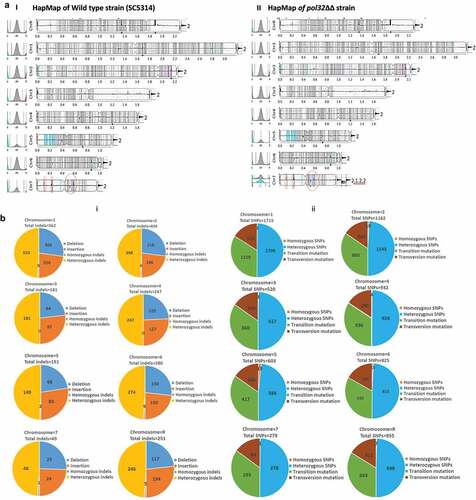
Figure 4. (Continued).
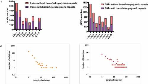
Over a period of time, our laboratory WT SC5314 strain accumulated 7942 indels (insertion + deletion) and 62995 SNPs when we compared it with the publicly available reference sequence (ASM18296v3). A detailed chromosome-wise accumulation of various genetic changes has been illustrated (Supplementary Figure 4 and Supplementary Tables 1 and 2). In addition to these genomic variations, 2125 indels and 6991 SNPs events occurred in the pol32ΔΔ genome which might have commenced most likely due to replication stress linked specifically to Pol32 function. This result is also supported by the fact that the loss of Pol32 enhanced 5FOAR LOH efficiency as shown in . A chromosome-wise accumulation of indels and SNPs specific to the pol32ΔΔ genome has been depicted ( and ). Among the chromosomes, as found in the YMAP pipeline Chr 3 accumulated the least number of indels and SNPs in both strains. About 181 indels and 520 SNPs, and 489 indels and 4311 SNPs were accumulated in Chr 3 of pol32ΔΔ and WT strain, respectively. Since Chr 3 has no major repeat sequence (MRS) region, the replication slippage events could be a bare minimum in this chromosome and thus, it is fairly stable. Contrarily, Chr 4 and Chr 6 despite being relatively smaller chromosome sizes (~1-1.6 MB) possessed a higher percentage of genetic variations. When we estimated the allelic variations, the WT strain had accumulated 179 homozygous indels, 7763 heterozygous indels, 366 homozygous SNPs, and 62629 heterozygous SNPs. Despite so much accumulation of genomic changes, these changes have not impacted the function of the WT genome as the WT strain has retained its phenotypes and virulence attributes as shown by the referred SC5314 strain. However, in the POL32 defective genome, 29 homozygous indels, 2098 heterozygous indels, 75 homozygous SNPs, and 6916 heterozygous SNPs additional variations were found that could influence the phenotypes of C. albicans.
Table 1. Accumulation of indels and SNPs only due to the loss of POL32 gene in C. albicans strain.
Fidelity and replication slippage associated with higher mutation rate in pol32ΔΔ strain
A mutant of Polδ that is defective in proofreading activity (L612M) frequently introduces AT to GC transitions with a preference for T•dG mispairs and CG to TA transition through G•dT mispairs.Citation28 Another class of Pol δ-specific mutations consists of GC to TA transversions formed mainly by C•dT rather than G•dA mispairs.Citation29 We observed ~2 folds more transition than transversion mutational signature in pol32ΔΔ C. albicans (). Out of 75 homozygous SNPs, 44 were transition and 31 were transversion mutations. Similarly, out of 6916 heterozygous SNPs, 4711 were transitions and 2205 transversions. Chromosomal features are known to govern mutational patterns in eukaryotic organisms. Repetitive regions, sub-telomeric regions, and gene families encoding cell surface proteins involved in host adhesion are the hotspots in the C. albicans genome where the mutation spectrum alters frequently.Citation25 Our NGS analyses revealed that about 67% of variations (1421 out of total 2125 indels) were seen in homo- or hetero-polymeric repeats regions of the chromosomes (, ). These repeats vary from 1 to 17 nucleotides of random combinations and are located across the chromosomes. However, the accumulation of SNPs was repeat-regions independent as an about equal percentage of SNPs were observed in both polymeric repeats (3364) and without any repeat regions (3627). This suggests that more than the fidelity, it is the processivity of Polδ that gets affected in the absence of Pol32 and contributed to high indel rates, probably due to the expansion or contraction of repeat sequences. Out of 15 homozygous deletions and 14 homozygous insertions, only 5 deletions and 1 insertion were frameshift mutations located in the uncharacterized protein encoding genes (). These deletions and insertions vary from 1 to ~100 bp, and the large-sized deletions were less frequently accumulated than the large-sized insertions ().
Table 2. Repeat regions specific accumulation of indels and SNPs only due to the loss of POL32 gene in C. albicans strain.
Pol32 regulates morphological switching in C. albicans
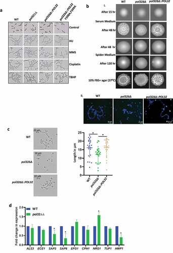
Under standard growth conditions, WT C. albicans cells are round-shaped. However, inadequate cellular dNTPs concentration and accumulation of DNA lesions inflict genomic stress that gets translated into filamentous growth in C. albicans.Citation30,Citation31 DNA replication defects, delay in cell cycle progression, and activation of checkpoints also regulate filamentation in C. albicans.Citation32 Since the deletion of POL32 caused genome instability and a significant delay in cell cycle progression under HU treatment, we investigated the association of CaPol32 function with morphological transition. C. albicans cells were subjected to susceptible but sub-lethal doses of HU, MMS, cisplatin, and TBHP in liquid cultures for 4 h, and their morphology was examined (). In the presence of genotoxic agents, cells of all the strains of C. albicans (WT, pol32ΔΔ, pol32ΔΔ::POL32, and pol32ΔΔ::POL32 F398A, F399A) switched their morphology to pseudohyphae, and only a very low percentage (<10%) of cells were either isolated or chained morphology. This result implicated that the loss of POL32 has no major effect on the genotoxins induced morphological switching of C. albicans. Further, we inspected colony morphology in spider and serum-containing solid media which are also known inducers of filamentation but do not inflict genomic stress.Citation23 On the serum-containing media, the colony of C. albicans appears rough and wrinkled; while on spider media prominent hairy outgrowths appear on the outer layer of the colony. We observed that at early time points, colonies of both Pol32 proficient and deficient strains looked very similar on both the media, however after prolonged incubation lesser wrinkled and hairy outgrowths colonies were found in pol32ΔΔ C. albicans cells than the POL32 proficient (WT and pol32ΔΔ::POL32) cells ( i). The wrinkleless of the colony was directly proportional to filamentation and the CFW staining of the colony from serum containing media substantiated reduced filamentation of pol32ΔΔ cells ( ii). In liquid media, supplementation of 10% serum to YPD leads to germ tube development in WT C. albicans.Citation33 In comparison to WT and pol32ΔΔ::POL32 cells, the Pol32 deficient cells produced stunted germ tubes (). All these results suggest that Pol32 could play an important role in genomic stress-independent morphology switching.
Figure 5. Morphogenesis of C. albicans strains. a. Cells from various strains of C. albicans were grown in liquid media for 4 hr in the presence or absence of various genotoxic agents as indicated and morphology was observed under a light microscope. b. Overnight cultures of various strains of C. albicans were diluted to 500 cells/ml, from which 200 µl cells were spreaded on YPD plates with 10% FBS, spider agar plates, and 10% FBS agar plates. All the plates were incubated at 37°C and images were taken at mentioned time points (i). Cells from YPD +10% FBS agar plates were taken, fixed, and stained with CFW, and images were captured under a fluorescent microscope (ii). c. Cells of WT, pol32ΔΔ and pol32ΔΔ::POL32 strains were grown for 1 hr in YPD liquid media containing 10% FBS and observed under the light microscope. The length of the germ tubes was measured using image j software and represented in dot plots. d. Expression of various virulence and hyphal associated genes were quantified by real-time PCR. GAPDH was used as a control. The results are presented as mean ± standard deviation. Asterisks indicate the statistically significant differences compared to the results of the WT using the student t-test (*P < .05).
Altered expression of hyphal and virulence genes in POL32 deficient strain of C. albicans
The morphological transition of C. albicans from round to filamentous shape is due to or renders altered expression of several genes such as transcription factors involved in filamentation, agglutinin-like sequence (Als) required for cell-surface adhesion, secreted aspartyl proteases (Sap), and Ece1 for membrane penetration, etc.Citation34–36 While EFG1 and CPH1 are the transcriptional activators, NRG1 and TUP1 are the transcriptional repressors of yeast to hyphal transition.Citation37 HWP1 is a downstream hyphal specific virulence gene. ECE1 encodes a cytolytic peptide toxin candidalysin that damages epithelial membranes, triggers a danger response-signaling pathway, and activates epithelial immunity.Citation38 To determine the effect of the absence of POL32 in virulence gene expression in C. albicans, differential expression of the above-mentioned genes was examined by both real-time and semi-quantitative end-point reverse transcription PCRs (, Supplementary Fig. 5A, and Supplementary Table 3). In both the assays, our expression analyses revealed that while we could not detect any noticeable difference in the mRNA levels of ALS3, ECE1, EFG1, CPH1, and TUP1 genes, expression of SAP3, SAP6, and HWP1 genes decreased marginally in pol32ΔΔ cells. Interestingly, hyper-expression of the NRG1 gene was noticed in Pol32 deficient cells, which is a well-known suppressor of filamentation.Citation37
Loss of Pol32 induces azoles drug resistance in C. albicans
Azoles are the frontline antifungal drugs, however, overuse of these drugs leads to their resistance.Citation39,Citation40 Azoles inhibit the C14α demethylation of lanosterol in fungi, which causes the depletion of ergosterol in the membrane. In C. albicans, the ERG11 gene encodes lanosterol demethylase, a target of azoles. The C-5 sterol desaturase gene (ERG3) is another essential enzyme required for ergosterol biosynthesis. Mutations in ERG3 and ERG11 that result in an amino acid substitution responsible for their non-functionality alter the membrane fluidity and result in drug resistance. C. albicans acquires azole resistance via structural alterations of the drug target, overexpression of the target gene products, an enhanced drug expulsion due to the overexpression of drug efflux transporters, and altering the cell wall and membrane architectures. The hyper-activation of TOR signaling is another important mechanism in C. albicans to bypass azole toxicity. Additionally, genome instability has been reported to be associated with multidrug resistance in C. albicans.Citation26,Citation41 Therefore, a correlation between Pol32 mediated genome stability and drug resistance in C. albicans was determined (). Interestingly, Pol32 defective cells exhibited resistance to all three azole drugs- fluconazole, miconazole, and ketoconazole when compared to WT C. albicans cells. Even in the liquid growth media, we found that the pol32ΔΔ strain was resistant to fluconazole and the growth rate was significantly high in comparison to treated POL32 proficient (WT and pol32ΔΔ::POL32) cells (). Interestingly, although Amphotericin B also targets ergosterol of fungal plasma membrane, pol32ΔΔ strain of C. albicans was hypersensitive. Similarly, in treatments with 5-fluorouracil (5FU) and 5-fluorocytosine (5FC) that target nucleotide metabolism and transcription, similar growth inhibition of both Pol32 proficient and deficient strains were observed. Since the re-integration of a single copy of the POL32 gene restored antimycotic susceptibility level similar to WT, mutation (/s) in the drugs target site or efflux pumps may not be a reason behind acquiring azole drug resistance and hypersensitivity to other antifungals by pol32ΔΔ strain. To understand a possible mechanism of azole drug resistance, first, we checked any modifications in the fungal cell wall and membrane fluidity in the pol32ΔΔ strain of C. albicans by spotting the cells on SDS, CaCl2, and CFW, and compared susceptibility with WT. Surprisingly, the growth of the pol32 null strain was significantly reduced in the presence of mentioned xenobiotics, while the growth of WT and POL32 revertant was not perturbed indicating certain modifications in the fungal cell wall and (or) membrane architecture (). To make sure that the plasma membrane localized drug transporters are not affected in POL32 deficient strains, retention ability of Congo red and berberine in the cells of WT, pol32ΔΔ, and pol32ΔΔ::POL32 strains of C. albicans was determined by spot analysis. Since these reagents are cytotoxic, retention of these will affect growth. Our spot analyses revealed that the growth of these strains was not affected suggesting the efficient functioning of drug efflux pumps. To strengthen our findings, cells were stained with fluorescent CFW, berberine, and Congo red compounds and analyzed by FACS ( and supplementary Figure 6). Since CFW stains the chitin of the fungal cell wall, a higher level of mean fluorescence intensity of pol32ΔΔ than the Pol32 expressing cells again confirmed altered cell wall composition. Similarly, the equal staining of WT, pol32ΔΔ, and pol32ΔΔ::POL32 cells by berberine and Congo red further ensured the efficient functioning of drug transporters and efflux pumps in these strains. Rapamycin inhibits Tor1 function, therefore, we compared the growth of various strains in the presence of rapamycin and found that the pol32 null strain was equally sensitive to rapamycin as WT and POL32-integrated strains. Thus, it ruled out any hyper-activation of Tor1 signaling in the POL32 deficient strain and its azole resistance is most likely due to the increased LOH events and modifications in the fungal cell wall and membrane structures.
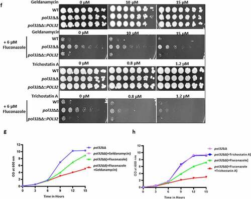
Figure 6. Antifungal drug susceptibility. a. Overnight cultures of WT, pol32ΔΔ and pol32∆∆::POL32 strains were serially diluted and spotted on YPD plates without or with the indicated concentration of various antifungal drugs. All the plates were incubated at 30°C for 48 hr and photographed. b. Overnight grown cultures of WT, pol32ΔΔ and pol32ΔΔ::POL32 strains were diluted in fresh YPD media and grown at 30°C in the absence or presence of fluconazole (6 μM). The absorbance was measured at OD600 for 15 hr at an interval of 3 hr and was plotted using Graph pad prism 8.0. The experiment was performed in biological duplicates and repeated twice. c. Serially diluted cultures of WT, pol32ΔΔ, and pol32∆∆::POL32 strains were spotted on YPD plates without or with indicated concentrations of SDS, CaCl2, CFW, rapamycin, berberine, and Congo red. Plates were incubated at 30°C for 48 hr and photographed. d. Cells from various strains of C. albicans were stained with CFW, berberine and Congo-red and analyzed by flow cytometry. The mean fluorescence intensity from 3 independent experiments were plotted using Graph Pad Prism 8.0. The results are presented as mean ± standard deviation. Asterisks indicate the statistically significant differences between WT, pol32ΔΔ, pol32ΔΔ::POL32 using the student t test (**P < .01, ***P < .001) e. Quantification of the expression of genes associated with azoles drug resistance by real-time PCR. GAPDH was used as a control. The results are presented as mean ± standard deviation. Asterisks indicate the statistically significant differences compared to the results of the WT using the student t-test (*P < .05; **P < .01). f. Serially diluted cultures of WT, pol32ΔΔ, and pol32ΔΔ::POL32 were spotted on YPD plates containing either indicated concentration of geldanamycin, trichostatin A, fluconazole, and in combinations. All the plates were incubated at 30°C for 48 hrs and photographed. g. The absorbance of pol32ΔΔ strain in YPD media in the presence or absence of fluconazole alone, geldanamycin alone or in a combination of both at 30°C was measured and plotted. h. The absorbance of pol32ΔΔ strain in YPD media in the presence or absence of fluconazole alone, trichostatin A alone or a combination of both at 30°C was measured and plotted.
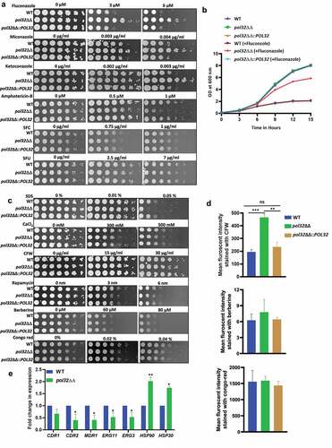
Figure 6. (Continued).
Heat-shock proteins but not drug transporters are involved in azoles resistance in the Pol32 defective strain of C. albicans
In addition to the drug efflux transporters (Cdr1, Cdr2, and Mdr1), the involvement of heat shock proteins in the drug resistance of C. albicans has been demonstrated.Citation42 Hsps are the ubiquitous proteins usually expressed in response to thermal stress; however, studies also show their activation to non-thermal stressors such as e.g., heavy metals and oxidative stress. Hsps in C. albicans confer resistance to antifungal drugs by regulating various signaling pathways, such as calcium-calcineurin, MAPK, Ras1-cAMP-PKA, and cell cycle control signaling.Citation43 To understand the mechanism of azole resistance of pol32ΔΔ strain at the molecular level, the expression of drug efflux pumps and Hsps was determined by using real-time and semi-quantitative RT-PCRs (, Supplementary Fig. 5B, and Supplementary Table 3). Our expression analyses revealed that while we could detect a marginal decrease in the mRNA levels of CDR1, CDR2, MDR1, ERG3, and ERG11 genes, expression of HSP30 and HSP90 genes increased significantly in pol32ΔΔ cells. Overexpression of CDR1, CDR2, MDR1, and ERG11 genes is usually found in drug-resistant clinical isolates. Since we observed lowered expression of these genes in the pol32ΔΔ strain of C. albicans, it suggested that these genes may not be responsible for azole resistance. However, decreased expression of the ERG3 gene has been reported to be one of the causative factors of azoles resistance in clinical isolates of C. albicans and could be a causative factor in pol32ΔΔ strain as well.Citation44 Inhibition of Hsp90 function results in azoles sensitivity in C. albicans planktonic cells and biofilms.Citation45,Citation46 Hsp30 is a plasma membrane heat shock protein, and its role in MDR has not been deciphered yet. Hsp90 inhibitors have been shown to have synergistic effects in combination with fluconazole (FLC) against FLC-resistant C. albicans.Citation47,Citation48 To confirm that the azoles resistance by pol32ΔΔ strain indeed occurred due to the overexpression of Hsp90, we performed spot and growth curve assays in the presence of Hsp90 inhibitors geldanamycin, trichostatin A, and fluconazole alone or a combination of these reagents (). Geldanamycin binds to the ADP/ATP-binding pocket of Hsp90, whereas trichostatin A blocks the acetylation of Hsp90 which is critical for Hsp90’s role in the emergence of azoles resistance.Citation42 Interestingly, in both assays, no growth reduction was observed for the pol32ΔΔ strain in the presence of geldanamycin, trichostatin A, and fluconazole alone. However, a significant reduction in the growth rate was observed in the presence of fluconazole with geldanamycin or trichostatin A. Thus, replication stress due to the loss of POL32 might be one of the reasons behind the hyper-expression of Hsp30 and Hsp90 and a possible cause of azoles resistance in C. albicans.
Ultrastructure of POL32 defective C. albicans strain reveals alteration in cell wall composition and thickness
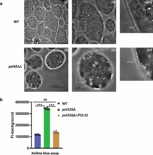
The cell wall of C. albicans is composed of three layers, the innermost layer is enriched in chitin, the middle layer contains 1–3 and 1–6 β-linked glucans, and the outermost fibrillar layer has mannoproteins. Chitin and glucan layers are well organized to determine the rigidity and shape of the cell wall, while the mannans layer is less structured with low permeability and porosity.Citation49 Since the susceptibility and effective staining of POL32 deficient cells to CFW indicated deformity of the cell wall with more chitin content, to confirm the finding, we determined and compared the ultrastructures of log phase cultured wild type and pol32ΔΔ C. albicans cells by transmission electron microscopy (). The microscopy of the ultra-thin sections (85 nm) of C. albicans cells revealed a differential morphology of both the cells. While the wild-type cells were slightly elongated round shaped, pol32ΔΔ cells were perfectly oval shaped. The pol32ΔΔ cells exhibited higher cell wall thickness and overall size compared to WT C. albicans. We found that the average thickness of the cell wall was 234 nm and 90 nm of pol32ΔΔ and WT cells, respectively. The higher thickness of the cell wall of pol32ΔΔ cell was due to the bulkiness of the middle glucan layer, and the aniline blue assay confirmed ~ 3-fold more amount of glucan content in the cell of pol32ΔΔ than that in WT and pol32ΔΔ::POL32 cells (). Since the cell wall of C. albicans acts as an interface between the fungus and host cells, the morphological modification of POL32 deficient cells could affect its pathogenicity in addition to its role in azoles drug resistance.
Figure 7. Ultrastructure of C. albicans cell. a. TEM images of ultra-thin sections of WT, and pol32ΔΔ depicting assembly of intact yeast cells, scale bar = 2 μm (i), an individual cell, scale bar = 1 μm (ii), and (iii) the thickness measurement of individual cell wall. Samples visualized up to a magnification of 14000X. Arrow indicates the zoom in image of the marked cell. b. Glucan content of the WT, pol32ΔΔ and pol32ΔΔ::POL32 cells as estimated by aniline blue assay.
Loss of POL32 induces biofilm formation of C. albicans
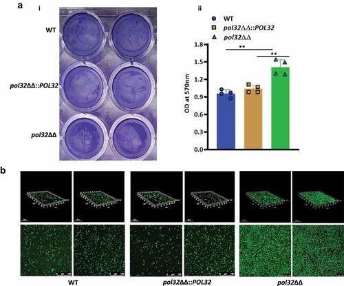
Biofilms are the microbial communities embedded in a matrix of extracellular polymers generally attached to a surface and a leading cause of chronic human fungal infection.Citation50 It has been observed that the mature biofilm is found to be less sensitive to antifungal drugs, and microbes released from the biofilm are found to be highly virulent as compared to free floating cells in a murine model of disseminated candidiasis.Citation51,Citation52 Hsp90 protein regulates both dispersions of biofilm and antifungal drug resistance mediated by biofilm.Citation42 Since we could find a role of Hsp90 in azole drug resistance in pol32ΔΔ, further we determined its ability to form biofilm on polystyrene surfaces. Biofilm formation was visualized by crystal violet staining and confocal scanning laser microscopy (CSLM) (). More presence of crystal violet-stained particles on polystyrene surface and followed by the optical density measurement at 570 nm suggested comparatively better biofilm formation ability of pol32ΔΔ than the WT and pol32ΔΔ::POL32 C. albicans cells (). We further characterized the morphology of the biofilm by confocal scanning laser microscopy (CSLM), using silicone squares as the base. As depicted in , the wild-type strains formed a uniform thin layer of biofilm, whereas the pol32 null strain developed compact but aggregated biofilm.
Figure 8. Biofilm estimation by crystal violet assay and Confocal Laser Scanning Microscopy (CLSM). a. The WT, pol32ΔΔ and pol32ΔΔ::POL32 strains of C. albicans (n = 4) were grown in YPD media with 10% FBS in 24 wells polystyrene plates for 24 hrs at 37°C. The wells were washed with PBS and stained with 0.05% crystal violet (i) and their absorbance was measured at 570 nm (ii). The obtained absorbance was plotted using Graph pad prism 8.0. The results are presented as mean ± standard deviation. Asterisks indicate the statistically significant differences compared to the results of the WT using the student t-test (**P < .01). b. WT, pol32ΔΔ and pol32ΔΔ::POL32 strains of C. albicans (n = 2) were grown on 6-wells polystyrene plates containing glass coverslips for 24 hrs at 37°C. The biofilm formed on the coverslip was stained with 1% acridine Orange and images were captured using Leica TCS SP8 confocal system with an excitation wavelength of 483 nm and emission wavelength of 510 nm. Upper panel is 3D image and lower panel is 2D image.
Pol32 protein is essential for C. albicans virulence and systemic candidiasis development
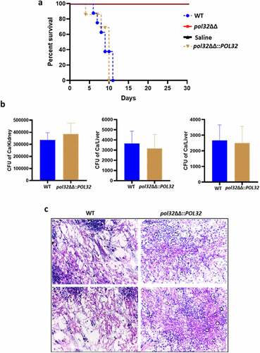
Although Pol32 is a non-essential subunit of Polδ in C. albicans, its deficiency caused genomic instability, altered cell shape and size, reduced filamentation, and lowered virulence gene expression. All of these observations suggest that Pol32 could be an important protein required for C. albicans pathogenesis. Additionally, the intra-species diversity among the clinical strains of C. albicans has been found to result in differential virulence.Citation18 To determine Pol32’s role in disease causing ability, we used a mouse model of systemic disseminated infections by the intravenous challenge of C. albicans cells. In this model, animals die due to severe sepsis with the highest fungal load in the kidneys; the phenotype mimics the severe cases of human fungal infections. The BALB/c male mice (n = 6) were injected with a fungal dose of 5 × 10⁶ CFU of pol32ΔΔ C. albicans cells per mouse via the lateral tail vein and monitored survival for 30 days (). Similarly, three sets of mice (n = 6 x2) were challenged with the same CFU of WT and pol32ΔΔ::POL32 C. albicans cells and the same volume of saline as controls. Mice who suffered due to severe candidiasis succumbed or were sacrificed based on the severity of humane endpoints. While the mice challenged with WT and pol32ΔΔ::POL32 C. albicans succumbed to infection within 11 days (5–11 days) of inoculation, all mice challenged with the pol32ΔΔ strain survived and did not show any noticeable symptoms (). The experiment of mice challenged with pol32ΔΔ C. albicans strain was repeated and the same result was obtained (Supplementary Figure 7). The CFU analysis of the vital organs such as the kidney, liver, and spleen excised out of the WT challenged group of mice confirmed the presence of C. albicans cells (). Fungal load in the kidney tissue of suffered mice was very high (~3-5 × 10⁵ cells) in comparison to that in the liver and spleen (~500 cells). Histopathology of PAS-stained kidney sections also showed a heavy load of C. albicans cells in the kidneys of mice infected with WT and pol32ΔΔ::POL32 C. albicans (). Our virulence-challenged analyses demonstrated that the pol32ΔΔ C. albicans strain is avirulent, thus the Pol32 subunit of Polδ contributes significantly to various aspects of the life cycle of C. albicans and its pathogenesis. This attenuated strain can be explored further to develop a whole-cell vaccine against C. albicans or any other fungal or microbial species infections.
Figure 9. Mice model of systemic candidiasis. a. Male BALB/c mice of 6–7 weeks of age were inoculated either with WT or pol32ΔΔ or pol32ΔΔ::POL32 (5x10⁶ CFU) C. albicans strains intravenously along with saline control and monitored their survival for 30 days. The survival curve was plotted using Graph pad prism 8.0 software. b. The murine kidney, liver, and spleen were collected and fungal burden was measured in all these organs by CFU determination. c. The kidneys of WT and pol32ΔΔ::POL32 mice (n = 2) were stained with PAS staining and images were captured in a light microscope with 40X magnification.
Discussion
Being Polδ an essential replicative DNA polymerase, its fidelity and processivity are critical for the proper functioning of a eukaryotic genome as well as that of a cell. Abnormal activity of Polδ increases mutagenesis in yeast and tumorigenesis in mice and humans.Citation5,Citation53 Despite Pol32 playing important roles in fidelity, stability, and processivity of the Polδ holoenzyme and it is indispensable for cell survival in certain organisms; incongruously, in comparison to other subunits, its role has not been appreciated, but rather considered to be a mere accessory subunit. Therefore, in this study by using a pathogenic yeast C. albicans as a model system, we further explored the critical roles of Pol32 in the life cycle of C. albicans and for the first time, we demonstrated that the activity of Pol32 links genome stability with virulence ().
Figure 10. Lack of Pol32 links genome instability with C. albicans pathogenesis. It is a pictorial representation of the consequences of the deletion of POL32 gene in C. albicans. Loss of Pol32 subunit renders replication stress by decreasing the processivity of Polδ, thereby inducing genome instability and delay in cell cycle progression. Critical hyphal and virulence genes get dysregulated to result in reduced filamentation, azoles resistance, enhanced biofilm formation, increased cell wall thickness with altered composition, and avirulency.

According to the Candida genome database, Polδ possesses genes encoding all three subunits: POL3 (C7_02790C_A/B and orf19.5182), POL31 (C5_04740C_A/B and orf19.3960), and POL32 (C1_05850W_A/B and orf19.2465). Similar to S. cerevisiae, human p12 homologue is absent in C. albicans as well. Thus, CaPolδ most likely is a holoenzyme of three subunits. Assuming that the Pol32 homologue of C. albicans will also be dispensable for cell survival, first, we generated a homozygous knockout by deleting both the alleles of POL32 gene in SC5314 strain and the derived pol32ΔΔ cells survived and grew similar to its parental isogenic strain (Supplementary Fig. 1C). But we were unsuccessful in generating heterozygous deletion of POL3 and POL31 genes. Thus, both the alleles of POL3 and POL31 could be essential for cell survival in C. albicans. The POL32 deficiency in C. albicans and S. cerevisiae caused growth retardation at high temperatures and in the presence of genotoxic agents (HU, UV, Cisplatin, MMS, and TBHP). Thus, consistent with earlier findings, Pol32 in yeasts plays a critical role in DNA replication and repair. To our surprise, unlike in S. cerevisiae where the N-terminal domain alone plays a vital role in the functioning of ScPol32,Citation7 this is the first direct evidence to show that the essentiality of CaPol32 function is also PIP motif dependent. Accordingly, more accumulation of fragmented DNA and delayed cell cycle progression in POL32 deficient and pol32ΔΔ::POL32 F398A, F399A strains of C. albicans upon HU treatment were observed. DNA Polδ is inefficient in carrying out DNA synthesis against UV damages; however, Polζ with Pol31-Pol32 (Polζ4) complex carries out an efficient bypass of UV lesions.Citation10 Since Polζ does not possess any PIP motif,Citation9,Citation12 the PIP motif of CaPol32 could be involved in the recruitment of Polζ4 to the stalled replication fork and essential for UV damage bypass or repair in C. albicans. Although the loss of pol32 reduces UV-induced mutagenesis by Polζ in S. cerevisiae,Citation54 the PIP motif of Pol32 during lesion bypass in S. cerevisiae is not critical, again it suggested a diverged mechanism of lesion bypass involving Pol32 in C. albicans. Intriguingly, contrary to S. cerevisiae pol32Δ,Citation11 we found an increased rate of LOH without any addition of genotoxic stressors in both C. albicans pol32ΔΔ and a strain carrying PIP mutant of POL32. Thus, Pol32 is mostly an antimutator in C. albicans. To get more insights into the antimutator phenotype of Pol32, whole-genome sequencing of WT and pol32ΔΔ strains of C. albicans was carried out and compared with the publicly available reference sequence of SC5314 strain. Over time, our laboratory WT strain has accumulated base substitutions and indels, however, these variations have not altered the native phenotypes such as morphological switching, sensitivity to fungal drugs, virulence, etc. Such microscale changes are very common and have already been demonstrated by whole-genome sequencing of C. albicans isolates passaged both in vitro and in vivo during laboratory culture and in the mammalian host.Citation25 YMAP clearly showed base substitutions but no aneuploidy in the WT strain. In addition to these genetic changes, the pol32ΔΔ strain accumulated 2125 indels and 6991 SNPs, and these could be specific to the Pol32 affecting Polδ’s function. To ensure that these genetic changes are specific to the function of Pol32 and were not obtained during the generation of deletion strain, we sequenced the whole genome of similarly generated mutant strains of C. albicans defective in DNA polymerase- zeta and epsilon along with the pol32ΔΔ strain. Our comparative analysis revealed that the rate and position of indels and SNPs are altogether different in different strains that suggest the specific role of respective DNA polymerases in genome instability (data not shown). Since we observed 2 folds more indels in homo- or hetero-polymeric repeat regions of the chromosome and as such regions are intrinsic replication blockers, it suggests that reduced processivity of Polδ in pol32ΔΔ could be the rationale behind the higher rate of such deletions and insertions. Genetic evidence suggests a division of labor at the replication fork where Polδ is mostly involved in lagging strand and Polε in leading strand DNA synthesis. Okazaki fragment maturation often leads to the generation of genetic variations.Citation55 Higher accumulation of heterozygous indels (2096 out of 2125) could be due to the role of Polδ in lagging strand DNA synthesis. We only observed a segmental aneuploidy in Chr 7 of pol32ΔΔ which may cause gene family contractions. Segmental aneuploidy is often associated with developmental abnormalities, reduced fitness, and occasionally a significant growth advantage over euploid cells under some conditions, most likely due to different gene dosages causing imbalances in protein stoichiometry.Citation56,Citation57 Among the chromosomes, Chr 7 and Chr R carry two MRS sequences flanking the centromere, thus they are vulnerable to being either contracted or expanded during a compromised replication condition. In budding yeast, deletion of the POL32 gene caused a 3.3-fold increase in the contraction rate of GAA repeats independently of fidelity of Polδ.Citation58
Since the loss of pol32 caused replication stress and genome instability that can impact the life cycle of C. albicans, various pathogenic attributes of the POL32 deficient cells were determined. Morphogenesis of C. albicans is one of the virulence determinants. In the presence of replication inhibitor and DNA damaging agents, we did not find any significant difference in the morphological transition of both Pol32 proficient and deficient strains of C. albicans; however, in the presence of serum containing media and spider media, reduced filamentation in pol32ΔΔ with smaller germ tube was found. This difference in morphology is also associated with somewhat reduced expression of virulence genes such as ECE1, SAP3, SAP6, and HWP1, and overexpression of transcriptional repressor NRG1. In fact, the role of NRG1 in the downregulation of ECE1 and HWP1 has been already deciphered.Citation59 Since NRG1 is belonged to Chr 7, whether the segmental aneuploidy in Chr 7 of pol32ΔΔ led to the up-regulation of NRG1 requires further investigation. However, the genome browser did not show any mutations in NRG1 of the pol32ΔΔ strain. The pol32 defective strain showed azole drug resistance but amphotericin B hypersensitivity. The mechanism of azoles drug resistance of pol32ΔΔ cells appears to be multifactorial. An altered cell shape, cell size, cell wall composition with more chitin and glucan content, lowered expression of ERG3, and hyper-expression of HSP90 and HSP30 could determine azoles drug resistance of pol32ΔΔ strain. A reduced level of ERG3 causing azole drug resistance in C. albicans has already been demonstrated.Citation44,Citation60 HSP90 functions via Tor1 signaling, and rapamycin inhibits the hyper-activation of Tor1. Since pol32ΔΔ is sensitive to rapamycin, we concluded that the Tor1 pathway is not involved in azoles drug resistance. However, hypoacetylated Hsp90 is involved in azoles drug resistance as geldanamycin and trichostatin A sensitized pol32ΔΔ strain to azole. Thus, we concluded that HSP90 could involve in azoles resistance in C. albicans independent of the Tor1 pathway. The pol32ΔΔ strain also formed a better biofilm than the wild type C. albicans and a link between HSP90, azole drugs resistance, and biofilm has been shown.Citation42 Replication and oxidative stresses are known to up-regulate HSP90 expression, although antifungal drugs enhance oxidative stress, this overexpression of HSP90 is most likely due to the replication stress in pol32ΔΔ strain.Citation61,Citation62 The stress response by Hsp90 is regulated by several factors like affinity of ATP binding and hydrolysis, interactions with co-chaperones, and post-translational modifications. A recent study suggested the role of lysine deacetylases (KDACs) like Hda1 and Rpd3 in regulating Hsp90 function in azole resistance in C. albicans and S. cerevisiae. Inhibition of lysine deacetylases by molecules like trichostatin A leads to the accumulation of hyperacetylated Hsp90. Hyperacetylation will prevent the binding of co-chaperons like calcineurin and other client proteins thereby inhibiting Hsp90’s function.Citation63 Since this study finds increasing efficacy of azole drugs by geldanamycin and trichostatin A, it again advocates the use of a combinatorial therapy by targeting the molecular chaperone Hsp90 and its upstream and downstream targets to enhance azoles drug efficacy. Further, we used an animal model to check the pathogenic potential of the pol32ΔΔ strain to develop systemic candidiasis, and it was completely attenuated and the infected mice survived. This attenuation of the pol32ΔΔ strain could be because of altered cell structure, reduced filamentation, and a lowering in the expression of certain virulence genes. We found hyperexpression of NRG1 in the pol32ΔΔ strain and previous studies have already described the efficacy of a genetically engineered C. albicans tet-NRG1 strain as an experimental live attenuated vaccine against hematogenously disseminated candidiasis.Citation64 ECE1 and SAP3 genes were shown to be involved in host cell adhesion and penetration; and decreased virulence of the respective null strains has already been demonstrated.Citation38,Citation65 We presume POL32 could be an important virulence gene and thus, the pol32ΔΔ strain could be a potential vaccine candidate. In summary, we conclude that although Pol32 is dispensable for cell survival, it plays a critical role in stabilizing the genome, morphological transition, and candidiasis development. Since the PIP motif of Pol32 is essential for its function in C. albicans, we propose to develop a small-molecule inhibitor to block its interaction with PCNA as an antifungal drug. In addition, as Pol32 is essential for C. albicans pathogenesis and no approved vaccine against any fungal diseases is available yet, we suggest exploring the pol32 null strain as a whole-cell vaccine against candidiasis and other fungal infections.
Materials and methods
Ethics statement
Mice experiments were conducted in strict accordance with the guidelines of the Institutional Animal Ethical Committee. Experimental animal protocols were fully approved by the committee and given the ethical Permit Number ILS/IAEC/133-26AH/AUG-18. Necessary precautions were taken to minimize animal suffering and to ensure the highest ethical and humane standards.
Animal, reagents, and chemicals
In-house breed strains of BALB/c male mice of 5–7 weeks old were maintained in individually ventilated cages under the standard condition with ad libitum. The oligonucleotides used in this study were procured from Integrated DNA Technologies (IDT, USA). C. albicans (SC5314) and S. cerevisiae (EMY74.7) were used as wild-type yeast strains and various knockouts were derived from these parental strains (). YPO69 is a pol32 null strain of S. cerevisiae that was derived from EMY74.7. Fetal bovine serum-South American origin (# P30-3302) from PAN Biotech, GmbH, Germany, YPD media (# G037) from HIMEDIA, India, enzymes required for cloning from NEB, genotoxic agents like HU (#102023), Cisplatin (#198872), TBHP (#190049), and MMS (#205518) from MP Biomedicals, and antifungal drugs such as fluconazole (Cat #F8929), ketoconazole (Cat# K1003), miconazole (#PHR1618), geldanamycin (#G3381), trichostatin A (#T8552), and Aniline blue (#B8563) from Sigma were procured. Periodic Acid Schiff (PAS) Stain Kit (Mucin Stain) (#ab150680) was obtained from Abcam, Cambridge, USA. The yeast strains were grown in YPD media without or with maltose or various synthetic drop-out media as required at 30°C.
Table 3. Various strains used for this study.
Bioinformatics analysis
The putative Pol32 ORF of C. albicans was aligned with ScPol32, SpCdc27, and Hsp68 by using Clustal Omega (https://www.ebi.ac.uk./Tools/msa/clustalo/). The online modeling database SWISS-MODEL (https://swisssmodel.expasy.org) was used to generate the model structure of CaPol32. The model structures of various PIP domains were predicted using the PEP-FOLD 3.0 server (https://bioserv.rpbs.univ-paris-diderot.fr/services/PEP-FOLD3. The model structures generated were aligned with the PIP peptide sequence from p68 (1U76) without or with PCNA.
Generation of Pol32 constructs
The CaPOL32 gene and its ORF were PCR amplified in a 50 μl reaction volume containing 50 ng of genomic DNA, 1.5 mM MgSO4, 1x buffer, 250 μM dNTPs, 2 U of Q5 DNA polymerase, and 10 pmol of each of the primer pairs NAP37 (5’-CCG GAA GCT TAG CTG CCC CTG TTG CTG CTC-3’) – NAP38 (5’-GGC CGA ATT CTA ACC ATA ATT TCA TTA GTA ATG TC-3’) and NAP39 (5’-GGC CAA GCT TGG ATC CAC ATA TGA CGA TGA GTA GTT CTG CTG ATG-3’) – NAP40 (5’-CCG GGA ATT CGG ATC CTC ACT TTT TCT TTC CAA AAA AAC-3’), respectively. The amplified PCR products were purified, digested with HindIII and EcoRI enzymes, and cloned into pUC19. The BamHI fragment containing Pol32 ORF was further subcloned into the BglII site of a yeast expression vector for protein purification. Similarly, the POL32 gene with PIP mutation (F398A, F399A) was amplified from SC5314 genomic DNA by using primers NAP404 (5’-CCG GGG TAC CCT GTT GCT GCT CAA CCT G-3’) and NAP463 (5-GGCC CTC GAG CAC TTT TTC TTT CCT GCA GCA CTC ATT AAT G-3’). The amplified product was purified, digested with KpnI and XhoI restriction enzymes, and cloned into the KpnI-XhoI site of a modified pSFS2 vector. The Pol32 ORF with PIP mutation (F398A, F399A) was further amplified from the resultant clone using primers NAP39 (5’-GGC CAA GCT TGG ATC CAC ATA TGA CGA TGA GTA GTT CTG CTG ATG-3’) and NAP336 (5’-GAA ATC CAG ACA GTC GAG-3’). The amplified product was digested with BamHI and cloned into the BglII site of a yeast expression vector. For S. cerevisiae, ScPol32 ORF and its PIP motif mutant (F344A, F345A) were amplified from EMY74.7 genomic DNA using forward primer NAP590 (5’-CCG GGG ATC CAC ATA TGG ATC AAA AGG CGT C-3’) and reverse primers NAP591 (5-CCG GGG TCC TTA TTT TGC CTT TCT TTT G-3’) and NAP813 (5’-CCG GGG ATC CTA TTT TGC CTT TCT TTT TGG CAG CGC TTT CC-3’), respectively. The amplified PCR products were cloned into the BamHI site of a 2µ-Sc-ADH1p plasmid vector. All the plasmid constructs were authenticated by DNA sequencing.
Protein expression and purification
Our yeast expression vector provides an option to express protein under the GAL-PGK promoter with an amino-terminal GST-fusion in a protease deficient S. cerevisiae strain.Citation66 While C. albicans Pol32 proteins were expressed in yeast, PCNA was expressed in E. coli BL21 DE3 cells as GST fusions and purified using glutathione-Sepharose 4B beads as described previously.Citation67,Citation68 To obtain untagged proteins, GST-fused proteins bound to glutathione-Sepharose beads were treated overnight at 4°C with PreScision protease to cleave the GST tag from the Pol32 or PCNA protein. All the purified proteins were stored at −80°C. Some of the uncleaved protein beads were saved for pull-down assay when required.
Generation of C. albicans knockout strains
The SAT1 flipper strategy was used to create a null mutant of the POL32 gene in the SC5314 strain of C. albicans as described before.Citation23,Citation69 Both copies of POL32 genes were deleted sequentially by transforming a linear DNA consisting of a SAT1 marker gene flanked by upstream and downstream sequences of the POL32 gene (Supplementary Figure 1A and B). Two deletion constructs were generated by PCR cloning each with the same downstream flanking region but differed in having upstream sequences. The downstream fragment was amplified with primers NAP311 (5’-GGC CGA GCT CCT CGC TCT GGA GAA CAA GAA G − 3’) and NAP407 (5’-CCG GGA GCT CGG TGA CCA AAT TTC ATT GC-3’) from the POL32 gene template construct and cloned into SacI site of pSFS2. The upstream flanking regions −342 to +109 bp and −342 to +249 bp of POL32 were PCR amplified with a common forward NAP404 (5’-CCG GGG TAC CCT GTT GCT GCT CAA CCT G-3’) and two reverse primers NAP405 5’-GGC CCT CGA GCA TGG ATG TTG AGT TG-3’) and NAP406 (5’-CCG GCT CGA GCA AAT CTG ACT CCA AAT C-3’), were digested with KpnI – XhoI and cloned into the construct containing downstream sequence to generate pNA1554 and pNA1559 deletion constructs, respectively. In the first round, the cassette was amplified by PCR from the deletion construct pNA1554 using forward primer NAP404 and reverse primer NAP407, and the amplified DNA was transformed into SC5314, and transformants were selected on a YPD-agar plate containing NAT (100 µg/ml nourseothricin). Integration of cassette at the correct locus was confirmed by PCR with primers NAP404 and NAP336. For recycling of the SAT1, PCR positive colonies were grown on YPM media for 48 hr, and the loss of the cassette was confirmed by their inability to grow on NAT containing plate. For generating homozygous deletion strain (pol32ΔΔ), the deletion cassette from pNA1559 was transformed to POL32/pol32Δ strain and a similar screening approach was used. The colonies were then cured for three days in YPM broth and streaked on YPM and YPM+NAT plates to confirm the absence of NAT cassette retention. Finally, the deletion of the POL32 gene was confirmed by PCR using the primers NAP39 and NAP40. Amino acids from 31 to 339 of CaPol32 orf (out of 403 amino acids) were disrupted in heterozygous deletant, whereas 31–339 and 81–339 amino acids were deleted in both the chromosomal copies in homozygous pol32 deletant. Therefore, we detected two PCR fragments of 308 and 448 base pairs corresponding to the deletion of both alleles in homozygous deletion in comparison to single-copy deletions. To integrate wild-type and PIP mutant of CaPOL32 into the pol32ΔΔ strain of C. albicans, full-length POL32 genes without or with F398A, F399A mutation was amplified by NAP404 forward and NAP462 (5’-GGCC CTC GAG ACA AAA CTA AAT TTC ACT TTT TCT TTC C-3’) or NAP463 (5’-GGCC CTC GAG C ACT TTT TCT TTC CTG CAG CAC TCA TTA ATG-3’) reverse primers. The PCR amplified products were digested with KpnI – XhoI and cloned into pNA1554 to generate integration constructs pNA1612 and pNA1620, respectively. The cassettes were amplified from the integration constructs pNA1612 and pNA1620 using forward primer NAP404 and reverse primer NAP407, and the linearized fragments were transformed into the pol32ΔΔ strain of C. albicans, the transformants were selected in YPD+NAT plates, and the same methodology was followed till NAT curing. The integration was confirmed by PCR using NAP39 and NAP40.
GST-pull down assay
GST-CaPol32 and CaPol32 (F398A, F399A) protein bound to glutathione sepharose beads were mixed with 0.5 µg CaPCNA and incubated overnight. The beads were then washed thrice with 0.3 ml of equilibration buffer (50 mM Tris-HCl pH 7.5, 150 mM NaCl, 5 mM dithiothreitol, 0.01% NP-40, 10% glycerol) to remove unbound proteins. The bound proteins were finally eluted in 40 µl of SDS loading buffer. Various fractions were resolved on 12.5% SDS-PAGE, followed by western blotting, and detection by using an in-house prepared anti-CaPCNA antibody (1:7000 dilution) and an HRP conjugated secondary antibody (Cat#69265, Novagen) (1:10000 dilution).
ITC assay
The purified CaPol32, CaPol32 PIP (F398A, F399A), and CaPCNA proteins were dialyzed overnight in a buffer containing 20 mM HEPES (pH7.4) and 150 mM NaCl. ITC experiments were performed using a MicroCal PEAQ-ITC system (Malvern Panalytical) at 25°C with a 20 μM concentration of CaPol32 or CaPol32 (F398A, F399A) loaded into the sample cell and 250 μM CaPCNA was injected into the cell by a given syringe. Twenty five consecutive injections of 1.5 μl each were made at an interval of 120 s. A control experiment was performed by monitoring any heat dilution of ScPol32 by buffer alone injection. Similarly, heat exchange by a buffer-buffer titration was also estimated and the value was subtracted from the respective individual experiment. Binding isotherms were fitted into a one-site binding model using the MicroCal PEAQ-ITC analysis software.
Growth curve
Various strains of C. albicans and S. cerevisiae were grown overnight in YPD broth. The overnight cultures of various strains were diluted to an OD600 = 0.01 with fresh 10 ml YPD media and allowed to grow at 30°C. Absorbance was measured at OD600 nm at every 2 hours interval for 14–15 hr. For antifungal drug susceptibility, a similar growth curve analysis was carried out in YPD medium containing 6 μM fluconazole. The OD values were plotted using a Graph pad prism v8.0. The experiments were performed twice with biological duplicates.
Sensitivity assays
Overnight grown cultures of various strains of C. albicans and S. cerevisiae were diluted in YPD liquid media to achieve an equal number of cells. They were further serially diluted by 10 folds and spotted on YPD plates containing the indicated concentration of HU, MMS, cisplatin, and TBHP. For UV sensitivity, the plates were exposed to different doses of UV and wrapped with aluminum foil. All the plates were incubated at 30°C for 2 days and images were captured. For the temperature sensitivity test, spotted plates were incubated at 16°C, 30°C, 37°C, and 42°C for 2 days and then photographed. For quantitative survival experiments, logarithmically growing cells were diluted to ~500 cells/ml, from which 200 μl was spread onto YPD plates with or without DNA-damaging agents at indicated concentrations. For UV treatment, the plates were exposed to UV irradiation at different doses. After 2- to 3-days of incubation, the number of colonies on each plate was counted and percent survival with respect to untreated was determined.
Pulsed-field gel electrophoresis
DNA plugs were prepared using Bio-Rad CHEF Yeast Genomic DNA Plug Kit as per the manufacturer’s instruction. The chromosomal separation was performed in 1.2% agarose gel with 0.5 X TBE buffer using a CHEF Mapper XA system. The running condition was voltage-6v×cm-1, switch time-60 to 120 s, angle-120° for 36 hr followed by voltage-4.5 v× cm-1, switch time-120s to 300s, angle- 120° for another 12 h. The gel was then stained with ethidium bromide and the image was captured in a ChemiDoc system.
Alkali agarose gel electrophoresis
Overnight grown cultures of various strains of C. albicans were diluted in 100 ml fresh YPD media to an OD600 = 1 and allowed to grow at 30°C for 2 hr. HU (100 mM) was then added and allowed to further grow for another 45 mins at 30°C. Cells were harvested and the pellet was washed twice with sterile distilled water, resuspended in 100 ml of fresh YPD media, and allowed to grow. Subsequently, cells were harvested at 0, 6, 12, and 24 hr of recovery periods, and the total genomic DNA was extracted. The genomic DNA was resolved by alkaline agarose gel electrophoresis as described previously.Citation68 For visualization, the gel was stained with ethidium bromide and the image was captured.
Loss of heterozygosity assay
To calculate chromosomal instability, a copy of the URA3 gene was deleted from the SC5314 and its derivative strains to generate heterozygous ura3Δ/URA3 strains. A similar method as described for POL32 gene knockout was used to delete a single copy of the URA3 gene. The upstream flanking region was amplified with primers NAP746 (5’-CCG GGG TAC CGT CAT TCC TCT TG-3’) and NAP747 (5’-GGC CCT CGA GAC TGG TGA GGC ATG AG −3’) from the genomic DNA and cloned into KpnI and XhoI sites of pSFS2. The downstream fragment was amplified by PCR with a forward NAP748 (5’-GGC CCC GCG GGA TGC TGG TTG GAA TG −3’) and reverse primers NAP749 (5’-CCG GGA GCT CGA AGA TTA TAA TGA TGT TC −3’), was digested with SacII-SacI, and cloned into the construct containing upstream sequence to generate pNA1760 URA3 gene deletion construct. The deletion cassette was excised out by KpnI–SacI digestion, and transformed into various C. albicans strains. The heterozygous deletion was confirmed by PCR using NAP746 and NAP749 primers. The strains were grown in YPD medium at 30°C overnight. The cells were then harvested, washed twice with sterile distilled water, and diluted to get an appropriate number of cells. Around 50–200 cells were spread on SD agar plates without or containing 100 mg/ml FOA. All the plates were incubated at 30°C for 3–4 days. The colonies of each strain were counted for SD and SD + FOA agar plates. The complete loss of URA3 in these strains in the presence of 5FOA was verified by growing them on media lacking uracil. The experiments were performed in biological triplicate with technical duplicates.
Cell cycle analysis
C. albicans cells were synchronized by inoculating an isolated colony in 5 ml of YPD broth from freshly streaked cells and incubated at 30°C for 16 hr in shaking condition. Synchronized cells were diluted in fresh YPD media without or with 4 mM HU and were grown at 30°C for 150 min. The cells were collected at various time points and washed twice with 700 μl of sterile distilled water. The cell pellet was resuspended in 300 μl of sterile distilled water and fixed with 700 μl of 100% ethanol at −20°C for 12 hr. Cells were harvested, washed twice with 1 ml of 50 mM of Sodium Citrate buffer, pH 7.4 for 10 mins at 25°C. The cell pellet was finally resuspended in 1 ml of 50 mM of Sodium Citrate containing 0.25 mg/ml RNase A and incubated at 37°C for 12 hr. Further, 25 μl of 20 mg/ml protease K was added and incubated for another 1 hr. The cell pellet was harvested and resuspended again in 500 μl of 50 mM sodium citrate buffer. The staining was done with 1 μM of SYTOX green by overnight incubation at 4°C in dark. The cells were transferred to FACS tubes, washed with 500 μl Sodium Citrate buffer, and analyzed by Flow cytometry. C. albicans cells were distinguished from debris by plotting SSC-A versus FSC-A, followed by singlet discrimination using FSC-H versus FSC-A, and finally singlet cells were visualized by blue laser excitation (488 nm).
Whole-genome sequencing
Genomic DNA libraries were constructed in alignment with microbial whole-genome sequencing recommendations of the NexteraTM DNA flex library preparation Kit from Illumina Inc. Briefly, 250 ng of DNA was used for tagmentation, followed by the addition of dual indices and adapters to generate the respective libraries. The cleaned libraries were quantitated on a QubitR fluorometer and appropriate dilutions were loaded on a D1000 screen tape to determine the size range of the fragments and the average library size. The average library size was 412 bp and 400 bp for WT and pol32ΔΔ strains of C. albicans, respectively. After QC clearance of the libraries, they were diluted to 4 nM, pooled, spiked with 5% PhiX pre-made library from Illumina, and loaded on a MiSeq v3 kit. Sequencing was performed for 2 × 150 cycles. The original raw data obtained from Illumina MiSeq as FASTQ files (containing paired-end reads sequences) was processed to obtain adapter free reads from MiSeq. The quality of the reads was checked using FastQC and the reads were trimmed using Trimmomatic. De-novo assembly of filtered data was performed using Shovill, which has SPAdes assembler at its core along with additional pre- and post-processing steps to improve the assembly. The raw reads were mapped to the publicly available reference genome of C. albicans SC5314 (assembly ASM18296 v3) using bowtie2 to calculate overall genome coverage. The overall alignment rate was found to be satisfactory to proceed with variant calling (94.91% for wild type and 95.68% for pol32ΔΔ). Variant calling was used to extract and filters SNPs and Indels. This was followed by filtering the variants to get the variants with high confidence. For filtering the variant, standard hard-filtering recommendations of GATK were used (https://gatk.broadinstitute.org/hc/en-us/articles/360035890471-Hard-filtering-germlineshortvariants). The filters used for SNPs are QD<2.0; FS>60; MQ<40; SOR>10 and for Indels, the filters used are QD<2.0; FS>200; SOR>10. Along with this, an additional filter of the depth-read (DP<50) was also applied. SnpEff database was used for variant annotation. The VCFPolyXutility of Jvarkit was used on the annotated variants to find out the number of repeated reference bases around the variant position
YMAP analysis
Copy number and allele status of various C. albicans strains were visualized using YMAP (https://lovelace.cs.umn.edu/Ymap/). The raw FASTQ files of both wild-type and pol32ΔΔ strains were uploaded into the YMAP server to map bonafide CNVs after applying filters to remove GC-content bias and chromosome-end bias. The reference strain used for this study is SC5314_A21-s02-m08-r09.
Morphology of C. albicans
Overnight grown C. albicans cells were diluted to an OD600 = 0.05 with 5 ml YPD medium with or without 5 mM of HU, 0.002% MMS, 0.5 mM cisplatin, and 0.004% TBHP and grown at 30°C. After 4 h of growth, cells were observed under the Leica microscope with 40X magnification. For colony morphology, 100 μl of the overnight grown culture of WT and pol32ΔΔ C. albicans containing an equal number of cells were spread on spider agar media (1% Nutrient Broth, 1% Mannitol, and 0.2% of K2HPO4) and YPD-FBS agar media (1% yeast extract, 1% peptone, 4% dextrose, and 10% FBS). Plates were incubated at 37°C, and images were taken after mentioned duration of incubation. Similarly, for germ tube development, C. albicans cells were cultured in a YPD medium containing 10% FBS at 37°C for 1 hr and observed under the microscope. The lengths of germ tubes (n = 24) were measured for each strain using Image J software and represented in a graph using Graph pad prism 8.0 software.
Antifungal drug susceptibility assay
The pre-cultures of the various strains of C. albicans were diluted to an OD600 of 1. The samples were further serially diluted by 10 to 10000 folds and spotted on YPD plates without or containing different concentrations of fluconazole, ketoconazole, miconazole, 5-flurocytosine, 5-fluorouracil, amphotericin B, SDS, CFW, CaCl2, berberine, rapamycin, and Congo red. For Hsp90 inhibition, cells were spotted on YPD plates containing geldanamycin or trichostatin A and without or with 6 μM fluconazole. All the plates were incubated at 30°C for two days and images were captured. Similarly, to check the sensitivity in the liquid medium, the strains were cultured in fresh YPD media with or without fluconazole (6 μM), geldanamycin (6 μM), trichostatin A (6 μM), and a combination of fluconazole and geldanamycin or trichostatin A. The OD values were plotted using Graph pad prism v8.0. The experiment was performed twice with biological duplicates.
Determination of chitin content by flow cytometer
The chitin content in the cell wall of C. albicans was measured using a methodology described previously with a minor modification.Citation70 Briefly, cells were harvested at their late logarithmic phase growth at 30°C, washed twice with sterile water, and re-suspended in sterile water to get about 106 number of cells per ml. About 2.5 µg CFW was added to 1 ml of cells and incubated at 25°C for 15 min. Next, the cells were washed twice with PBS, resuspended in PBS, transferred to FACS tubes, and acquired in BD LSRFortessa™ Cell Analyzer – Flow Cytometers using UV laser (350 nm) with bandpass filter (450/50 nm). The mean fluorescence intensity obtained from stained and unstained yeast cells were analyzed and processed in the flowjo software.
Berberine and Congo-red accumulation assay
The uptake and accumulation of berberine and Congo red compounds in various strains of C. albicans was measured by using FACS.Citation71 The log phase cells were harvested, washed in PBS, and diluted to O.D.600 of 0.1 in PBS containing 2% glucose. A final concentration of 25 µM berberine or Congo-red was added to the cell suspension, and incubated at 30°C for 1 hr in shaking condition. The cells were pelleted, washed with PBS, and resuspended in PBS. The cell suspension was taken up in FACS tubes and analyzed in BD LSRFortessa™ Cell Analyzer – Flow Cytometers using UV laser (350 nm) with bandpass filter (450/50 nm) or Blue laser (488 nm) with long pass mirror (610 nm). The mean fluorescence intensity obtained from stained and unstained yeast cells were analyzed and processed in the flowjo software
Gene expression analysis by PCR
Overnight cultures of WT and pol32ΔΔ strains were harvested and total RNA was isolated using the Gene JET RNA purification kit as per the manufacturer’s instruction. About 2 µg of RNA was used to synthesize cDNA using high capacity cDNA reverse transcription kit from Invitrogen with the provided random primers. A total volume of 20 µl of qRT PCR reaction mixture was setup containing 50ng of cDNA, 10 pmol of forward and reverse primers, and 2x SYBR green qPCR Master mix. The qRT PCR cycling was carried out in Quant Studio 3 with fast cycle conditions including 95°C for 2 min followed by 40 cycles of 95°C for 5 sec denaturation and 60°C for 30 sec for annealing and extension. All the experiments were performed in biological duplicates with technical triplicates. The data obtained were analyzed using the 2−ΔΔCT method. The gene expression of WT and pol32ΔΔ strain was normalized with GAPDH, the housekeeping gene and graphs were plotted using a Graph Pad Prism 8 software. Similarly, for semi-quantitative real-time PCR, 20 µL of reaction mixture was setup containing 50 ng of cDNA, 10 pmol of respective forward and reverse primers, 250 µM dNTPs, 1x Taq DNA polymerase buffer, and 1 U of Taq DNA polymerase. The PCR conditions used were initial denaturation at 95°C for 1 min, followed by 30 cycles of 95°C for 30 sec, 60°C for 30 sec and 72°C for 30 sec. The amplified products were resolved in 1.2% and images were captured in a gel doc system. The band intensity of the amplified products were analyzed using image J software. The band intensity of WT and pol32ΔΔ strain was normalized by the house keeping gene GAPDH and graphs were plotted using Graph Pad Prism 8 software. The primer sequences of various genes used for real-time PCR were given in .
Table 4. Primers used for real-time PCR.
Transmission electron microscopy (TEM)
Ultrastructure of WT and pol32ΔΔ C. albicans cells were examined by TEM using a protocol described before.Citation72 Briefly, similarly grown log-phase cultures were centrifuged at 5000 g for 5 min followed by washing with 1 ml of 0.1 M cacodylate buffer. The pellet so obtained was fixed with 1 ml of 2.5% glutaraldehyde for 30 min at 25°C. The fixed cells were washed again using cacodylate buffer followed by post-fixation using 200 μl of 1% osmium tetraoxide for 3 hr. The dehydration process was carried out with 50%, 70%, 90%, and absolute alcohol, each with 10 min of incubation. Next, the pellet was first resuspended in 500 μl of propylene oxide and then again resuspended in 1 ml of a mixture of propylene oxide and Spurr’s resin (1:1 ratio) for 3 hrs. The impregnation of samples was carried out in pure resin and incubated overnight at RT. Prior to sample embedment, the overnight pellet was centrifuged at 5000 g, the resin was discarded, and again resuspended in a resin mixed hardening reagent DMAE (Ted Pella, Inc.). The mixture was allowed to polymerize at 60°C for 48 hrs. The hardened sample was sectioned using Leica EM UC7 microtome. The sections were collected on the copper grid, stained with uranyl acetate for 30 min, followed by 3 times washings with distilled water, and allowed to dry for 2 h. Samples on the grid were visualized under JEM-2100Plus JEOL TEM imaging machine.
Aniline blue assay
The β-glucan level of the fungal cell wall was measured by aniline blue assay.Citation73 Briefly, 1 ml of overnight grown cells was harvested and washed two times with 1xTE buffer. The cells were resuspended in 1xTE buffer to obtain an OD600 of 0.2. About 500 μl cell suspension was treated with 100 μl of 6 M NaOH at 80°C for 30 min, followed by the addition of 2.1 ml of aniline blue mix (0.03% aniline blue, 0.18 M HCl, and 0.49 M glycine-NaOH, pH 9.5), and then vortexed. The tubes were again incubated at 50°C for 30 min followed by cooling at 25°C for 30 min. The fluorescence intensity (FI) was measured by taking the mixture in a 96 well black microplate using a plate reader at an excitation wavelength of 400 nm/slit and an emission wavelength of 460 nm/slit with a cutoff of 455 nm. The fluorescence intensity obtained from both the samples was plotted using Graph pad Prism v 8.0.
Biofilm growth
C. albicans cells were grown in 24-well polystyrene plates containing YPD media with 10% serum for 24 hours at 37°C. Supernatants were discarded and plates were washed two times gently with 1x PBS. The biofilm was then treated with 0.1% crystal violet stain for 20–30 minutes at room temperature, followed by a wash with distilled water, and allowed to dry at room temperature for 5 hours. The plates were then photographed. For quantification, the bound dye was then re-suspended after 1 h of addition of 1 ml of 33% glacial acetic acid at 25°C. The absorbance was recorded at 570 nm using a plate reader.
CLSM for biofilm detection
C. albicans cells were grown as described above in a 6-well cell culture plate with glass coverslips at 37°C for 24 hr. After 24 hr, the supernatant was discarded and washed with 1x PBS followed by staining with 1% acridine orange for 20 min in dark. Excess stain was removed by washing with 1x PBS, followed by fixation with 4% formaldehyde at 25°C for 30 min. Excess fixative was removed by washing thrice with 1X PBS. Finally, the glass coverslips were placed gently on the glass slide, and images were captured using Leica TCS SP8 confocal scanning system with an excitation wavelength of 483 nm and emission wavelength of 500 to 510 nm bandpass filter.
Mouse model of systemic candidiasis
The overnight cultures of WT, pol32ΔΔ, and pol32ΔΔ::POL32 strains of C. albicans were washed thoroughly with sterile distilled water, followed by a saline wash, and finally, the inoculum was prepared in saline. For inoculum preparation, overnight grown strains were washed twice with sterile distilled water followed by washing with PBS and finally dissolved in PBS. The cells were counted using the Neubauer chamber slide in the central area (5 × 5 squares) and 5 × 10⁶ cells/ml cell suspension was prepared for each strain. To reconfirm the inoculum size, CFU analyses for each strains were carried out by spreading a diluted culture of the inoculum on YPD plates. Male BALB/c mice of 5–7 weeks were injected intravenously with 100 μl of 5 × 10⁶ CFU of C. albicans cells or saline and survivability were monitored for 30 days. The mice were humanly sacrificed as described earlier.Citation33 The kidney, liver, and spleen organs were extracted from each of the mice. Half of these organs were used for CFU determination while the other half of these organs were stored in formalin for tissue sectioning. The mouse kidney paraffin sections were stained with periodic acid Schiff reagents as per manufacturer instructions, and images were captured under a light microscope with 40X magnification. The survival curve was prepared using Graph Pad Prism v8.0 software.
CFU determination
The kidney, liver, and spleen organs were homogenized in 1x PBS using a tissue homogenizer. An appropriate dilution of the homogenate of each organ was prepared by diluting the sample with 1X PBS and plated on YPD agar plates containing chloramphenicol (50 μg/ml). The colonies were counted for each organ and CFU was determined.
Availability of data and materials
The data that support the findings of this study are available from the corresponding author upon reasonable request. The whole-genome sequence data of pol32ΔΔ and WT SC5314 strains can be retrieved from the NCBI database using the accession number PRJNA851971 and PRJN862296.
Authors’ contributions
NA conceptualized, designed, supervised, and analyzed the study; SKP, SRS, BU, and SB generated materials, conducted experiments, analyzed the data, and prepared an initial draft of the manuscript. NA and SKP wrote the final draft. NA obtained the funding. All the authors reviewed the results and approved the final version of the manuscript.
Supplemental Material
Download PDF (1.7 MB)Acknowledgments
We thank Mr. Sitendra Prasad Panda and Mr. Paritosh Nath for their technical assistance. Mr. Biswajit Patro for his technical assistance and nurturing of the animals was highly appreciated. We thank our other laboratory colleagues for their thoughtful discussion. For genomics, miBiome therapeutics is highly acknowledged. SRS and BU are grateful for CSIR/UGC-Senior Research Fellowships. ILS animal house, biophysical characterization, Imaging, and FACS facilities are highly acknowledged.
Disclosure statement
NA, SRS, and SKP are listed as inventors in a related patent application. The remaining authors declare no competing interests.
Supplementary material
Supplemental data for this article can be accessed online at https://doi.org/10.1080/19490976.2022.2163840
Correction Statement
This article has been corrected with minor changes. These changes do not impact the academic content of the article.
Additional information
Funding
References
- Burgers PMJ, Kunkel TA. Eukaryotic DNA replication fork. Annu Rev Biochem. 2017;86(1):417–35. doi:10.1146/annurev-biochem-061516-044709.
- Guilliam TA, Yeeles JTP. An updated perspective on the polymerase division of labor during eukaryotic DNA replication. Crit Rev Biochem Mol Biol. 2020;55(5):469–481. doi:10.1080/10409238.2020.1811630.
- Acharya N, Klassen R, Johnson RE, Prakash L, Prakash S. 2011. PCNA binding domains in all three subunits of yeast DNA polymerase delta modulate its function in DNA replication. Proceedings of the National Academy of Sciences of the United States of America; 108, 17927–17932. doi:10.1073/pnas.1109981108.
- Acharya N, Khandagale P, Thakur S, Sahu JK, Utkalaja BG. Quaternary structural diversity in eukaryotic DNA polymerases: monomeric to multimeric form. Curr Genet. 2020;66(4):635–655. doi:10.1007/s00294-020-01071-1.
- Khandagale P, Peroumal D, Manohar K, Acharya N. Human DNA polymerase delta is a pentameric holoenzyme with a dimeric p12 subunit. Life Sci Alliance. 2019;2(2):e201900323. doi:10.26508/lsa.201900323.
- Zuo S, Gibbs E, Kelman Z, Wang TS, O’Donnell M, MacNeill SA, Hurwitz J. (1997). DNA polymerase delta isolated from Schizosaccharomyces pombe contains five subunits. Proceedings of the National Academy of Sciences of the United States of America; 94, 11244–11249. doi:10.1073/pnas.94.21.11244.
- Johansson E, Garg P, Burgers PM. The Pol32 subunit of DNA polymerase delta contains separable domains for processive replication and proliferating cell nuclear antigen (PCNA) binding. J Biol Chem. 2004;279(3):1907–1915. doi:10.1074/jbc.M310362200.
- MacNeill SA, Moreno S, Reynolds N, Nurse P, Fantes PA. The fission yeast Cdc1 protein, a homologue of the small subunit of DNA polymerase delta, binds to Pol3 and Cdc27. EMBO J. 1996;15(17):4613–4628. doi:10.1002/j.1460-2075.1996.tb00839.x.
- Makarova AV, Stodola JL, Burgers PM. A four-subunit DNA polymerase zeta complex containing Pol delta accessory subunits is essential for PCNA-mediated mutagenesis. Nucleic Acids Res. 2012;40(22):11618–11626. doi:10.1093/nar/gks948.
- Johnson RE, Prakash L, Prakash S. 2012. Pol31 and Pol32 subunits of yeast DNA polymerase delta are also essential subunits of DNA polymerase zeta. Proceedings of the National Academy of Sciences of the United States of America; 109, 12455–12460. doi:10.1073/pnas.1206052109.
- Huang ME, Rio AG, Galibert MD, Galibert F. Pol32, a subunit of Saccharomyces cerevisiae DNA polymerase δ, suppresses genomic deletions and is involved in the mutagenic bypass pathway. Genetics. 2002;160(4):1409–1422. doi:10.1093/genetics/160.4.1409.
- Acharya N, Johnson RE, Pages V, Prakash L, Prakash S. 2009. Yeast Rev1 protein promotes complex formation of DNA polymerase zeta with Pol32 subunit of DNA polymerase delta. Proceedings of the National Academy of Sciences of the United States of America; 106, 9631–9636
- Hirota K, Yoshikiyo K, Guilbaud G, Tsurimoto T, Murai J, Tsuda M, Phillips LG, Narita T, Nishihara K, Kobayashi K, et al. The POLD3 subunit of DNA polymerase delta can promote translesion synthesis independently of DNA polymerase zeta. Nucleic Acids Res. 2015;43(3):1671–1683. doi:10.1093/nar/gkv023.
- Lancey C, Tehseen M, Raducanu VS, Rashid F, Merino N, Ragan TJ, Savva CG, Zaher MS, Shirbini A, Blanco FJ, et al. Structure of the processive human Pol delta holoenzyme. Nat Commun. 2020;11(1):1109. doi:10.1038/s41467-020-14898-6.
- Zheng F, Georgescu RE, Li H, O’Donnell ME. 2020. Structure of eukaryotic DNA polymerase delta bound to the PCNA clamp while encircling DNA. Proceedings of the National Academy of Sciences of the United States of America; 117, 30344–30353. doi:10.1073/pnas.2017637117.
- Wenzel RP. Nosocomial candidemia: risk factors and attributable mortality. Clin Infect Dis. 1995;20(6):1531–1534. doi:10.1093/clinids/20.6.1531.
- Sahu SR, Bose S, Singh M, Kumari P, Dutta A, Utkalaja BG, Patel SK, Acharya N. Vaccines against candidiasis: status, challenges and emerging opportunity. Front Cell Infect Microbiol. 2022;12:1002406. doi:10.3389/fcimb.2022.1002406.
- Hirakawa MP, Martinez DA, Sakthikumar S, Anderson MZ, Berlin A, Gujja S, Zeng Q, Zisson E, Wang JM, Greenberg JM, et al. Genetic and phenotypic intra-species variation in Candida albicans. Genome Res. 2015;25(3):413–425. doi:10.1101/gr.174623.114.
- Pavlov YI, Zhuk AS, Stepchenkova EI. DNA polymerases at the eukaryotic replication fork thirty years after: connection to cancer. Cancers (Basel). 2020;12(12):3489. doi:10.3390/cancers12123489.
- Rustchenko E. Chromosome instability in Candida albicans. FEMS Yeast Res. 2007;7(1):2–11. doi:10.1111/j.1567-1364.2006.00150.x.
- Kumari P, Sundaram R, Manohar K, Vasudevan D, Acharya N. Interdomain connecting loop and J loop structures determine cross-species compatibility of PCNA. J Biol Chem. 2021;297(1):100911. doi:10.1016/j.jbc.2021.100911.
- Huang ME, Le Douarin B, Henry C, Galibert F. The Saccharomyces cerevisiae protein YJR043C (Pol32) interacts with the catalytic subunit of DNA polymerase alpha and is required for cell cycle progression in G2/M. Mol Gen Genet. 1999;260(6):541–550. doi:10.1007/s004380050927.
- Manohar K, Peroumal D, Acharya N. TLS dependent and independent functions of DNA polymerase eta (Polη/Rad30) from pathogenic yeast Candida albicans. Mol Microbiol. 2018;110(5):707–727. doi:10.1111/mmi.14004.
- Forche A, Abbey D, Pisithkul T, Weinzierl MA, Ringstrom T, Bruck D, Petersen K, Berman J. Stress alters rates and types of loss of heterozygosity in Candida albicans. mBio. 2011;2. doi:10.1128/mBio.00129-11.
- Ene IV, Farrer RA, Hirakawa MP, Agwamba K, Cuomo CA, Bennett RJ. 2018. Global analysis of mutations driving microevolution of a heterozygous diploid fungal pathogen. Proceedings of the National Academy of Sciences of the United States of America; 115, E8688–E8697. doi:10.1073/pnas.1806002115.
- Ford CB, Funt JM, Abbey D, Issi L, Guiducci C, Martinez DA, Delorey T, Li BY, White TC, Cuomo C, et al. The evolution of drug resistance in clinical isolates of Candida albicans. eLife. 2015;4:e00662. doi:10.7554/eLife.00662.
- Abbey DA, Funt J, Lurie-Weinberger MN, Thompson DA, Regev A, Myers CL, Berman J. YMAP: a pipeline for visualization of copy number variation and loss of heterozygosity in eukaryotic pathogens. Genome Med. 2014;6(11):100. doi:10.1186/s13073-014-0100-8.
- Nick Mcelhinny SA, Stith CM, Burgers PM, Kunkel TA. Inefficient proofreading and biased error rates during inaccurate DNA synthesis by a mutant derivative of Saccharomyces cerevisiae DNA polymerase delta. J Biol Chem. 2007;282(4):2324–2332. doi:10.1074/jbc.M609591200.
- Lujan SA, Clausen AR, Clark AB, MacAlpine HK, MacAlpine DM, Malc EP, Mieczkowski PA, Burkholder AB, Fargo DC, Gordenin DA, et al. Heterogeneous polymerase fidelity and mismatch repair bias genome variation and composition. Genome Res. 2014;24(11):1751–1764. doi:10.1101/gr.178335.114.
- Bachewich C, Thomas DY, Whiteway M. Depletion of a polo-like kinase in candida albicans activates cyclase-dependent hyphal-like growth. Mol Biol Cell. 2003;14(5):2163–2180. doi:10.1091/mbc.02-05-0076.
- Shi QM, Wang YM, Zheng XD, Lee RT, Wang Y. Critical role of DNA checkpoints in mediating genotoxic-stress–induced filamentous growth in Candida albicans. Mol Biol Cell. 2007;18(3):815–826. doi:10.1091/mbc.e06-05-0442.
- Loll-Krippleber R, d’Enfert C, Feri A, Diogo D, Perin A, Marcet-Houben M, Bougnoux ME, Legrand M. A study of the DNA damage checkpoint in Candida albicans: uncoupling of the functions of Rad53 in DNA repair, cell cycle regulation and genotoxic stress-induced polarized growth. Mol Microbiol. 2014;91(3):452–471. doi:10.1111/mmi.12471.
- Peroumal D, Manohar K, Patel SK, Kumari P, Sahu SR, Acharya N. Virulence and pathogenicity of a Candida albicans mutant with reduced filamentation. Cell Microbiol. 2019;21(12):e13103. doi:10.1111/cmi.13103.
- Tsuchimori N, Sharkey LL, Fonzi WA, French SW, Edwards JE Jr., Filler SG. Reduced virulence of HWP1 -deficient mutants of Candida albicans and their interactions with host cells. Infect Immun. 2000;68(4):1997–2002. doi:10.1128/IAI.68.4.1997-2002.2000.
- Correia A, Lermann U, Teixeira L, Cerca F, Botelho S, da Costa RM, Sampaio P, Gartner F, Morschhauser J, Vilanova M, et al. Limited role of secreted aspartyl proteinases sap1 to sap6 in Candida albicans virulence and host immune response in murine hematogenously disseminated candidiasis. Infect Immun. 2010;78(11):4839–4849. doi:10.1128/IAI.00248-10.
- Lo HJ, Kohler JR, DiDomenico B, Loebenberg D, Cacciapuoti A, Fink GR. Nonfilamentous C. albicans mutants are avirulent. Cell. 1997;90(5):939–949. doi:10.1016/S0092-8674(00)80358-X.
- Murad AM, Leng P, Straffon M, Wishart J, Macaskill S, MacCallum D, Schnell N, Talibi D, Marechal D, Tekaia F, et al. NRG1 represses yeast-hypha morphogenesis and hypha-specific gene expression in Candida albicans. EMBO J. 2001;20(17):4742–4752. doi:10.1093/emboj/20.17.4742.
- Moyes DL, Wilson D, Richardson JP, Mogavero S, Tang SX, Wernecke J, Hofs S, Gratacap RL, Robbins J, Runglall M, et al. Candidalysin is a fungal peptide toxin critical for mucosal infection. Nature. 2016;532(7597):64–68. doi:10.1038/nature17625.
- Cowen LE, Anderson JB, Kohn LM. Evolution of drug resistance in Candida Albicans. Annu Rev Microbiol. 2002;56(1):139–165. doi:10.1146/annurev.micro.56.012302.160907.
- Marie C, White TC. Genetic basis of antifungal drug resistance. Curr Fungal Infect Rep. 2009;3(3):163–169. doi:10.1007/s12281-009-0021-y.
- Dunkel N, Blass J, Rogers PD, Morschhauser J. Mutations in the multi-drug resistance regulator MRR1, followed by loss of heterozygosity, are the main cause of MDR1 overexpression in fluconazole-resistant Candida albicans strains. Mol Microbiol. 2008;69(4):827–840. doi:10.1111/j.1365-2958.2008.06309.x.
- Robbins N, Uppuluri P, Nett J, Rajendran R, Ramage G, Lopez-Ribot JL, Andes D, Cowen LE. Hsp90 governs dispersion and drug resistance of fungal biofilms. PLoS Pathog. 2011;7(9):e1002257. doi:10.1371/journal.ppat.1002257.
- LaFayette SL, Collins C, Zaas AK, Schell WA, Betancourt-Quiroz M, Gunatilaka AA, Perfect JR, Cowen LE. PKC signaling regulates drug resistance of the fungal pathogen Candida albicans via circuitry comprised of Mkc1, calcineurin, and Hsp90. PLoS Pathog. 2010;6(8):e1001069. doi:10.1371/journal.ppat.1001069.
- Regan J, DeJarnette C, Luna-Tapia A, Parker JE, Reitler P, Barnett S, Tucker KM, Kelly SL, Palmer GE. Titration of C-5 sterol desaturase activity reveals its relationship to Candida albicans virulence and antifungal susceptibility is dependent upon host immune status. mBio. 2022;13(2):e0011522. doi:10.1128/mbio.00115-22.
- Robbins N, Caplan T, Cowen LE. Molecular evolution of antifungal drug resistance. Annu Rev Microbiol. 2017;71(1):753–775. doi:10.1146/annurev-micro-030117-020345.
- Shapiro RS, Uppuluri P, Zaas AK, Collins C, Senn H, Perfect JR, Heitman J, Cowen LE. Hsp90 orchestrates temperature-dependent Candida albicans morphogenesis via Ras1-PKA signaling. Curr Biol. 2009;19(8):621–629. doi:10.1016/j.cub.2009.03.017.
- Cowen LE, Singh SD, Kohler JR, Collins C, Zaas AK, Schell WA, Aziz H, Mylonakis E, Perfect JR, Whitesell L, et al. 2009. Harnessing Hsp90 function as a powerful, broadly effective therapeutic strategy for fungal infectious disease. Proceedings of the National Academy of Sciences of the United States of America; 106, 2818–2823. doi:10.1073/pnas.0813394106.
- Li Y, Chang W, Zhang M, Li X, Jiao Y, Lou H. Synergistic and drug-resistant reversing effects of diorcinol D combined with fluconazole against Candida albicans. FEMS Yeast Res. 2015;15(2). doi:10.1093/femsyr/fov001.
- Garcia-Rubio R, de Oliveira HC, Rivera J, Trevijano-Contador N. The fungal cell wall: candida, cryptococcus, and aspergillus species. Front Microbiol. 2019;10:2993. doi:10.3389/fmicb.2019.02993.
- Desai JV, Mitchell AP. Candida albicans biofilm development and its genetic control. Microbiol Spectr. 2015;3(3). doi:10.1128/microbiolspec.MB-0005-2014.
- Finkel JS, Mitchell AP. Genetic control of Candida albicans biofilm development. Nat Rev Microbiol. 2011;9(2):109–118. doi:10.1038/nrmicro2475.
- Gulati M, Nobile CJ. Candida albicans biofilms: development, regulation, and molecular mechanisms. Microbes Infect /Institut Pasteur. 2016;18(5):310–321. doi:10.1016/j.micinf.2016.01.002.
- Jain R, Rice WJ, Malik R, Johnson RE, Prakash L, Prakash S, Ubarretxena-Belandia I, Aggarwal AK. Cryo-EM structure and dynamics of eukaryotic DNA polymerase delta holoenzyme. Nat Struct Mol Biol. 2019;26(10):955–962. doi:10.1038/s41594-019-0305-z.
- Gerik KJ, Li X, Pautz A, Burgers PM. Characterization of the two small subunits of Saccharomyces cerevisiae DNA polymerase delta. J Biol Chem. 1998;273(31):19747–19755. doi:10.1074/jbc.273.31.19747.
- Reijns MAM, Kemp H, Ding J, de Proce SM, Jackson AP, Taylor MS. Lagging-strand replication shapes the mutational landscape of the genome. Nature. 2015;518(7540):502–506. doi:10.1038/nature14183.
- Torres EM, Williams BR, Amon A. Aneuploidy: cells losing their balance. Genetics. 2008;179(2):737–746. doi:10.1534/genetics.108.090878.
- Pavelka N, Rancati G, Zhu J, Bradford WD, Saraf A, Florens L, Sanderson BW, Hattem GL, Li R. Aneuploidy confers quantitative proteome changes and phenotypic variation in budding yeast. Nature. 2010;468(7321):321–325. doi:10.1038/nature09529.
- Khristich AN, Armenia JF, Matera RM, Kolchinski AA, Mirkin SM. 2020. Large-scale contractions of Friedreich’s ataxia GAA repeats in yeast occur during DNA replication due to their triplex-forming ability. Proceedings of the National Academy of Sciences of the United States of America; 117, 1628–1637. doi:10.1073/pnas.1913416117.
- Braun BR, Kadosh D, Johnson AD. NRG1, a repressor of filamentous growth in C.albicans, is down-regulated during filament induction. EMBO J. 2001;20(17):4753–4761. doi:10.1093/emboj/20.17.4753.
- Sanglard D, Ischer F, Parkinson T, Falconer D, Bille J. Candida albicans mutations in the ergosterol biosynthetic pathway and resistance to several antifungal agents. Antimicrob Agents Chemother. 2003;47(8):2404–2412. doi:10.1128/AAC.47.8.2404-2412.2003.
- Belenky P, Camacho D, Collins JJ. Fungicidal drugs induce a common oxidative-damage cellular death pathway. Cell Rep. 2013;3(2):350–358. doi:10.1016/j.celrep.2012.12.021.
- Sottile ML, Nadin SB. Heat shock proteins and DNA repair mechanisms: an updated overview. Cell Stress Chaperones. 2018;23(3):303–315. doi:10.1007/s12192-017-0843-4.
- Robbins N, Leach MD, Cowen LE. Lysine deacetylases Hda1 and Rpd3 regulate Hsp90 function thereby governing fungal drug resistance. Cell Rep. 2012;2(4):878–888. doi:10.1016/j.celrep.2012.08.035.
- Saville SP, Lazzell AL, Chaturvedi AK, Monteagudo C, Lopez-Ribot JL. Efficacy of a genetically engineered Candida albicans tet-NRG1 strain as an experimental live attenuated vaccine against hematogenously disseminated candidiasis. Clin Vaccine Immunol. 2009;16(3):430–432. doi:10.1128/CVI.00480-08.
- Hube B, Sanglard D, Odds FC, Hess D, Monod M, Schafer W, Brown AJ, Gow NA. Disruption of each of the secreted aspartyl proteinase genes SAP1, SAP2, and SAP3 of Candida albicans attenuates virulence. Infect Immun. 1997;65(9):3529–3538. doi:10.1128/iai.65.9.3529-3538.1997.
- Acharya N, Haracska L, Johnson RE, Unk I, Prakash S, Prakash L. Complex formation of yeast Rev1 and Rev7 proteins: a novel role for the polymerase-associated domain. Mol Cell Biol. 2005;25(21):9734–9740. doi:10.1128/MCB.25.21.9734-9740.2005.
- Manohar K, Acharya N. Characterization of proliferating cell nuclear antigen (PCNA) from pathogenic yeast Candida albicans and its functional analyses in S. cerevisiae. BMC Microbiol. 2015;15(1):257. doi:10.1186/s12866-015-0582-6.
- Manohar K, Khandagale P, Patel SK, Sahu JK, Acharya N. The ubiquitin-binding domain of DNA polymerase eta directly binds to DNA clamp PCNA and regulates translesion DNA synthesis. J Biol Chem. 2022;298(2):101506. doi:10.1016/j.jbc.2021.101506.
- Sasse C, Morschhauser J. Gene deletion in Candida albicans wild-type strains using the SAT1-flipping strategy. Methods Mol Biol. 2012;845:3–17.
- Oliveira Carvalho V, Okay TS, Melhem MS, Walderez Szeszs M, Del Negro GM. The new mutation L321F in Candida albicans ERG11 gene may be associated with fluconazole resistance. Rev Iberoam Micol. 2013;30(3):209–212. doi:10.1016/j.riam.2013.01.001.
- Khandelwal NK, Chauhan N, Sarkar P, Esquivel BD, Coccetti P, Singh A, Coste AT, Gupta M, Sanglard D, White TC, et al. Azole resistance in a Candida albicans mutant lacking the ABC transporter CDR6/ROA1 depends on TOR signaling. J Biol Chem. 2018;293(2):412–432. doi:10.1074/jbc.M117.807032.
- Grigor’eva A, Bardasheva A, Tupitsyna A, Amirkhanov N, Tikunova N, Pyshnyi D, Kleshev M, Ryabchikova E. Changes in the ultrastructure of candida albicans treated with cationic peptides. Microorganisms. 2020;8. doi:10.3390/microorganisms8040582.
- Suwunnakorn S, Wakabayashi H, Rustchenko E. Chromosome 5 of human pathogen Candida albicans carries multiple genes for negative control of caspofungin and anidulafungin susceptibility. Antimicrob Agents Chemother. 2016;60(12):7457–7467. doi:10.1128/AAC.01888-16.