
ABSTRACT
Bifidobacterium longum subsp. infantis is a prevalent member of the gut microbiota of breastfed infants. In this study, the effects of human breastmilk-derived B.longum subsp. infantis CCFM1269 on bone formation in developing BALB/c mice were investigated. Newborn female and male mice were assigned to control group (administered saline), CCFM1269 group (administered B. longum subsp. infantis CCFM1269, 1 × 10⁹ CFU/mouse/day) and I5TI group (administered B. longum subsp. infantis I5TI, 1 × 10⁹ CFU/mouse/day) from 1-week-old to 3-, 4- and 5-week old. B. longum subsp. infantis I5TI served as a negative control in this study. The results demonstrated that B. longum subsp. infantis CCFM1269 promoted bone formation in growing mice by modulating the composition of the gut microbiota and metabolites. The expression of genes and proteins in the PI3K/AKT pathway was stimulated by B. longum subsp. infantis CCFM1269 through the GH/IGF-1 axis in growing mice. This finding suggests B. longum subsp. infantis CCFM1269 may be useful for modulating bone metabolism during growth.
1. Introduction
Bone formation is influenced by various factors such as genetics, nutrition, health, and the environment.Citation1 The formation of bones starts from embryonic development. During childhood and adolescence, bone mass increases with age until it reaches its peak in young adulthood when skeletal development is mature.Citation2
During infancy, childhood, and adolescence, improving bone growth through breast milk and dietary nutrition can have significant implications for bone health later in life.Citation3 Breastfeeding is the recommended feeding method by the World Health Organization for the first six months of life.Citation4 Breast milk is an important source of intestinal microbiota for infants.Citation5 and breastfeeding enables the vertical transmission of microorganisms from mother to infants.Citation6,Citation7 Thus, breastfeeding enables infants to acquire strains of beneficial bacteria such as Bifidobacterium, Lactobacillus, and Bacteroides from breast milk and this transfer process helps infants establish a well-functioning gut microbiota system early on.Citation6 The gut microbiota is an essential component of the host and its composition and functionality influence host health. Gut microbiota colonization during infancy is a critical event that can have long-term effects on the individual’s growth, development, and overall health.Citation8 Among the infant gut microbiota, B. longum subsp. infantis is one of the dominant bacterial species, and the abundance changes of Bifidobacterium can have important implications for the infant’s early and future physiological status and health.Citation9
In recent years, an increasing amount of evidence in small animal models has shown that Bifidobacterium has the potential to influence the skeletal system through the gut-bone axis. For example, supplementing a single strain of B. longum accelerated the formation of callus, thereby improving the mechanical performance of fractured limbs and promoting bone fracture repair in 18-month-old female mice.Citation10 B. longum UBBL-64 significantly enhanced bone mass and strength in osteoporotic mice, regulated the ratio of immune cells in lymphocytes, and improved bone microstructure.Citation11 B. longum 35624® inhibited osteoclast formation through the production of extracellular polysaccharides, thereby activating the TLR2-dependent pathway.Citation12 In addition, the combination of snow lotus fruit powder and B. longum ATCC 15,707 helped increased the mineral concentrations of calcium (Ca), phosphorus (P), and magnesium (Mg) in the tibia of rats.Citation13 Also in rats, B. animalis subsp. lactis HN019 reduced bone and connective tissue loss and increased bone-protecting content.Citation14 In high-fat diet-fed mice, B. pseudocatenulatum CECT 7765 reversed the changes in bone volumetric fraction, trabecular number, and trabecular pattern factor, and reversed the decreased volumetric bone mineral density of trabeculae, all caused by obesity.Citation15 B. adolescentis ATCC 15,703 enhanced intestinal barrier integrity, suppressed systemic inflammation response following bone fracture, accelerated fracture healing and cartilage remodeling, and strengthened the protective ability of the skeleton after fracture in mice.Citation16 However, there are limited studies available on the impact of B. longum subsp. infantis derived from breast milk on bone health. Furthermore, B. longum subsp. infantis derived from breast milk may have a greater influence on bone growth and development. Therefore, in our previous study, we screened B. longum subsp. infantis CCFM1269 from human breast milk with the ability to regulate the bone formation.
This study primarily evaluated the potential of CCFM1269 and I5TI in improving bone growth and development in 3-, 4-, and 5-week-old mice through analyzing bone length, pathological sections, and serum biochemical indicators. Then the impact of CCFM1269 and I5TI on the diversity of gut microbiota and metabolites in growing mice was explored using fecal microbiota 16S rRNA amplicon sequencing and bifidobacterial groEL sequencingas well as untargeted metabolome analysis. Moreover, the potential mechanisms for these two strains of B. longum subsp. infantis regulating bone growth and development were further validated from the transcription and translation of PI3K/AKT pathway using RT-qPCR and Western blot analysis. I5TI was chosen as a negative control due to its weaker ability in adjusting the intestinal barrier compared to CCFM1269 in our previous study.
2. Materials and methods
2.1 Animal experiment
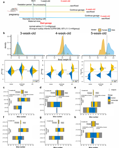
The animal experiment was approved by the Jiangnan University Animals Ethics Committee (JN.No20220315b0900725[008]). Six-week-old female- and male-specific pathogen-free (SPF) BALB/c mice from Charles River Laboratories Inc. (Hangzhou, China) were used. Thirty-six female mice and eighteen male mice were caged together at a ratio of 2:1 after one week of acclimatization. The constant temperature of 22 ± 2°C and 40%-70% humidity was maintained in the breeding environment with a light-to-dark cycle of 12:12 h. The males were removed from the cage after the females were pregnant. The females were untreated during pregnancy. The mice was fed with Co60 irradiation experimental mouse feed. Pups from the same pregnant mice were all in the same intervention group. And pups from multiple litters were grouped if there were less than 8 pups per litter for both genders. Pups from different litters were still fed by their mothers even though they were combined into one group. Groups of more than eight mice after litter merging were randomly selected to eight mice for each group at the time of testing. The dams were gavaged with saline (control group), B. longum subsp. infantis CCFM1269 (1 × 10⁹ CFU/day/mice, CCFM1269 group) or I5TI (1 × 10⁹ CFU/day/mice, I5TI group) from one-week-old to three-, four-, and five-week-old, respectively (n = 8 per group) (). The female and male dams were separated at three weeks old and supplementation was continued until four and five weeks old (). The specific culture information of the two strains of B. longum subsp. infantis was in supplementary materials. Feces were collected before sacrifice and stored at −80°C. All the dams were sacrificed, and blood samples were collected. The left leg of each animal was fixed in 4% formalin solution for tartrate-resistant acid phosphatase (TRAP) and alizarin red staining. The right leg was measured along with the length of the femur and tibia before being stored at −80°C. Other samples were stored at −80°C for further analysis.
Figure 1. Experimental design and change of body weight and bone length.
2.2 Trap and alizarin red staining
Bone tissue was fixed in 4% paraformaldehyde for 24 hours and then placed in an EDTA decalcifying solution, which was changed every three days until the bone tissue was soft. It was then dehydrated in gradient ethanol and fixed in an embedding frame. The fixed bone tissue was cut into 5 µm slices by the slicer and fixed on slides for subsequent staining. The procedure for TRAP and alizarin red staining followed the instructions (supplemental materials).
2.3 Blood and colonic biochemical analysis
Serum insulin-like growth factor 1 (IGF-1), osteoclastogenesis inhibitory factor (OPG), Osteocalcin/Bone bone-γ- carboxyglutamic acid-containing protein (OT/BGP), procollagen I N-terminal tripeptide (PINP), C-telopeptide of type I collagen (CTX-1), growth hormone (GH), and insulin-like growth factor binding protein 3 (IGFBP3) were measured by ELISA kits according to the manufacturer’s instructions (supplementary material). 20 mg colon was homogenized by PBS (mass: volume = 1:9) and centrifuged at 4°C, 4,000 g for 10 min. The supernatant was collected and the BCA kit was used to measure concentrations. Then the concentration of IGF-1 in the colon was measured by the ELISA kit through instruction (Supplemental materials).
2.4 High throughput 16S rRNA gene and bifidobacterial groEL gene sequencing
The methods for 16S rRNA amplicon sequencing and bifidobacterial groEL gene sequencing of DNA extracted from mice feces were performed according to previous studies.Citation9,Citation17,Citation18 Briefly, bacterial DNA was extracted from feces according to the manufacturer’s instructions (supplementary material). DNA was used as a template with the 16S rRNA V3-V4 primers (341F, 806 R) and bifidobacterial-specific primers (supplemental material). PCR reaction for 16S rRNA sequencing contained 25 μL 2× Taq master Mix, 1 μL 10 μM former and reverse primer, 1 μL DNA, and 22 μL ddH2O. The PCR procedures were as follows: pre-denaturation at 95°C for 5 min, followed by 30 cycles consisting of denaturation at 95°C for 30 s, annealing at 60°C for 30 s, and extension at 72°C for 1 min, and a final extension of 72°C for 10 min. PCR reaction for bifidobacterial groEL sequencing contained 25 μL 2× Taq master Mix, 1 μL 10 μM former and reverse primer, 2 μL DNA, and 21 μL ddH2O. The PCR procedures were as follows: pre-denaturation at 95°C for 8 min, followed by 30 cycles consisting of denaturation at 95°C for 40 s, annealing at 60°C for 40 s, and extension at 72°C for 40 s, and a final extension of 72°C for 8 min. Negative controls were included using deionized sterile water as a template. The PCR products were purified and their concentrations were determined with the Qubit 3.0 (Thermofisher, CA, U.S.A.). Then DNA libraries of specific quality were sequenced on the Illumina MiSeq (San Diego, CA, U.S.A) platform. Qiime 2 was used to analyze the raw sequences of each sample. Αlpha diversity was calculated using the MicrobiomeAnalyst platform (https://www.microbiomeanalyst.ca/).Citation19 Beta diversity was shown through R packages (“Vanga”, “ggplot2”) and PERMANOVA was used to evaluate beta diversity based on Bray-Curtis distance from ASV-tables. The sequence data was rarefied before alpha and beta diversity analyses.
2.5 Metabolome analysis
The feces were lyophilized and weighed to 20 mg, added to 200 µl of ultrapure water and zirconium strain, and then crushed at 65 Hz for 1 min. 800 µl of the methanol-acetonitrile mixture (v/v = 1:1) was added and the sample was precipitated for 1 hour at 20°C, and then centrifuged for 4°C, 15000 g for 15 min. 700 µl from the supernatant was added to a new tube and spun dry until free of liquid. The precipitate was resuspended by adding 200 µl of acetonitrile-water mixture (v:v = 1:1) before being centrifuged at 4°C, 15000 g for 15 min. After centrifugation, the supernatant was filtered through a 0.22 µm membrane and 10 µl of each sample was mixed as quality control to ensure stability and reproducibility. The acetonitrile-water mixture (v:v = 1:1) was used as a blank sample. Metabolites in feces were analyzed in full scan mode using an ItiMateU3000 UPLC system (2.1 × 100 mm T3 column) and a high-resolution Q-Exactive Plus mass spectrometer. A non-targeted metabolism workflow using Compound Discovery 3.3 software was used to process the raw data from the mass spectra, including data extraction, background feature filtering, peak identification, deconvolution, peak alignment and integration, and metabolite identification, comparison with databases. Orthogonal partial least squares discriminant analysis (OPLS-DA) was performed using SIMCA software and VIP > 1 and p-value <0.05 were considered significant for metabolite differences.
2.6 Quantitative real-time polymerase chain reaction analysis
Quantitative real-time polymerase chain reaction analysis (qRT-PCR) was performed according to a previous study.Citation20 Briefly, the RNA of femur and tibia was extracted and quantified, and then reverse transcribed. The expression of specific genes was detected using the CFX Opus 384 Real-Time PCR System from Bio-Rad (Hercules, CA, U.S.A). PCR reaction contained 5 μL 2× Taq master Mix,0.2 μL 10 μM former and reverse primer, and 1 μg cDNA, making up to 10 μL with ddH2O. The PCR procedures were as follows: pre-denaturation at 95°C for 5 min, followed by 30 cycles consisting of denaturation at 95°C for 30 s, annealing at 60°C for 30 s, and extension at 72°C for 2 min, and a final extension of 72°C for 10 min. Data processing used the2−ΔΔCt method. The primers and conditions used were listed in Table S1.
2.7 Western blot analysis
Forty milligram of femur and tibia were homogenized by adding RIPA lysate (containing 2% phosphatase inhibitors and protease inhibitors), the supernatant was centrifuged at 4°C, 12000 g for 5 min and the BCA kit was used to determine the concentration of the supernatant which was then added to SDS loading buffer to preserve the protein. Sample proteins were separated by SDS-PAGE and transferred to nitrocellulose membranes, incubated with the same specific antibody, and then protein expression was measured.Citation21,Citation22 The target protein bands were exposed by Tanon 46,000 chemiluminescence imager (Shanghai, China). Image J was used to evaluate the grayscale of the band.Citation23
2.8 Statistical analysis
All data are shown as mean ± standard error of the mean (SEM). Comparisons between two or more groups were made by one-way ANOVA followed by Tukey multiple comparison test on GraphPad (version 8.0). If the distribution was not normal, Mann-Whitney tests were performed. A p-value <0.05 was considered significant. Simca (v14.0) was used for metabolite analysis. VIP > 1 and p < 0.05 of metabolites represented differential metabolites between the two groups.Citation24 The significant differential metabolites between the two groups were performed by ranking dotplot (https://hiplot.cn/basic). Tbtools was used to display an upset plot.Citation25 Redundancy analysis (RDA) was performed with R packages (“Vegan”, “ggrepel”, “ggplot2”).
3. Results
3.1 B. longum subsp. infantis regulated bone length and the number of osteoblasts and osteoclasts
The macroscopic growth parameters (weight) were not significantly altered after supplementation with B. longum subsp. infantis CCFM1269 and I5TI regardless of age and gender compared to the control group (p < 0.05, ). Differences in body weight between males and females become progressively more pronounced with increasing age (p < 0.05, ). However, supplementation with CCFM1269 significantly increased femur length in male and female mice at 3-, 4-, and 5-week of age compared to the corresponding control group (p < 0.05, ). Additionally, tibia length was significantly increased in female and male mice at 5-week-old in the CCFM1269 group compared to the corresponding control group (p < 0.05, ) while no significant change in tibia length in 3- and 4-week-old females and males was observed (). Supplementation with I5TI showed no significant influence on the femur and tibia length.
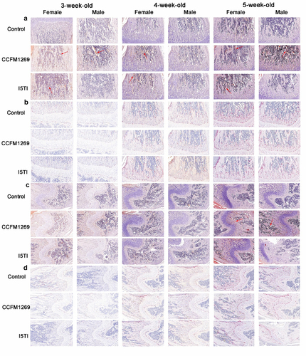
The femur and tibia were stained with Alizarin Red and TRAP to observe the number of osteoblasts and osteoclasts, respectively. It was observed that the number of both osteoblasts and osteoclasts increased with age (, Figure S1 and S2). CCFM1269 increased the number of calcium nodules in the femur which represented osteoblast formation but showed less effects on osteoclast number in the femur of females and males regardless of age (, Figure S1A,B). For osteoblasts and osteoclasts in the tibia, CCFM1269 affected the number of calcium nodules and osteoclasts of 3- and 4-week-old females and males slightly while it increased the number of calcium nodules (, Figure S2A,B).
Figure 2. Alizarin Red and TRAP staining of femur and tibia.
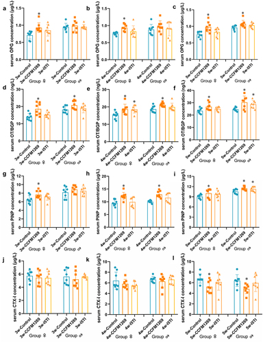
3.2 B. longum subsp. infantis CCFM1269 regulated osteoclast and osteogenic factors in serum
Serum osteoclast and osteogenic factors were also detected to evaluate the impact of CCFM1269 and I5TI on bone formation. CCFM1269 supplementation increased the level of OPG of 3- and 4-week-old female mice (p < 0.05, ) and OPG of 5-week-old female and male mice compared to the corresponding control group (p < 0.05, ). Similarly, the level of OT/BGP was significantly higher in the CCFM1269 group compared to the corresponding control group regardless of age and gender (p < 0.05, ). Additionally, the level of PINP was only significantly higher in the CCFM1269 group of 3-week-old female mice while significantly higher in 4- and 5-week-old mice both female and male compared to the corresponding control group (). However, CCFM1269 only significantly decreased the level of CTX-1 when mice were 5-week-old (p < 0.05, ) and there were no significant effects on 3- and 4-week-old female and male mice (). I5TI supplementation only significantly increased the level of OT/BGP of 4-week-old female mice (p < 0.05, ) and OT/BGP and PINP of 5-week-old male mice (p < 0.05, ).
Figure 3. Effects of B. longum subsp. infantis on the serum osteogenic factors.
3.3 B. longum subsp. infantis regulated gut microbiota
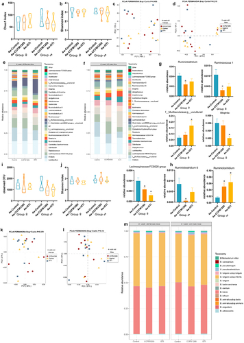
Chao 1 and Shannon indexes were used to evaluate the effects of CCFM1269 and I5TI on α diversity of gut microbiota. For 3-week-old mice, CCFM1269 supplementation significantly increased the Chao1 and Shannon indices of both female and male mice compared to the corresponding control group (p < 0.05, ). I5TI supplementation also significantly increased the Chao 1 index of both female and male mice but only significantly increased the Shannon index of female mice compared to the corresponding control group (p < 0.05, ). PCoA analysis also showed that the microbiota structure was significantly different among control, CCFM1269, and I5TI groups of both female (p = 0.001, ) and male mice (p = 0.001, ). The dominant genus in female mice in the control, CCFM1269, and I5TI groups was f__Muribaculaceae;g__uncultured (21.04%), Lachnospiraceae NK4A136 group (20.39%) and f__Muribaculaceae;g__uncultured (15.50%), respectively (). And the dominant genus in male mice in control, CCFM1269 and I5TI groups was Lachnospiraceae NK4A136 group (18.33%), Lachnospiraceae NK4A136 group (19.73%) and f__Muribaculaceae;g__uncultured (12.98%), respectively (). In contrast, CCFM1269 significantly increased the relative abundance of Alistipes (p < 0.05), Bifidobacterium (p < 0.05), Roseburia (p < 0.05), and Lachnospiraceae NK4A136 group (p < 0.05) and decreased the relative abundance of Odoribacter (p < 0.05) and Staphylococcus in female mice compared to the control group (p < 0.05, ). I5TI significantly increased the relative abundance of Alistipes (p < 0.05), Bifidobacterium (p < 0.05), and Roseburia (p < 0.05), and decreased the relative abundance of Odoribacter (p < 0.05) and Staphylococcus in female mice compared to the control group (p < 0.05, ). For male mice, CCFM1269 significantly increased the relative abundance of Bifidobacterium (p < 0.05) and lowered the relative abundance of Ruminiclostridium 5 (p < 0.05), Staphylococcus (p < 0.05) and Bacteroides (p < 0.05) compared to the control group (). I5TI significantly increased the relative abundance of Bifidobacterium (p < 0.05) and lowered the relative abundance of Ruminiclostridium 5 (p < 0.05) and Staphylococcus (p < 0.05) compared to the control group (p < 0.05, ). The composition of Bifidobacterium at the species level was also determined. CCFM1269 significantly increased the observed index of female mice and significantly lowered the Shannon index of male mice (p < 0.05, ). Interestingly, the Bifidobacterium composition at the species level was significantly different among control, CCFM1269, and I5TI groups of female mice (p = 0.001, ) whereas no significant difference was found in male mice (p = 0.26, ). The dominant Bifidobacterium in control, CCFM1269, and I5TI groups were B. breve (40.95%), and B. longum subsp. infantis (81.43%) and B. breve (57.2%) of female mice, respectively (). Additionally, the dominant Bifidobacterium in control, CCFM1269, and I5TI groups were B. breve (53.11%), and B. longum subsp. infantis (51.56%) and B. longum subsp. infantis (52.02%) of male mice, respectively (). CCFM1269 significantly increased the relative abundance of B. longum subsp. infantis in both females and males (p < 0.05, ) while it decreased the relative abundance of B. breve compared to the corresponding control group in females (p < 0.05, ). However, I5TI decreased the relative abundance of B. longum subsp. infantis significantly compared to the control group in females (p < 0.05, ).
Figure 4. Effects of B. longum subsp. infantis on the gut microbiota of 3-week-old mice.
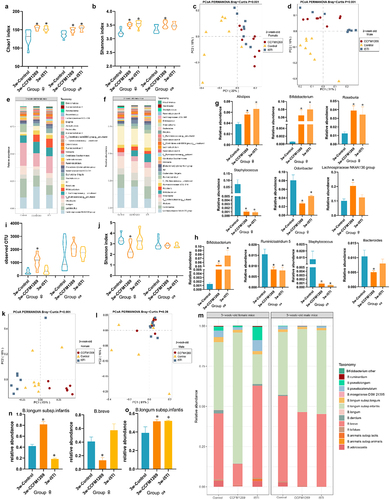
The same methods were used to analyze the gut microbiota composition of 4-week-old mice. In contrast to the results of 3-week-old mice, CCFM1269 and I5TI did not significantly influence the α and β diversity indices of neither females nor males (). The dominant genus in control, CCFM1269, and I5TI groups was Alistipes (16.17%), Lactobacillus (15.63%), and f__Muribaculaceae;g__uncultured (16.87%) of female mice, respectively (). Furthermore, the dominant genus in the control, CCFM1269, and I5TI groups were Alistipes (20.64%, 12.61%, 19.47%) in male mice, respectively (). CCFM1269 significantly decreased the relative abundance of Ruminiclostridium (p < 0.05), Ruminococcus 1 (p < 0.05), and Lachnospiraceae FCS020 group (p < 0.05) in female mice (). I5TI only significantly decreased the relative abundance of Lachnospiraceae FCS020 group (p < 0.05) in female mice (). For the male mice, CCFM1269 decreased the relative abundance of Ruminiclostridium 6 (p < 0.05 and increased the relative abundance of Ruminiclostridium (p < 0.05) and I5TI increased the relative abundance of Ruminiclostridium (p < 0.05, ). The bifidobacterial community of 4-week-old female and male mice was not significantly affected by CCFM1269 or I5TI in terms of α and β diversity, and the relative abundance of bifidobacterial species (). B. longum subsp. infantis remained the predominant Bifidobacterium in female mice (50.79%, 51.37%, and 51.02%, respectively) whereas B. breve, and B. longum subsp. infantis, B. longum subsp. infantis were dominant in the control (49.43%), CCFM1269 (49.28%), and I5TI (48.76%) groups, respectively ().
Figure 5. Effects of B. longum subsp. infantis on the gut microbiota of 4-week-old mice.
For the changes in the gut microbiota community of the 5-week-old mice, CCFM1269 and I5TI did not significantly influence α diversity (). Interestingly, β diversity showed that the microbiota structure among control, CCFM1269, and I5TI groups was significantly different in males (p = 0.002) while there was no significant influence in female mice (). f_Muribaculaceae;g__uncultured was predominant in control (26.12%), CCFM1269 (33.09%), and I5TI (29.93%) groups of females (). And f_Muribaculaceae;g__uncultured was also predominant in control (26.04%), CCFM1269 (29.17%), and I5TI (37.71%) groups of males (). CCFM1269 significantly decreased the relative abundance of Candidatus Arthromitus (p < 0.05), Ruminiclostridium (p < 0.05), and increased the relative abundance of Desulfovibrio (p < 0.05), and Ruminococcaceae UCG-010 (p < 0.01) in female mice (). I5TI only decreased the relative abundance of Candidatus Arthromitus (p < 0.05) in female mice (). For the male mice, CCFM1269 significantly increased the relative abundance of Ruminococcaceae UCG-010 (p < 0.01) and decreased the relative abundance of Lachnospiraceae NK4A136 group (p < 0.05), Ruminiclostridium 6 (p < 0.05) and Ruminiclostridium (p < 0.05, ). I5TI significantly increased the relative abundance of Ruminococcaceae UCG-010 (p < 0.01) and decreased the relative abundance of Lachnospiraceae NK4A136 group (p < 0.05) and Ruminiclostridium compared to the control group (p < 0.05, ).
Figure 6. Effects of B. longum subsp. infantis on the gut microbiota of 5-week-old mice.
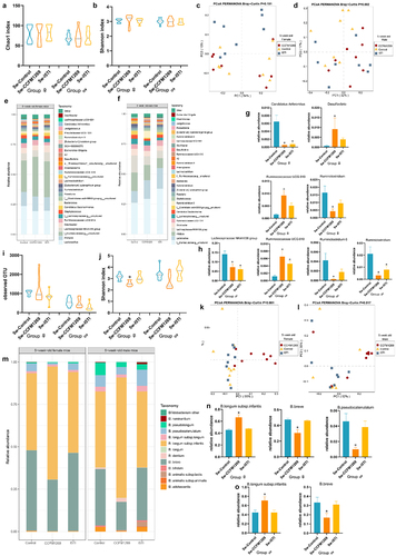
For bifidobacterial community, CCFM1269 significantly decreased the Shannon index of male mice (p < 0.05) but only had a slight influence on the Shannon index of female mice (). However, the bifidobacterial composition was significantly different among control, CCFM1269, and I5TI groups of females (p = 0.001, ) and male mice (p = 0.017, ). B. longum subsp. infantis remained the dominant Bifidobacterium species in control (45.37%, 44.78%), CCFM1269 (66.56%, 70.95%), and I5TI (47.54%, 44.84%) groups regardless of gender (). CCFM1269 significantly increased the relative abundance of B. longum subsp. infantis and decreased the relative abundance of B. breve in both females and males compared to the corresponding control group (p < 0.05, ). Furthermore, the relative abundance of B. pseudocatenulatum was only significantly decreased in the CCFM1269 group in female mice compared to the control group (p < 0.05, ).
3.4 B. longum subsp. infantis regulated gut metabolites
Non-target metabolome analysis of the effects of CCFM1269 and I5TI on fecal metabolites was performed. According to Vip and p-value, significantly different metabolites were observed (). For female mice, CCFM1269 showed 28, 12, and 13 different metabolites in 3-, 4- and 5-week-old mice, respectively (). In terms of metabolic pathway enrichment for differential metabolites, CCFM1269 affected the metabolic function, for instance, lactose degradation, tryptophan metabolites, and glucose-alanine cycle (Figure S3A-C). I5TI showed 7, 17, and 5 different metabolites in 3-, 4- and 5-week-old mice, respectively (Table S2-S4). For male mice, CCFM1269 displayed 9, 9, and 8 different metabolites of 3-, 4- and 5-week-old mice, respectively (). CCFM1269 had less effect on metabolic pathways in male rats than in female mice. However, CCFM1269 still affected tryptophan metabolites in male mice (Figure S3D-F). I5TI showed 2, 12, and 2 different metabolites of 3-, 4- and 5-week-old mice, respectively (Table S5-S7). RDA analysis revealed the metabolites between CCFM1269 and control groups were different and the effect of differential bacteria on differential metabolites was equivalent for 3-, 4- and 5-week-old females and males ().
Figure 7. Effects of B. longum subsp. infantis CCFM1269 on fecal metabolites.
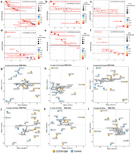
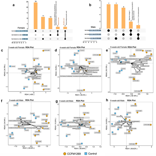
The different metabolites between CCFM1269 and the control group were subjected for further analysis. Differential metabolites were both unique and shared between male and female mice at 3-, 4- and 5–5-week-old. 3-week-old females shared 5-(4-Carboxy-3-methylbutyl)-1,4a-dimethyl-6-methylenedecahydro-1-naphthalenecarboxylic acid and oleanolic acid with 4-week-old female and nicotinic acid and methylimidazoleacetic acid with 5-week-old females, respectively (). Male mice were different from female mice. 3-week-old males shared 3-amino-2-phenyl-2 H-pyrazolo[4,3-c]pyridine-4,6-diol and disperse orange 3 with 4-week-old males and 4-week-old males shared 2-amino-1,3,4-octadecanetriol with 5-week-old female, respectively (). Differential metabolites acted on serum bone formation indicators (OPG, PINP, OC/BGP) and bone length (Femur and tibia) were limited. Only 10, 11, and 11 metabolites among 28, 12, and 13 different metabolites of 3-, 4- and 5-week-old females showed effects on serum bone formation indicators (OPG, PINP, OC/BGP) and bone length (). For males, 9, 8, and 8 metabolites among 9, 9 and 8 different metabolites of 3-, 4- and 5-week-old males showed effects on serum bone formation indicators (OPG, PINP, OC/BGP) and bone length ().
Figure 8. Co-existing metabolites of different age groups and RDA analysis between differential metabolites and OPG, OT/BGP, PINP, femur length, and tibia length.
3.5 B. longum subsp. infantis CCFM1269 regulated serum growth factors
Compared to the corresponding control group, CCFM1269 increased the level of GH in 3- and 4-week-old females and males significantly (p < 0.05, ) but did not influence GH levels in 5-week-old females and males (). I5TI only increased the GH level in 3- and 4-week-old male mice (p < 0.05, ). Furthermore, the level of IGF-1 was significantly higher in the CCFM1269 group of 4- and 5-week-old female and male mice (p < 0.05, ), and the level of IGFBP3 were significantly higher in the CCFM1269 group of 3-, 4- and 5-week-old female and male mice (p < 0.05, ) compared to the corresponding control group, whereas I5TI only increased the IGF-1 level of 5-week-old females and males (p < 0.05, ). Additionally, supplementation with CCFM1269 increased the level of 1,25(OH)2D3 of 4- and 5-week-old female mice (p < 0.05) but had no effect on male mice regardless of age ().
Figure 9. Effects of B. longum subsp. infantis on serum growth factors.
3.6 B. longum subsp. infantis regulated GH/IGF-1 pathway
At the transcription level, CCFM1269 supplementation significantly upregulated the relative expression of GHR, IGF-1 R, IGFBP3, IRS1, and RUNX2 (p < 0.05, ), decreased the relative expression of mTOR (p < 0.05, ) in the femur of 3-, 4- and 5-week-old male and female mice compared to the corresponding control group. CCFM1269 supplementation also decreased the expression of Grb10 in the femur of 3-week-old male and female mice (p < 0.05, ). I5TI supplementation significantly increased the relative expression of GHR in the femur of 3-week-old males and females (p < 0.05, ), increased the relative expression of GHR, IGFR, IGFBP3, and IRS1 in the femur of 4- and 5-week-old males and females (p < 0.05, ), and increased the expression of RUNX2 in the femur of 5-week-old males and females (p < 0.05, ). Additionally, I5TI supplementation decreased the relative expression of mTOR in the femur of 4-week-old males and females and 5-week-old females (p < 0.05, ).
Figure 10. Effects of B. longum subsp. infantis on the PI3K/AKT pathway at the transcript level.
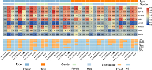
The effect of CCFM1269 on the tibia was different from that of the femur, as supplementation with CCFM1269 only significantly increased the expression of GHR, IGFR, and IRS1 in 3-week-old male and female mice (p < 0.05, ) and increased the expression of GHR, IGFR, IGFBP3, and IRS1 in 4-week-old male and female mice (p < 0.05, ). Additionally, the expression of GHR, IGFR, IGFBP3, IRS1, and RUNX2 was significantly higher and mTOR was significantly lower in the CCFM1269 group of 5-week-old male and female mice compared to the control group (p < 0.05, ). I5TI supplementation significantly increased the expression of GHR in 4-week-old female mice and GHR, IGFR, and IGFBP3 in 4-week-old male mice compared to the control group (p < 0.05, ). Furthermore, I5TI supplementation increased the relative expression of IGFR in 5-week-old females (p < 0.05, ), and increased the relative expression of GHR, IGFR, and IRS1 expression in 5-week-old male mice compared to the control group (p < 0.05, ).
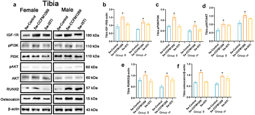
At the transcriptiontranslational level, supplementation with CCFM1269 significantly increased the relative expression of IGFR, pPI3K, pAKT, RUNX2, and osteocalcin in the femur of 3-, 4- and 5-week-old male and female mice (p < 0.05, ) and the relative expression of IGFR, pPI3K, pAKT, RUNX2 and osteocalcin in the tibia of 5-week-old male and female mice (p < 0.05, ). Supplementation with I5TI significantly increased the relative expression of osteocalcin in the femur of 3-week-old females, pAKT in the femur of 4-week-old males, and RUNX2 in 5-week-old males (p < 0.05, ).
Figure 11. Effects of B. longum subsp. infantis on the PI3K/AKT pathway at the translation level of the femur.
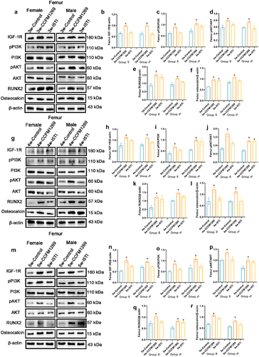
Figure 12. Effects of B. longum subsp. infantis on PI3K/AKT pathway at translation level of tibia.
4. Discussion
In the current study, the length of the femur and tibia, the levels of osteogenesis and osteoclasis markers, growth factors, and changes in the gut microbiota, as well as the expression of critical genes and proteins pertaining to the GH/IGF-1 signaling pathway in the femur and tibia of adolescent mice across various weeks of tests, had all been assessed in growing mice. B. longum subsp. infantis CCFM1269 can alter the gut microenvironment of growing mice, which in turn affected the composition of related metabolites and markers in the gut and serum, ultimately regulating bone growth via the GH/IGF-1 signaling pathway.
As key players in bone production and resorption, osteoblasts (OB) and osteoclasts (OC) can interact with one another through cytokine paracrine.Citation26 B. longum subsp. infantis CCFM1269 altered bone length and the indicators of bone formation and bone metabolites including PINP, OPG, OT/BGP, and CTX-1. Interestingly, the change in bone length did not cause a significant change in body weight, but there was still a slight increase after CCFM1269 supplementation. Additionally, the difference in body weight between males and females became progressively larger regardless of age at week. CCFM1269 mainly affects osteoblasts and to a lesser extent osteoclasts as confirmed by serum indicators and morphological staining of bone tissue. Only OT/BGP was enhanced by CCFM1269 supplementation irrelevant of age or gender among PINP, OPG, OT/BGP, and CTX-1. Osteocalcin is mainly synthesized by osteoblasts and odontoblasts and plays an important role in the regulation of bone calcium metabolism.Citation27 Therefore, CCFM1269 mainly promoted osteoblast development, both in terms of serum indicators and in terms of osteoblast and osteoclast staining in the femur and tibia. OPG is well known in the OPG/RANKL/RANK signaling pathway and can bind to RANKL which inhibits bone resorption.Citation28 However, CCFM1269 did not significantly inhibit the bone resorption according to the effect on CTX-1 levels. The level of CTX-1 only decreased significantly at 5 weeks of age which suggested that CCFM1269 supplementation did not affect bone formation rather than bone resorption during the adult phase. In human subjects, serum PINP has proven to be a trustworthy indicator of bone turnover, and it is frequently used to track bone development.Citation29 In this study, CCFM1269 showed the effects on PINP of 3-, 4- and 5-week-old mice but CTX-1 was only influenced by CCFM1269 when mice were 5-week-old. This may indicate PINP and CTX-1 are not interdependent as observed in another study where milk oligosaccharides supplementation reduced CTX-1 levels in malnourished mice but did not affect PINP levels.Citation30
Previous preclinical studies in adult germ-free and conventionally reared mice had linked the gut microbiota to the regulation of bone mass, although the effects were context dependent, with sex and genetic background influencing the observed phenotypes.Citation31,Citation32 However, the influence of intestinal microbiota on bone development is undeniable.Citation31,Citation33 For instance, malnutrition can lead to severe growth retardation and underweight and supplementation with Lactobacillus plantarum WJL can increase linear growth.Citation33,Citation34 B. longum improves ovariectomy-induced bone loss and enhances the anti-osteoclastogenic and immunomodulatory potential of regulatory B cells (Bregs).Citation11 In the present study, time- and sex-dependent changes were observed in intestinal microbiota following CCFM1269 supplementation, especially around weaning. Before weaning, the diversity of the intestinal bacteria was affected by foreign species and the relative abundance of Bifidobacterium and B. longum subsp. infantis increased significantly. In contrast, at 4 weeks of age, the first week after weaning, continuous supplementation did not affect the diversity of the intestinal microbiota in males and females and the differential bacterial genera were altered. Interestingly, continuous supplementation with CCFM1269 also did not affect the relative abundance of B. longum subsp. infantis in male and female mice at four weeks of age. After continuous supplementation up to 5 weeks of age, it was found that the level of differential genera was both similar and different from 4 weeks of age, while the bifidobacterial species level remained the same in 3-week-old males and females. Differences in diet before and after weaning may lead to different responses in the host when receiving intervention with foreign bacterial strains.Citation35 The shift in microbiota around weaning is a critical period for host development.Citation36 This change also affects the metabolites of the colon, which in turn affects downstream host metabolism.
The effects of CCFM1269 on fecal metabolites were equally age- and sex-dependent. RDA analysis can present the degree of fit of the corresponding variables to the explanatory variables. The different genera were all factors contributing to the significant differences in metabolites between the two groups by RDA analysis. However, when the effect of differential metabolites on serum metrics (OPG, PINP, OT/BGP) and bone length (femur and tibia) was analyzed by RDA, it was found that not all differential metabolites affected bone-related metrics. Additionally, enrichment by the RDA and KEGG metabolic pathways revealed that CCFM1269 affected tryptophan metabolism in both male and female mic. For instance, the differential metabolite 5-hydroxyindole-3-acetic-acid at 3 weeks of age and the differential metabolite nicotinic acid which applied NAD+/NADH for kynurenine at 5 weeks of age were all involved in tryptophan metabolism.Citation37 Males differed slightly from females, producing 5-hydroxyindole-3-acetic acid at 4 weeks of age and xanthurenic acid at 5 weeks of age. These products in the tryptophan-related metabolic pathway have also been shown to promote IGF-1 levels.Citation38,Citation39 However, in the present study, CCFM1269 not only affected tryptophan metabolism but also increased fecal isobutyric acid and oleanolic acid in 3-week-old female mice and fecal oleanolic acid in 4-week-old female mice. Short-chain fatty acids (SCFAs) are the main metabolites produced by gut microorganisms after the fermentation of dietary fiber in the gut, and they affect local and systemic immune function. Treatment of mice with SCFAs, as well as feeding with a high-fiber diet, significantly increased bone mass.Citation31 The protective effect of SCFAs on bone mass was associated with the inhibition of osteoclast differentiation and bone resorption both in vitro and in vivo, while bone formation was unaffected. SCFAs are a potential regulators of osteoclast metabolism and bone homeostasis.Citation40 Similar to isobutyric, oleanolic acid, a triterpenoid, showed bone anti-resorption activities.Citation41 In contrast, our results found that CCFM1269 supplementation primarily affected bone formation rather than bone resorption. This may be because bone formation is greater than bone resorption in adult mice themselves. In addition, short-chain fatty acids were also able to promote IGF-1.Citation42 However, isobutyric acid changed significantly only in 3-week-old females, suggesting that the effect of CCFM1269 on isobutyric acid was in a time- and sex-dependent manner. Therefore, the effects of intestinal microbiota and their metabolites on bone development in adult mice are synergistic. In brief, probiotics and their metabolites (extracellular vesicles, SCFA, hydrogen sulfide, and polyamines) influence hormones (estrogen and parathyroid hormone), cytokines (IL-4 and IL-10), and immune cells (Th17 cells and Treg cells) in the organism to regulate bone remodeling has gradually become a new therapy to promote bone health, for example, promotion of bone growth, and the alleviation of diseases including osteoporosis, arthritis, and periodontitis.Citation43
Changes in both gut microbiota and metabolites showed gender-dependence after CCFM1269 intervention. The current study found by inoculating germ-free mice with male and female intestinal microbiota that the differences they ultimately remained dependent on host gender.Citation44 Our results found greater adjustment of gut microbiota and metabolites by CCFM1269 in female mice. Research indicated that females showed higher diversity of gut microbiota,Citation45 which may explain the more pronounced response to CCFM1269 in female mice. Gender differences in butyrate-producing gut microbiota have been reported.Citation46 The gut microbiota of males and females administered a diet containing oligofructose were characterized by different SCFAs,Citation47 in which SCFAs-producing bacteria were increased in females but were not changed in males which was consistent with our results.
B. longum subsp. infantis promoted bone formation through the GH/IGF-1 axis via the PI3K-AKT pathway. Osteoclasts, osteoblasts, and osteocytes operate autocratically or paracrinely to generate IGF-1 which is a key effector in the growth hormone signaling pathway affecting bone growth and postnatal development.Citation30,Citation48 The results of a previous study strengthened the link between gut microbiota and IGF-1 by demonstrating that manipulation of the microbiota in adult mice alters serum IGF-1 levels, with durable effects observed after long-term colonization.Citation31 In addition to acting as a GH mediator to control bone formation, the IGF-1, which has effects through its receptor (IGF-1 R), also promotes the creation of extracellular matrix and raises bone mineral density.Citation49 At the transcriptional and translational levels, CCFM1269 was found to promote bone IGF-1 R levels, although there was no significant change in serum IGF-1 levels at 3 weeks of age, maybe because the effect of CCFM1269 on IGF-1 levels was not only produced endocrinologically, but may also be produced parasitologically, thus affecting bone development.Citation50 IGF-1 must be coupled to IGFBP3 to function and IGFBP3 also extends the half-life of IGF-1.Citation51 Thus, IGFBP3 expression was increased by CCFM1269, which also encouraged IGF-1 activity. Osteogenic differentiation requires activation of PI3K/AKT and CCFM1269 can activate PI3K/AKT signaling pathway, increase downstream RUNX2 expression, and increase osteocalcin expression, thus promoting bone development.Citation52,Citation53 Activation of the PI3K/AKT signaling pathway promotes osteoblast differentiation which was consistent with previous research.Citation54 It has been reported that IGF-1/PI3K/Akt protein expression is age- and tissue-dependent.Citation55 In our study, femur length was more likely to be increased compared to tibial length after supplementation with CCFM1269. Similar results were observed in other studies, where the femur was more susceptible to alteration.Citation56 However, the mechanism needs to be further investigated.
5. Conclusion
B. longum subsp. infantis CCFM1269 can promote bone formation in growing mice by modulating the composition of gut microbiota and their metabolites, as well as the expression of genes and proteins related to the GH/IGF-1 axis via PI3K/AKT signaling pathways and downstream osteogenic differentiation factors. This shows that B. longum subsp. infantis CCFM1269 may have prospective uses in regulating bone metabolism during growth, even if the effects of B. longum subsp. infantis CCFM1269 are gender-dependent.
renamed_96453.docx
Download MS Word (4.7 MB)Disclosure statement
No potential conflict of interest was reported by the author(s).
Supplementary material
Supplemental data for this article can be accessed online at https://doi.org/10.1080/19490976.2023.2290344
Correction Statement
This article has been corrected with minor changes. These changes do not impact the academic content of the article.
Additional information
Funding
References
- Cronin O, Lanham-New SA, Corfe BM, Gregson CL, Darling AL, Ahmadi KR, Gibson PS, Tobias JH, Ward KA, Traka MH, et al. Role of the Microbiome in Regulating Bone Metabolism and Susceptibility to Osteoporosis. Calcif Tissue Int. 2022;110(3):273–24. doi:10.1007/s00223-021-00924-2.
- Monjardino T, Silva P, Amaro J, Carvalho O, Guimarães JT, Santos AC, Lucas R. Bone formation and resorption markers at 7 years of age: relations with growth and bone mineralization. PLoS ONE. 2019;14(8):e0219423. doi:10.1371/journal.pone.0219423.
- Givens DI. Review: dairy foods, red meat and processed meat in the diet: implications for health at key life stages. Animal. 2018;12(8):1709–1721. doi:10.1017/S1751731118000642.
- Taha Z, Wikkeling-Scott L. Review of Kangaroo mother Care in the Middle East. Nutrients. 2022;14(11):2266. doi:10.3390/nu14112266.
- Notarbartolo V, Giuffrè M, Montante C, Corsello G, Carta M. Composition of human breast milk microbiota and its role in Children’s health. Pediatr Gastroenterol Hepatol Nutr. 2022;25(3):194–210. doi:10.5223/pghn.2022.25.3.194.
- Ding M, Yang B, Ross RP, Stanton C, Zhao J, Zhang H, Chen W. Crosstalk between sIgA-Coated bacteria in infant gut and early-life health. Trends Microbiol. 2021;29(8):725–35. doi:10.1016/j.tim.2021.01.012.
- Yang B, Ding M, Chen Y, Han F, Yang C, Zhao J, Malard P, Stanton C, Ross RP, Zhang H, et al. Development of gut microbiota and bifidobacterial communities of neonates in the first 6 weeks and their inheritance from mother. Gut Microbes. 2021;13(1):1–13. doi:10.1080/19490976.2021.1908100.
- Wang S, Ryan CA, Boyaval P, Dempsey EM, Ross RP, Stanton C. Maternal vertical transmission affecting early-life microbiota development. Trends Microbiol. 2020;28(1):28–45. doi:10.1016/j.tim.2019.07.010.
- Ding M, Chen H, Yu R, Ross RP, Stanton C, Zhang H, Yang B, Chen W. Shared and non-shared sIgA-Coated and -uncoated bacteria in intestine of mother–infant pairs. IJMS. 2022;23(17):9873. doi:10.3390/ijms23179873.
- Roberts JL, Golloshi M, Harding DB, Conduah M, Liu G, Drissi H. Bifidobacterium longum supplementation improves age-related delays in fracture repair. Aging Cell. 2023;22(4):e13786. doi:10.1111/acel.13786.
- Sapra L, Shokeen N, Porwal K, Saini C, Bhardwaj A, Mathew M, Mishra PK, Chattopadhyay N, Dar HY, Verma B, et al. Bifidobacterium longum Ameliorates Ovariectomy-Induced Bone Loss via Enhancing Anti-Osteoclastogenic and Immunomodulatory Potential of Regulatory B Cells (Bregs). Front Immunol. 2022;13:875788. doi:10.3389/fimmu.2022.875788.
- Wallimann A, Hildebrand M, Groeger D, Stanic B, Thompson K, Zeiter S, Richards RG, Moriarty TF, O’Mahony L, Thompson K. An exopolysaccharide produced by Bifidobacterium longum 35624® inhibits osteoclast formation via a TLR2-dependent mechanism. Calcif Tissue Int. 2021;108(5):1–13. doi:10.1007/s00223-020-00790-4.
- Rodrigues FC, Castro ASB, Rodrigues VC, Fernandes SA, Fontes EAF, de Oliveira TT, Martino HSD, de Luces Fortes Ferreira CL. Yacon flour and Bifidobacterium longum modulate bone health in rats. J Med Food. 2012;15(7):664–70. doi:10.1089/jmf.2011.0296.
- Maia LP, de Almeida Silva Levi YL, Henrique Félix Silva P, Carla Wons L, Pizzo Pitelli L, Goulart de Castro J, da Silva Dólens E, Gregorio D, Gouveia Straioto F, dos Santos Santinoni C, et al. Impact of Bifidobacterium animalis subsp. lactis HN019 probiotic in the prevention of periodontitis associated with immunosuppression. J Periodontol. 2023;94(3):389–404. doi:10.1002/JPER.22-0146.
- Fernández-Murga ML, Olivares M, Sanz Y. Bifidobacterium pseudocatenulatum CECT 7765 reverses the adverse effects of diet-induced obesity through the gut-bone axis. Bone. 2020;141:115580. doi:10.1016/j.bone.2020.115580.
- Roberts JL, Liu G, Darby TM, Fernandes LM, Diaz-Hernandez ME, Jones RM, Drissi H. Bifidobacterium adolescentis supplementation attenuates fracture-induced systemic sequelae. Biomed Pharmacother. 2020;132:110831. doi:10.1016/j.biopha.2020.110831.
- Ding M, Yang B, Khine WW, Lee Y-K, Rahayu ES, Ross RP, Stanton C, Zhao J, Zhang H, Chen W, et al. The species-level composition of the fecal Bifidobacterium and Lactobacillus genera in Indonesian children differs from that of their mothers. Microorganisms. 2021;9(9):1995. doi:10.3390/microorganisms9091995.
- Ding YZ M, Tian RP, Liu F, Ross F, Stanton4 C, Yu J, Zhao R, Zhang B, Yang H, Chen1 W. 8. Lactation time influences the composition of Bifidobacterium and Lactobacillus at species level in human breast milk. Benef Microbes. 2022;13(4):319–30. doi:10.3920/BM2021.0119.
- Qi C, Ding M, Li S, Zhou Q, Li D, Yu R, Sun J. Sex-dependent modulation of immune development in mice by secretory IgA–coated Lactobacillus reuteri isolated from breast milk. J Dairy Sci. 2021;104(4):3863–75. doi:10.3168/jds.2020-19437.
- Li B, Ding M, Liu X, Zhao J, Ross RP, Stanton C, Yang B, Chen W. Bifidobacterium breve CCFM1078 Alleviates collagen-induced arthritis in rats via modulating the gut microbiota and repairing the intestinal barrier damage. J Agr Food Chem. 2022;70(46):14665–78. doi:10.1021/acs.jafc.2c04602.
- Ge Y, Yang Y, Jiang Y, Feng C, Li B, Sun J, Tang X, Shi Y, Le G. Oxidized pork induces hepatic steatosis by impairing thyroid hormone function in mice. Mol Nutr Food Res. 2022;66(1):e2100602. doi:10.1002/mnfr.202100602.
- Ge Y, Li B, Yang Y, Feng C, Tang X, Shi Y, Le G, Sun J. Oxidized pork induces disorders of glucose metabolism in mice. Mol Nutr Food Res. 2021;65(6):e2000859. doi:10.1002/mnfr.202000859.
- Feng C, Jiang Y, Wu G, Shi Y, Ge Y, Li B, Cheng X, Tang X, Zhu J, Le G, et al. Dietary Methionine Restriction improves Gastrocnemius Muscle Glucose metabolism through improved insulin secretion and H19/IRS-1/Akt pathway in middle-aged mice. J Agr Food Chem. 2023;71(14):5655–5666. doi:10.1021/acs.jafc.2c08373.
- Yang Y, Zhang Y, Xu Y, Luo T, Ge Y, Jiang Y, Shi Y, Sun J, Le G. Dietary methionine restriction improves the gut microbiota and reduces intestinal permeability and inflammation in high-fat-fed mice. Food Funct. 2019;10(9):5952–68. doi:10.1039/C9FO00766K.
- Chen C, Xia R, Chen H, He Y. Tbtools, a toolkit for biologists integrating various HTS-data handling tools with a user-friendly interface. Cold Spring Harbor Lab. 2018;23:56–58.
- Ma Z, Tang X, Chen Y, Wang H, Li Y, Long Y, Liu R. Promote Osteoblastogenesis and Inhibit Osteoclastogenesis against Osteoporosis via Acting on Osteoblast-Osteoclast Communication. Oxidative Medicine And Cellular Longevity. 2023;2023:1–24. doi:10.1155/2023/7212642.
- Saugandhika S, Sapra L, Kumari K, Srivastava RK. High Salt Diet Impairs Male Fertility in Mice via Modulating the Skeletal Homeostasis. Reprod Sci. 2023;30(11):3339–3352. doi:10.1007/s43032-023-01278-w.
- Marcadet L, Bouredji Z, Argaw A, Frenette J. The roles of RANK/RANKL/OPG in cardiac, skeletal, and smooth muscles in health and disease. Front Cell Dev Biol. 2022;10. doi:10.3389/fcell.2022.903657.
- Hale LV, Galvin RJS, Risteli J, Ma YL, Harvey AK, Yang X, Cain RL, Zeng Q, Frolik CA, Sato M, et al. PINP: a serum biomarker of bone formation in the rat. Bone. 2007;40(4):1103–9. doi:10.1016/j.bone.2006.11.027.
- Cowardin CA, Ahern PP, Kung VL, Hibberd MC, Cheng J, Guruge JL, Vinaik S, Richard DH, Daniela B, David AM, et al. Mechanisms by which sialylated milk oligosaccharides impact bone biology in a gnotobiotic mouse model of infant undernutrition. Proc Natl Acad Sci USA. 2019;116(24):11988–96. doi:10.1073/pnas.1821770116.
- Yan J, Herzog JW, Tsang K, Brennan CA, Bower MA, Garrett WS, Sartor BR, Aliprantis AO, Charles JF. Gut microbiota induce IGF-1 and promote bone formation and growth. Proc Natl Acad Sci. 2016;113(47):E7554–E63. doi:10.1073/pnas.1607235113.
- Li J-Y, Chassaing B, Tyagi AM, Vaccaro C, Luo T, Adams J, Darby TM, Weitzmann MN, Mulle JG, Gewirtz AT, et al. Sex steroid deficiency–associated bone loss is microbiota dependent and prevented by probiotics. J Clin Invest. 2016;126(6):2049–63. doi:10.1172/JCI86062.
- Schwarzer M, Gautam UK, Makki K, Lambert A, Brabec T, Joly A, Šrůtková D, Poinsot P, Novotná T, Geoffroy S, et al. Microbe-mediated intestinal NOD2 stimulation improves linear growth of undernourished infant mice. Sci. 2023;379(6634):826–833. doi:10.1126/science.ade9767.
- Blanton LV, Charbonneau MR, Salih T, Barratt MJ, Venkatesh S, Ilkaveya O, Subramanian S, Manary MJ, Trehan I, Jorgensen JM, et al. Gut bacteria that prevent growth impairments transmitted by microbiota from malnourished children. Sci. 2016;351(6275):aad3311. doi:10.1126/science.aad3311.
- Makki K, Deehan EC, Walter J, Bäckhed F. The impact of dietary fiber on gut microbiota in host health and disease. Cell Host & Microbe. 2018;23(6):705–715. doi:10.1016/j.chom.2018.05.012.
- Lubin J-B, Green J, Maddux S, Denu L, Duranova T, Lanza M, Wynosky-Dolfi M, Flores JN, Grimes LP, Brodsky IE, et al. Arresting microbiome development limits immune system maturation and resistance to infection in mice. Cell Host & Microbe. 2023;31(4):554–70.e7. doi:10.1016/j.chom.2023.03.006.
- Braidy N, Liu Y. NAD+ therapy in age-related degenerative disorders: A benefit/risk analysis. Exp Gerontol. 2020;132:110831. doi:10.1016/j.exger.2020.110831.
- Lanes R, Lunar L, Carrillo E, Villaroel O, Gunczler P, Palacios A. Acipimox, a nicotinic acid analog, stimulates growth hormone secretion in Short healthy prepubertal children. J Pediatr Endocrinol Metab. 2000;13(8):1115–1120. doi:10.1515/JPEM.2000.13.8.1115.
- Qiu X, Lu P, Zeng X, Jin S, Chen X. Study on the mechanism for SIRT1 during the process of exercise improving depression. Brain Sci. 2023;13(5):719. doi:10.3390/brainsci13050719.
- Lucas S, Omata Y, Hofmann J, Böttcher M, Iljazovic A, Sarter K, Albrecht O, Schulz O, Krishnacoumar B, Krönke G, et al. Short-chain fatty acids regulate systemic bone mass and protect from pathological bone loss. Nat Commun. 2018;9(1):55. doi:10.1038/s41467-017-02490-4.
- Cao S, Dong X-L, Ho M-X, Yu W-X, Wong K-C, Yao X-S, Wong M-S. Oleanolic acid exerts osteoprotective effects and modulates vitamin D metabolism. Nutrients. 2018;10(2):247. doi:10.3390/nu10020247.
- Yan J, Takakura A, Zandi-Nejad K, Charles JF. Mechanisms of gut microbiota-mediated bone remodeling. Gut Microbes. 2018;9(1):84–92. doi:10.1080/19490976.2017.1371893.
- Lyu Z, Hu Y, Guo Y, Liu D. Modulation of bone remodeling by the gut microbiota: a new therapy for osteoporosis. Bone Research. 2023;11(1):31. doi:10.1038/s41413-023-00264-x.
- Fransen F, van Beek AA, Borghuis T, Meijer B, Hugenholtz F, Jongh C, Savelkoul HF, de Jonge MI, Faas MM, Boekschoten MV, et al. The impact of gut microbiota on gender-specific differences in immunity. Front Immunol. 2017;8. doi:10.3389/fimmu.2017.00754.
- Sinha T, Vich Vila A, Garmaeva S, Jankipersadsing SA, Imhann F, Collij V, Bonder MJ, Jiang X, Gurry T, Alm EJ, et al. Analysis of 1135 gut metagenomes identifies sex-specific resistome profiles. Gut Microbes. 2019;10(3):358–366. doi:10.1080/19490976.2018.1528822.
- Vemuri R, Sylvia KE, Klein SL, Forster SC, Plebanski M, Eri R, Flanagan KL. The microgenderome revealed: sex differences in bidirectional interactions between the microbiota, hormones, immunity and disease susceptibility. Semin Immunopathol. 2019;41(2):265–75. doi:10.1007/s00281-018-0716-7.
- Shastri P, McCarville J, Kalmokoff M, Brooks SPJ, Green-Johnson JM. Sex differences in gut fermentation and immune parameters in rats fed an oligofructose-supplemented diet. Biol Sex Differ. 2015;6(1):13. doi:10.1186/s13293-015-0031-0.
- Wang Y, Bikle DD, Chang W. Autocrine and paracrine actions of IGF-I signaling in skeletal development. Bone Res. 2013;1(3):249–59. doi:10.4248/BR201303003.
- Dixit M, Poudel SB, Yakar S. Effects of GH/IGF axis on bone and cartilage. Mol Cell Endocrinol. 2021;519:111052. doi:10.1016/j.mce.2020.111052.
- Lu W, Xiao W, Xie W, Fu X, Pan L, Jin H, Yu Y, Zhang Y, Li Y. The role of Osteokines in Sarcopenia: therapeutic directions and application prospects. Front Cell Dev Biol. 2021;9:735374. doi:10.3389/fcell.2021.735374.
- Jogie-Brahim S, Feldman D, Oh Y. Unraveling insulin-like growth factor binding protein-3 actions in human disease. Endocr Rev. 2009;30(5):417–437. doi:10.1210/er.2008-0028.
- Ochiai H, Okada S, Saito A, Hoshi K, Yamashita H, Takato T, Azuma T. Inhibition of insulin-like growth factor-1 (IGF-1) expression by prolonged transforming growth factor-β1 (TGF-β1) administration suppresses osteoblast differentiation. J Biol Chem. 2012;287(27):22654–61. doi:10.1074/jbc.M111.279091.
- Youssef A, Han VKM. Regulation of osteogenic differentiation of placental-derived mesenchymal stem cells by insulin-like growth factors and low oxygen tension. Stem Cells Int. 2017;2017:4576327. doi:10.1155/2017/4576327.
- Qiu ML, Xie Y, Tu LD, Zhao MJ, Yang MC, Fang LK, Gu J. Overexpressed RAD51 promoted osteogenic differentiation by activating IGF1R/PI3K/AKT pathway in osteoblasts. Acta Biochim Pol. 2023. doi:10.18388/abp.2020_5971.
- Hadem IKH, Sharma R. Age- and tissue-dependent modulation of IGF-1/PI3K/Akt protein expression by dietary restriction in mice. Horm Metab Res. 2016;48(3):201–6. doi:10.1055/s-0035-1559770.
- Yakar S, Rosen CJ, Beamer WG, Ackert-Bicknell CL, Wu Y, Liu J-L, Ooi GT, Setser J, Frystyk J, Boisclair YR, et al. Circulating levels of IGF-1 directly regulate bone growth and density. J Clin Invest. 2002;110(6):771–781. doi:10.1172/JCI0215463.