
ABSTRACT
Helicobacter pylori strains can be broadly classified into two groups based on whether they contain or lack a chromosomal region known as the cag pathogenicity island (cag PAI). Colonization of the human stomach with cag PAI-positive strains is associated with an increased risk of gastric cancer and peptic ulcer disease, compared to colonization with cag PAI-negative strains. The cag PAI encodes a secreted effector protein (CagA) and components of a type IV secretion system (Cag T4SS) that delivers CagA and non-protein substrates into host cells. Animal model experiments indicate that CagA and the Cag T4SS stimulate a gastric mucosal inflammatory response and contribute to the development of gastric cancer. In this review, we discuss recent studies defining structural and functional features of CagA and the Cag T4SS and mechanisms by which H. pylori strains containing the cag PAI promote the development of gastric cancer and peptic ulcer disease.
H. pylori cag pathogenicity island
Helicobacter pylori are Gram-negative bacteria highly adapted for colonization of the human stomach. About half of the world’s population is persistently colonized by these bacteria.Citation1 Most individuals never develop adverse consequences attributable to H. pylori colonization, but the presence of H. pylori confers an increased risk of peptic ulcer disease and gastric cancer.Citation2,Citation3
H. pylori strains isolated from unrelated individuals exhibit a high level of genetic diversity.Citation4-6 One of the most striking differences among H. pylori strains is the presence or absence of a 40-kb chromosomal region known as the cytotoxin-associated gene pathogenicity island (cag PAI).Citation7,Citation8 The cag PAI has a guanine-cytosine content substantially lower than the rest of the H. pylori chromosome, which suggests that it was acquired through a horizontal transfer event. Since cag PAI-positive strains are geographically dispersed in human populations throughout the world, the cag PAI was probably acquired by an ancestral strain prior to human migrations out of Africa.Citation9
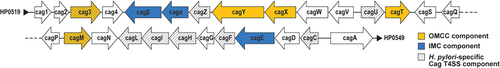
In most cag PAI-positive H. pylori strains, the entire 40-kb cag PAI is localized between a Sel1-like gene (HP0519 in prototype strain 26695) and a gene encoding glutamate racemase (glr, corresponding to HP0549 in strain 26695). The gene content and gene order within the cag PAI are relatively well-conserved among unrelated H. pylori strainsCitation9 (). Variations can result from gene deletions, gene insertions (sometimes associated with IS605 or IS606 elements), genomic rearrangements, or gene inversions. In some strains, fragments of the cag PAI are distributed in separate chromosomal loci.Citation7,Citation8
Figure 1. Organization of genes within the cag PAI. Five genes encoding proteins localized to the T4SS OMCCCitation10,Citation11 and three genes encoding putative ATPases localized to the T4SS IMC are indicated. Genes required for Cag T4SS activity that lack homologs in other bacterial species are indicated with diagonal stripes.
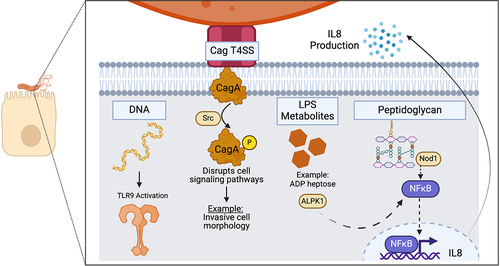
Early studies noted that one gene within the cag PAI encodes an immunodominant antigenic protein (CagA) recognized by human serum antibodies.Citation12,Citation13 Subsequent studies showed that CagA is a secreted effector protein delivered into host cells by a type IV secretion system (Cag T4SS), components of which are encoded by genes within the cag PAI.Citation14-19 The Cag T4SS also delivers several types of non-protein substrates into host cells ().Citation20 Epidemiologic studies, coupled with cell biology and animal model experiments, have demonstrated important roles of CagA and the Cag T4SS in the pathogenesis of both gastric cancer and peptic ulcer disease, and recent studies have provided important insights into functional and structural properties of CagA and the Cag T4SS. In this review, we provide an overview of CagA and the Cag T4SS, and we discuss mechanisms by which products of the cag PAI contribute to the pathogenesis of gastric cancer and peptic ulcer disease.
Figure 2. Cag T4SS-mediated delivery of CagA and non-protein substrates into host cells. The Cag T4SS is required for delivery of CagA, LPS metabolites, peptidoglycan and DNA into host cells. Each substrate elicits a cellular response. CagA is phosphorylated by tyrosine kinases (such as Src), and phosphorylated CagA can cause disruptions to a variety of signaling pathways. LPS metabolites and peptidoglycan elicit NF-κB activation, leading to IL-8 production. DNA translocation causes TLR9 activation. Created with BioRender.com.
Epidemiologic links between cag PAI-positive H. pylori strains and gastric disease
Early serologic studies noted that serum antibodies or gastric mucosal IgA antibodies to CagA were detected more commonly in individuals with peptic ulcer disease than in H. pylori-positive control patients without ulcers or H. pylori-negative individuals.Citation12,Citation13,Citation21–24 Serum antibodies to CagA are also detected more commonly in individuals with gastric adenocarcinoma or gastric premalignant conditions (such as atrophic gastritis or intestinal metaplasia) than in H. pylori-positive control patients or H. pylori-negative individuals.Citation25-34
The presence of anti-CagA serum antibodies is correlated with the presence of cagA-positive H. pylori strains in the stomach.Citation23 Accordingly, genetic analyses of H. pylori strains or gastric tissue samples have shown that cagA-positive strains are detected more commonly in individuals with gastric adenocarcinoma or premalignant lesions than in H. pylori-positive individuals with non-atrophic gastritis only.Citation35-46 In a large study of 2145 patients from Venezuela, there was a strong association between the presence of cagA-positive strains and premalignant gastric lesions.Citation36 Specifically, the odds ratio for gastric dysplasia (a statistical assessment of relative risk) was 15.5 (95% confidence interval 6.42 to 37.2) in individuals colonized with cagA-positive strains compared with H. pylori-negative individuals, and 0.90 (95% confidence interval 0.37 to 2.17) in individuals colonized with cagA-negative strains compared with H. pylori-negative individuals. The proportion of cagA-positive H. pylori strains is also higher among individuals with duodenal or gastric ulcers than among individuals with non-atrophic gastritis,Citation37,Citation41,Citation47-50 especially if cases of ulcers caused by non-steroidal anti-inflammatory drugs are excluded. Similarly, the severity of gastric inflammation is typically higher among individuals colonized with cagA-positive strains than among those colonized with cagA-negative strains.Citation40,Citation51,Citation52
The detection of cagA in H. pylori strains or gastric samples suggests that the cag PAI is present, but further analyses of H. pylori strains, such as whole genome sequencing or functional assays to assess T4SS-dependent phenotypes, are required to verify the presence of an intact cag PAI. Genetic detection of an “empty site locus” in the H. pylori chromosomal region between HP0519 (sel1-like gene) and HP0549 (glr) can provide evidence that the cag PAI is absent.Citation53 Most analyses of H. pylori strains or clinical samples have assessed the presence or absence of cagA in relation to gastric disease states, without assessing whether an intact cag PAI is present. Notably, a genome-wide association study of 173 H. pylori isolates from European patients with defined disease strains demonstrated an association between multiple genes in the cag PAI and gastric cancer.Citation54
Isolation of H. pylori strains from the stomach for genetic analysis or detection of cag PAI genes in gastric specimens typically requires sampling of the stomach using endoscopic biopsies, which evaluate only small portions of the stomach. Therefore, CagA serologic tests are potentially more sensitive methods for detecting cagA-positive H. pylori strains, compared to genetic methods. The detection of anti-CagA antibodies can reflect either active H. pylori colonization or previous colonization.Citation30
Most studies analyzing relationships between gastric disease states and CagA (or the cag PAI) have been cross-sectional or case-control analyses, comparing groups of symptomatic patients with different disease states who underwent upper gastrointestinal endoscopy. Further insights have come from studies of serum samples collected decades prior to the development of gastric disease. Analysis of the stored serum samples demonstrated an association between CagA seropositivity and the subsequent development of gastric cancer.Citation31 Additional insights have come from the analysis of serial gastric biopsies collected over time. In a large prospective study of Venezuelan patients (mean follow-up 3.5 years), gastric biopsies were analyzed to detect progression or regression of premalignant lesions.Citation36 Individuals colonized with cagA-positive H. pylori strains were more likely to exhibit progression of premalignant lesions than those colonized with cagA-negative strains, but the differences were not statistically significant. Similarly, a longitudinal study of patients in Spain showed that colonization with cagA-positive strains was associated with progression of preneoplastic lesions.Citation44
Most studies demonstrating a positive correlation between CagA or the cag PAI and disease states have been conducted in geographic regions where both cagA-positive and cagA-negative H. pylori strains are commonly isolated. Such relationships have been less frequently detected in East Asia or other geographic regions where nearly all H. pylori isolates are cagA-positive.Citation55,Citation56
Individuals with a history of duodenal ulcer disease have a reduced incidence of gastric cancer compared to matched control patients without a history of duodenal ulceration.Citation57 Therefore, the association of cag PAI-positive strains with an increased risk of both gastric cancer and duodenal ulcer disease is somewhat surprising. One possible explanation is that the cag PAI contributes to the pathogenesis of both diseases (for example, by stimulating gastric mucosal inflammation), and host-specific traits related to levels of gastric acid production determine whether an individual is predisposed to develop duodenal ulceration or gastric cancer.
Properties of the CagA effector protein
CagA is an immunodominant H. pylori protein that was originally identified based on its antigenic properties.Citation12,Citation13,Citation21,Citation22 CagA is recognized by both human serum antibodies and gastric mucosal IgA antibodies.Citation12,Citation13,Citation21,Citation22 The molecular masses of CagA proteins produced by unrelated H. pylori strains range from about 120 kDa to 150 kDa. The sequence of CagA does not exhibit relatedness to sequences of proteins in other bacterial species. The structure of the amino-terminal portion of CagA (residues 1–829) has been determined by X-ray crystallography,Citation58,Citation59 and three structurally distinguishable domains within this portion of CagA have been described. The relatively unstructured carboxy-terminal portion of CagA contains motifs important for CagA activity and a C-terminal secretion signal, discussed further in subsequent sections.
The first evidence for CagA entry into host cells came from experiments in which gastric epithelial cells were co-cultured with cagA-positive H. pylori strains. A tyrosine-phosphorylated ~130 kDa band was detected in lysates of the co-culture mixtures but not in lysates of uninfected gastric cells, and this band was shown to be a tyrosine-phosphorylated form of CagA.Citation14-19 Subsequent studies have detected CagA entry into host cells using a translocation reporter assay in which β-lactamase (TEM-1) is fused to CagA,Citation60 or by use of a split luciferase (HiBiT) translocation reporter assay.Citation61 Upon delivery into gastric cells, CagA localizes to the inner leaflet of the plasma membrane in a multimeric state.Citation62,Citation63
Tyrosine phosphorylation of CagA within host cells occurs on tyrosine residues within CagA EPIYA motifs (glutamate-proline-isoleucine-tyrosine-alanine), located within the C-terminal unstructured portion of CagA,Citation64,Citation65 and is mediated by tyrosine kinases (including c-Src, Fyn, Lyn, YES, and Abl).Citation66,Citation67 Tyrosine-phosphorylated CagA can interact with a large number of intracellular proteins, resulting in alterations of protein function.Citation68-70 Non-phosphorylated CagA can also interact with host cell proteins and alter their activity.Citation71-75 The interactions involving non-phosphorylated CagA are mediated by one or more sites designated “conserved repeat responsible for phosphorylation-independent activity” (CRPIA) motifs, located within the C-terminal unstructured portion of CagA.Citation73
Unrelated H. pylori strains contain variable numbers and types of CagA EPIYA motifs. Different EPIYA motifs are selectively phosphorylated by different kinases in a stepwise process; therefore, CagA is phosphorylated on only one or two EPIYA motifs.Citation76 Variation among H. pylori strains in the types of EPIYA motif sequences, the number of copies of EPIYA motifs, and the arrangement of the motifs contributes to variation in CagA activity among strains in vitro.Citation77,Citation78 Moreover, differences among strains in the number and type of EPIYA motifs have been correlated with differences in gastric disease risk.Citation79 Geographic variations in CagA EPIYA motifs and the associated impacts on CagA activity and gastric disease are discussed further in a subsequent section.
Properties of the Cag T4SS
At least 16 genes within the H. pylori cag PAI are required for delivery of CagA into host cells.Citation19,Citation20,Citation80 Several of these genes encode proteins exhibiting sequence relatedness to the components of T4SSs in other bacterial species. T4SSs are a versatile group of nanomachines that can transport an assortment of substrates, including protein and DNA.Citation81–84 T4SSs are widespread among both Gram-negative and Gram-positive bacterial species and are also present in Archaea. Two of the most common actions of T4SSs are horizontal transfer of DNA among bacteria (conjugation) and delivery of effector proteins into target cells.Citation81-84
Prototype T4SSs in Gram-negative bacteria (conjugation systems and the Agrobacterium tumefaciens VirB/VirD4 system) are composed of 12 protein components, designated VirB1–11 and VirD4.Citation81-84 Most of these components are organized into two large subassemblies known as the outer membrane core complex (OMCC) and inner membrane complex (IMC).Citation84-86 The OMCC is localized within the periplasm and includes proteins that interact with the outer membrane and/or inner membrane. The IMC spans the inner membrane and includes proteins projecting into the cytoplasm and periplasm. Three protein components (VirB7, VirB9, and VirB10) compose the OMCC in prototype T4SSs. The IMC is composed of three ATPases (VirB4, VirB11, and VirD4), along with VirB3 and VirB8. Additional proteins (VirB5 and VirB6) localize to a stalk connecting the OMCC and IMC.Citation86 An extracellular pilus structure (composed of two protein components, VirB2, and VirB5) is present in T4SSs from some bacterial species.Citation84
The overall structural organization of the H. pylori Cag T4SS has multiple features resembling those of prototype T4SSs, including the presence of an OMCC and IMCCitation85 (). Cryo-electron tomography (cryo-ET) analyses of intact H. pylori has allowed visualization of these Cag T4SS subassemblies in situ (in intact bacteria).Citation87,Citation88 The Cag T4SS OMCC is a large mushroom-shaped complex localized between the inner membrane and the outer membrane. The portion of the OMCC visualized by cryo-ET has 14-fold symmetry.Citation87,Citation88 The IMC has 6-fold symmetry and consists of three concentric rings surrounding a central channel.Citation88 The OMCC and IMC are connected by a stalk region. Other Cag T4SS-associated features visualized by cryo-ET (with low resolution) include periplasmic elements designated as “wings” or a “collar”.Citation87,Citation88 These probably correspond to regions of prototype T4SSs designated as “arches”.Citation86
Figure 3. Structural organization of the Cag T4SS OMCC. The 14-fold-symmetric OMC and 17-fold-symmetric PR are illustrated.Citation10,Citation11 (a, b, c) conserved Cag T4SS components. Purple = CagY, yellow = CagX, pink = CagT. (d, e, f) H. pylori-specific T4SS components. Blue = Cag3, green = CagM.
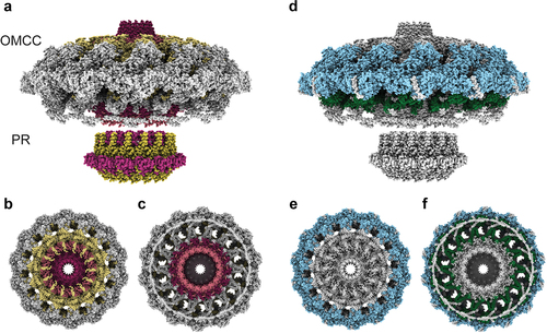
The H. pylori Cag T4SS OMCC remains intact in the presence of detergent, which has facilitated isolation and detailed structural analysis of this subassembly, using single-particle cryo-electron microscopy (cryo-EM).Citation10,Citation11,Citation89 The OMCC is about 41 nm in diameter and is composed of three main subassemblies: an outer membrane cap (OMC), a periplasmic ring (PR) and a stalk. The OMC has 14-fold symmetry and contains 5 proteins (CagY, CagX, CagT, CagM, and Cag3) in a 1:1:2:2:5 stoichiometric ratio.Citation10,Citation11 In total, the OMC portion of the OMCC contains 154 polypeptide chains.Citation11 The periplasmic ring (PR) has 17-fold symmetry and contains only CagX and CagY.Citation11 CagY, CagX, and CagT are homologous to VirB10, VirB9 and VirB7 components of T4SSs in other bacterial species, whereas CagM and Cag3 are species-specific components of the Cag T4SS. CagY (like VirB10 components in other bacterial T4SSs) is predicted to span from the outer membrane to the inner membrane,Citation90 but thus far, only the C-terminal portion of CagY has been structurally defined. Both cryo-ET analysis of intact H. pylori and cryo-EM analysis of isolated OMCCs have detected a stalk-like structure that connects the OMCC with the IMC,Citation10,Citation88 but the molecular composition of the stalk has not yet been defined.
A high-resolution structural model is not yet available for the Cag T4SS IMC, but insights into its composition have been provided by cryo-ET analysis of the T4SS in intact bacteria,Citation88 combined with comparisons to the structural organization of the IMC of a conjugation system.Citation86 Similar to prototypical T4SSs, the Cag T4SS IMC is predicted to contain three putative ATPases (Cagα, Cagβ, CagE), corresponding to VirB11, VirD4, and VirB3/VirB4, respectively. Each of these putative ATPases is required for CagA secretion.Citation19,Citation91 Cryo-ET analysis of H. pylori mutant strains with deletions of genes encoding individual ATPases revealed that CagE constitutes the IMC density closest to the inner membrane, followed by Cagα, and then Cagβ, which makes up the cytoplasmic density furthest from the inner membrane.Citation88 The Cag T4SS IMC contains additional densities that were unable to be assigned to the three ATPases. These might correspond to CagV (a VirB8 homolog) and CagU, which are predicted components of the IMC.
Among the cag PAI-encoded proteins required for CagA translocation, five are components of the OMCCCitation10,Citation11 and at least five are known or predicted to be components of the IMCCitation88 (). The localization of several species-specific Cag proteins required for CagA translocation (lacking sequence relatedness to T4SS components in prototype systems) remains unclear. CagF is proposed to be a cytoplasmic protein, functioning as a CagA chaperone, but it might also be associated with the inner membrane.Citation92-94 CagW (a VirB6 homolog) might be a component of the stalk, based on the observed localization of VirB6 to the stalk in a conjugation system.Citation86 CagH, CagI, and CagL physically interact to form a complex,Citation95,Citation96 but the subcellular localization of these proteins remains unclear. CagC, CagL, and CagI have been detected on the surface of H. pylori,Citation97-99 and it has been proposed that these proteins might interact with receptors on host cells.
Table 1. cag PAI-encoded proteins that contribute to Cag T4SS activity.
T4SSs in some bacterial species include extracellular pilus structures, composed of VirB2 and VirB5 components.Citation84 It has been proposed that H. pylori CagC and CagL might be VirB2- and VirB5-like components, respectively,Citation98,Citation100 but there is no sequence or structural relatedness between H. pylori CagL and VirB5 proteins from other bacterial species. Several papers have reported the production of extracellular pilus-like structures by H. pylori,Citation97,Citation101 but the relationship between these structures and the Cag T4SS remains unclear. In a cryo-ET analysis of H. pylori co-cultured with AGS gastric cells, the pilus-like structures were described as “membranous tubes with lateral ports”.Citation87 These pilus- or tube-like structures were never visualized in association with the Cag T4SS OMCC, and similar outer membrane protrusions, unrelated to T4SSs, have been visualized in many bacterial species.Citation102
In comparison to prototype T4SSs (classified as minimized T4SSs), the Cag T4SS is much larger in size. For example, the diameter of the Cag T4SS OMCC (approximately 41 nm) is nearly double the diameters of OMCCs in prototypical T4SSs.Citation85 Among the cag PAI genes required for delivery of CagA into host cells, 10 (CagY, CagX, CagT, Cagα, Cagβ, CagE, CagV, CagW, Cag4, and CagC) exhibit relatedness to VirB/VirD4 proteins in prototype systems, and 8 (Cag3, CagM, CagH, CagI, CagL, CagF, CagU, and CagZ) are found uniquely in H. pylori (). Therefore, the H. pylori Cag T4SS has been classified as an “expanded T4SS”, along with the Legionella pneumophila Dot/Icm T4SS and the Coxiella burnetii T4SS.Citation82,Citation84,Citation85
In addition to the classification of T4SSs into two groups (minimized and expanded) based on the number of components and physical size of the membrane-spanning structures, T4SSs have been classified into two groups (type IVA and type IVB) based on phylogenetic analysis of conserved components.Citation103,Citation104 Most type IVA systems, including the prototypical Agrobacterium tumefaciens VirB/VirD4 system, contain the minimum 12 proteins required for T4SS function, whereas type IVB systems, such as the Legionella Dot/Icm T4SS and the Coxiella T4SS, contain multiple additional species-specific components. Despite having limited sequence homology to the VirB/VirD4 system and containing multiple species-specific components, the H. pylori Cag T4SS has been classified as a type IVA secretion system.Citation103
Cag T4SS-mediated delivery of CagA into host cells
H. pylori contact with gastric epithelial cells triggers CagA secretion and delivery into host cells. In contrast, CagA is not secreted into the extracellular milieu if H. pylori is cultured in vitro under routine conditions. The stimuli that trigger CagA secretion have not yet been defined. The carboxy-terminal portion of H. pylori CagA contains a 20-amino-acid secretion signal, similar to secretion signals found in T4SS effector proteins in other bacterial species.Citation105 Both the carboxy-terminal motif and a large ~ 350-amino-acid segment within the amino-terminal portion of CagA (Domains I and II) are required for CagA secretion.Citation94 Current evidence suggests that CagA is secreted in an unfolded state. Specifically, a CagA-dihydrofolate reductase (DHFR) fusion protein is unable to be translocated when DHFR folding is stabilized with methotrexate.Citation61 Similarly, a CagA-GFP fusion protein is not delivered into host cells and can exert a dominant-negative inhibitory effect on secretion of wild-type CagA.Citation105
CagA physically interacts with CagF, a H. pylori-specific protein proposed to be a cytoplasmic chaperone that stabilizes CagA prior to its recruitment to the T4SS apparatus.Citation92-94 Cagβ (a VirD4 ATPase homolog) is predicted to be required for CagA recruitment to the T4SS apparatus, based on the role of VirD4 as a coupling protein in prototypical T4SSs. Cagβ interacts with a species-specific Cag protein known as CagZ.Citation106 It has been proposed that Cagβ does not incorporate into the IMC when bound to CagZ.Citation106 The stimuli promoting CagA binding or release from CagF, CagZ binding or release from Cagβ, and CagA recruitment to the T4SS apparatus have not yet been defined.
The precise mechanisms by which CagA is transported through the T4SS apparatus are not known. Yeast two-hybrid experiments and other in vitro analyses suggest that CagA can interact not only with CagF and Cagβ, but also with multiple additional T4SS components.Citation107,Citation108 In addition to Cagβ, two other putative ATPases (Cagα and CagE) are required for CagA delivery into host cells.Citation19,Citation91 The specific secretory steps powered by these individual ATPases are not known.
Interactions between the Cag T4SS and the surface of host cells are poorly understood. Several Cag proteins (including CagC, which exhibits weak sequence relatedness to VirB2 pilins)Citation98 are reported to be present on the H. pylori surface, and these can potentially interact with host cells. CagL, CagI, and CagY have been reported to interact with various integrins.Citation97,Citation109,Citation110 These interactions are presumed to be important in mediating interactions between the T4SS and host cells. Integrins are localized to the basolateral surface of gastric epithelial cells and are predicted to be inaccessible to H. pylori bound to the apical surface of gastric epithelial cells.Citation111 Experiments with polarized epithelial monolayers indicate that the activity of H. pylori proteases such as HtrA promotes H. pylori access to the basolateral surface of epithelial cells, thereby facilitating interactions of Cag T4SS components with integrins.Citation111,Citation112
The mechanisms by which CagA is translocated across the plasma membrane of host cells also remain poorly understood. Thus far, there is not any evidence indicating that Cag T4SS components directly insert into the plasma cell of host cells (analogous to the translocon of type III secretion systems). Therefore, CagA might enter host cells through endocytic processes. CagA can physically interact with β1 integrin,Citation59,Citation109 which provides a potential route for CagA binding and entry into host cells. As an additional or alternate route for CagA entry into host cells, H. pylori contact with epithelial cells induces externalization of phosphatidylserine to the outer leaflet of the plasma membrane, and CagA can interact with phosphatidylserine.Citation113
Intracellular actions of CagA
Early studies noted that the entry of CagA into gastric epithelial cells was associated with an alteration of cellular morphology known as the “hummingbird phenotype”, characterized by an elongation of cell shape and cytoskeletal alterations.Citation14 Subsequent studies showed that CagA causes numerous additional alterations in host cells, including loss of cell polarity,Citation114 increased cell motility,Citation75,Citation115,Citation116 cell scattering,Citation71,Citation115,Citation117 cell proliferation,Citation71,Citation73 cell invasiveness,Citation114 an epithelial to mesenchymal transition-like phenotype,Citation114 disruption of intercellular junctions, and disruption of epithelial barrier functions.Citation118 These cellular changes are the consequences of CagA interactions with multiple intracellular proteins. CagA (in either phosphorylated or non-phosphorylated forms) is reported to interact with at least a dozen proteins in host cells, leading to cellular alterations that are relevant for oncogenesis (discussed in previous reviews).Citation68,Citation69 Several examples are presented here.
One of the most extensively studied CagA interactions is the binding of tyrosine-phosphorylated CagA to Src homology region 2 (SH2)-containing protein tyrosine phosphatase 2 (SHP2).Citation119 Under physiologic conditions, SHP2 is in an enzymatically inactive conformation. The interaction of phosphorylated CagA EPIYA motifs with SHP2 triggers a conformational change, resulting in an active form of SHP2 that stimulates signaling pathways involved in cell morphology, motility, and proliferation.Citation68 Phosphorylated CagA interacts with Src-homology 2 domains in several additional proteins, including C-terminal SRC kinase (CSK).Citation120 CagA interactions with SHP2 contribute to the “hummingbird phenotype” observed following H. pylori co-culture with gastric epithelial cells.Citation119
Another extensively studied CagA interaction is its binding to the partitioning defective 1 (PAR1) family of serine/threonine kinases [known as microtubule affinity-regulating kinases (MARKs)], which are important in maintaining the polarization of epithelial cells and tight junctions, as well as microtubule dynamics.Citation69,Citation74,Citation121 CagA interactions with PAR1/MARK proteins require the CagA multimerization (CM) motif but are not dependent on the phosphorylation state of CagA.Citation69,Citation74,Citation121 Inhibition of PAR1 function by CagA contributes to the hummingbird phenotypeCitation122 and results in mislocalization of tight junction proteins (e.g., ZO-1) and basolateral proteins (such as E-cadherin), leading to defects in cell polarity and impaired tight junction barriers.Citation74
CagA also interacts with E-cadherin (a component of adherens junctions).Citation123,Citation124 This interaction results in the destabilization of the E-cadherin/β-catenin complex, translocation of β-catenin to the nucleus,Citation125 and the activation of Wnt signaling. CagA may also activate Wnt signaling through additional mechanisms.Citation68,Citation126
CagA interacts with apoptosis-stimulating protein of p53 (ASPP2),Citation127 which promotes the proteasomal degradation of the tumor suppressor p53.Citation128 The interaction of CagA with ASPP2 leads to inhibition of apoptosis (resistance to cell death). CagA can also negatively regulate p53 through additional mechanisms.Citation129,Citation130
Another important consequence of CagA intracellular activity is stimulation of DNA damage and double-strand DNA breaks in host cells.Citation131-133 One mechanism involves inactivation of PAR1b, leading to alteration of PAR1b-dependent BRCA1 phosphorylation and impaired nuclear localization of BRCA1.Citation134 Another mechanism involves upregulation of spermine oxidase and resulting oxidative stress.Citation131 CagA is also implicated in the downregulation of several genes involved in DNA repair.Citation133,Citation135
Cag T4SS-mediated delivery of non-protein substrates into host cells
Early studies noted that co-culture of cag PAI-positive H. pylori strains with gastric epithelial cells stimulated the production and secretion of interleukin-8 (IL-8), a proinflammatory cytokine that promotes recruitment and activation of neutrophils.Citation7,Citation8,Citation19,Citation136 This phenotype was dependent on multiple cag PAI genes encoding Cag T4SS components but did not require CagA.Citation7,Citation8,Citation19,Citation136 The IL-8 phenotype, a consequence of NF-κB activation, is now known to result primarily from the entry of non-protein H. pylori substrates into host cells. Although CagA is not required for IL-8 induction,Citation19 CagA can contribute to the capacity of some H. pylori strains to stimulate IL-8 production in gastric epithelial cells.Citation137
Mutagenesis of several H. pylori genes required for LPS inner core heptose biosynthesis (gmhA, hldE, and rfaE) leads to a marked reduction in the capacity of H. pylori to stimulate NF-κB activation and IL-8 production.Citation138,Citation139 In contrast, these mutations do not inhibit T4SS-mediated delivery of CagA into host cells. These findings suggested that H. pylori LPS intermediates might be mediators of the IL-8 phenotype. Initial studies concluded that H. pylori heptose 1,7-bisphosphate (HBP) was the relevant pathogen-associated molecular pattern (PAMP),Citation138-140 similar to what had been reported previously in studies of other bacterial species.Citation141-143 Subsequent studies found that H. pylori lysates contain very low concentrations of heptose 1,7-bisphosphate (HBP) and identified ADP-glycero-β-D-manno-heptose (ADP heptose), a derivative of HBP, as a more active H. pylori PAMP.Citation144 Cag T4SS-dependent activation of NF-κB and IL-8 production by LPS metabolites is a consequence of the activation of alpha kinase 1 (ALPK1), which activates the TRAF-interacting protein with forkhead domain (TIFA).Citation139,Citation145 TIFA then forms large complexes (TIFAsomes) composed of TIFA and other cellular proteins, including TRAF2, leading to the activation of NF-κB.Citation145
Co-culture of H. pylori with gastric epithelial cells results in activation of Nod1 through a Cag T4SS-dependent process, which provides an additional mechanism for NF-κB activation and IL-8 production.Citation146 Peptidoglycan from many bacterial species is known to be a stimulus for Nod1 activation. In the case of H. pylori, meso-diaminopimelate (mDAP)-containing N-acetylglucosamine-N-acetylmu-ramic acid (GM-tripeptide) is the peptidoglycan moiety that is specifically recognized by Nod1. The results of one study suggested that Nod1 activation has a minimal role in IL-8 activation compared to the activation of the TIFA pathway.Citation139
Another consequence of H. pylori co-culture with gastric epithelial cells is the activation of Toll-like receptor 9 (TLR9).Citation147 This phenotype is dependent on Cag T4SS activity and is attributed to the entry of H. pylori DNA into host cells.Citation147,Citation148 TLR9 receptors recognize unmethylated CpG motifs on DNA, which are predominantly found on bacterial or viral DNA but not mammalian DNA. Activation of TLR9 may lead to a dampening of the inflammatory response by suppressing IL-17-mediated responses.Citation149 The anti-inflammatory effect of TLR9 activation contrasts with the pro-inflammatory effects resulting from entry of LPS metabolites and peptidoglycan into host cells and might promote persistent H. pylori colonization.
Relatively little is known about the T4SS-dependent processes by which non-protein substrates are delivered into host cells. In contrast to CagA secretion, which requires three ATPases (CagE, Cagα, and Cagβ), delivery of non-protein substrates into host cells requires CagE and Cagα but not Cagβ.Citation19,Citation91 Similarly, CagF is required for CagA secretion but not delivery of non-protein substrates.Citation92,Citation93
To systematically identify H. pylori genes required for T4SS-dependent processes, one study screened a H. pylori transposon mutant library to identify mutants unable to activate NF-κB in gastric epithelial cells.Citation150 As expected, this analysis identified numerous cag PAI genes and also identified three non-cag PAI genes: hopQ (which encodes an outer membrane protein), a gene encoding a predicted LPS glycosyltransferase (HP0159), and a gene encoding a predicted flagellar-associated protein (HP1029/1028). Subsequent studies showed that HopQ is an outer membrane protein that interacts with carcinoembryonic antigen-related cell adhesion molecules (CEACAMs) on host cells, thereby promoting T4SS-mediated delivery of substrates into host cells.Citation151-153
Two additional H. pylori outer membrane proteins (BabA and AlpA/B) are reported to contribute to Cag T4SS-dependent processes.Citation154,Citation155 Other non-cag PAI genes reported to contribute to T4SS-dependent processes include hyd (encoding hydrogenase) and HP1564 (encoding a protein of unknown function).Citation156,Citation157
In addition to cellular responses resulting from T4SS-dependent entry of CagA and non-protein substrates into host cells, there is evidence that components of the T4SS can directly cause cellular responses.Citation158 One mechanism involves interactions of Cag proteins with Toll-like receptor 5 (TLR5), a receptor that typically recognizes bacterial flagellins. H. pylori flagellin is adapted to avoid recognition by TLR5,Citation159,Citation160 but two T4SS components (CagL and CagY) can directly interact with TLR5 and activate this receptor, leading to downstream signaling that triggers the production of specific cytokines and chemokines.Citation161,Citation162
In summary, CagA is the only protein known to be secreted and translocated by the Cag T4SS. The Cag T4SS can also deliver multiple types of non-protein substrates into host cells, resulting in proinflammatory signaling (ADP-heptose and peptidoglycan) or anti-inflammatory signaling (DNA). Unusual features of the Cag T4SS include its capacity to trigger cellular alterations through the properties of T4SS components, independent of the translocation of the effector molecule.
Activities of CagA and the T4SS in animal models
Several transgenic animal models have been used to evaluate the consequences of intracellular CagA activity in vivo. Transgenic mice engineered to express CagA developed gastric epithelial hyperplasia, gastric polyps, adenocarcinomas of the stomach and small intestine, myeloid leukemias, and B cell lymphomas.Citation163 Similarly, transgenic zebrafish expressing CagA developed hyperplasia of the adult intestinal epithelium.Citation164 Intestinal hyperplasia was detected in zebrafish following long-term transgenic expression of wild-type CagA, but not a phosphorylation-resistant form of CagA.Citation164 Transgenic expression of CagA in a Drosophila model resulted in an assortment of abnormalities in morphogenesis, including alterations in ocular photoreceptor development.Citation165,Citation166 Expression of CagA within Drosophila intestinal stem cells promoted excess cell proliferation and led to alterations in host microbiota.Citation167 Ectopic expression of CagA in Xenopus laevis embryos resulted in impaired gastrulation, neural tube formation, and axis elongation.Citation126 Therefore, transgenic CagA expression in vivo results in extensive cellular alterations and oncogenic effects, consistent with studies of CagA action in vitro. Notably, transgenic expression of CagA in mice did not lead to a prominent inflammatory response.Citation163
Experimental intragastric administration of H. pylori to mice results in H. pylori colonization of the stomach and detectable gastric inflammation, but gastric ulceration and gastric cancer do not develop in wild-type mice infected with H. pylori. H. pylori colonization of the mouse stomach does not require the cag PAI, and during colonization of the mouse stomach, cag PAI-positive H. pylori strains commonly acquire mutations leading to inactivation of Cag T4SS function.Citation168 Moreover, mouse gastric epithelial cells are resistant to the actions of CagA due to the inability of HopQ to bind to mouse CEACAMs.Citation169 The loss of Cag T4SS activity in vivo, combined with resistance of mouse cells to CagA activity, may account at least in part for the absence of gastric cancer or gastric ulceration in H. pylori-infected wild-type mice.
The apparent selective advantage of strains lacking Cag T4SS function in mice complicates efforts to study a potential contribution of the cag PAI to gastric inflammation or gastric disease in wild-type mouse models. Nevertheless, several studies have reported that CagA or the T4SS contribute to gastric inflammation and gastric disease in wild-type mice.Citation170,Citation171 Two studies have shown that the activation of gastric stem cell populations (Lgr5- or Lrig-1-positive cells) is dependent on Cag T4SS activity.Citation171,Citation172 In a transgenic hypergastrinemic mouse model of gastric carcinogenesis (INS-GAS), there was a trend toward delayed development of gastric cancer in animals infected with a cagE mutant strain compared to a wild-type strain.Citation173
Administration of cag PAI-positive H. pylori to Mongolian gerbils commonly results in severe gastric inflammation, often accompanied by gastric ulceration, premalignant changes, and gastric adenocarcinoma.Citation174-183 In contrast, cagA mutant strains and mutant strains defective in Cag T4SS activity cause only mild gastric inflammation and do not cause ulceration or gastric cancer in the gerbil model.Citation174-181,Citation183 Similarly, an H. pylori strain in which expression of Cag T4SS components is controlled by the TetR/tetO system caused more severe gastric inflammation and disease under conditions in which the expression of relevant genes (cagU and cagT) was de-repressed than under conditions in which expression was repressed.Citation184 One study reported that the H. pylori colonization density was higher in gerbil stomachs colonized with wild-type H. pylori strains than in gerbil stomachs colonized with cagA mutants.Citation177
Non-human primate models have also been used to assess the effects of H. pylori CagA and the Cag T4SS in vivo. Rhesus macaques experimentally infected with a wild-type H. pylori strain developed increased gastric mucosal inflammation compared to animals infected with a cag PAI mutant strain.Citation185
Geographic variations in prevalence of cag PAI-positive strains and features of CagA EPIYA motifs
Estimates of the prevalence of cag PAI-positive H. pylori strains within populations have been based on analysis of symptomatic patients who underwent endoscopic procedures because of gastric symptoms. The results of such studies may not accurately reflect the prevalence of cag PAI-positive strains within asymptomatic populations. Nevertheless, the available data indicate that there are geographic variations in the prevalence of cag PAI-positive strains. Within the U.S. and Western Europe, the prevalence of cag PAI-positive strains and cag PAI-negative strains is similar. In contrast, >90% of H. pylori strains isolated in many parts of East Asia (including Japan, Korea, and parts of China) are cag PAI-positive.Citation55,Citation56 The predominance of cag PAI-positive strains in Japan, Korea, and parts of China correlates with a high rate of gastric cancer incidence in these geographic regions.Citation186
The factors that determine the relative abundance of cag PAI-positive strains within populations are not known. In populations with high levels of H. pylori transmission, human stomachs can be colonized by multiple H. pylori strains, potentially leading to competition and eventual selection of strains that have the highest level of fitness. cag PAI-positive strains might have a selective advantage compared to cag PAI-negative strains in such settings. Conversely, cag PAI-negative strains might have a selective advantage in settings with low levels of H. pylori transmission and acquisition. Geographic variations in human genetic characteristics, diet, or other environmental factors might also influence the relative abundance of cag PAI-positive strains within geographic regions.
H. pylori strains isolated in different parts of the world are genetically distinct and have been classified into several different groups based on multi-locus sequencing typing (MLST) or genome-based analyses.Citation187 For example, H. pylori strains isolated from East Asian, European, and African human populations can be readily differentiated by genetic analysis. Among genes in the cag PAI, cagA sequences exhibit the highest level of geographic diversity.Citation9,Citation188
Some of the most striking geographic differences in CagA sequences are variations in the number and type of EPIYA phosphorylation site motifs.Citation189 CagA proteins produced by East Asian strains contain a type of EPIYA phosphorylation site motif (EPIYA-D) not detected in CagA proteins in other parts of the world.Citation78,Citation190,Citation191 Conversely, EPIYA-C motifs are commonly detected in CagA proteins produced by Western strains but not in CagA proteins produced by East Asian strains.Citation78,Citation190,Citation191 In vitro studies indicate that East Asian CagA proteins harboring the EPIYA-D motif exhibit activities that are different from those of CagA proteins lacking this motif. Specifically, CagA proteins containing a phosphorylated EPIYA-D motif bind SHP-2 with markedly higher affinity than CagA proteins containing the phosphorylated Western EPIYA-C motif, resulting in increased cellular morphologic alterations.Citation78,Citation190,Citation191 In contrast to the properties of CagA from East Asian H. pylori strains, Cag proteins from Amerindian strains have a relatively low level of activity in vitro.Citation192
Geographic differences in the prevalence of cag PAI-positive H. pylori strains likely contribute to geographic differences in gastric cancer incidence. Similarly, geographic differences in the characteristics of CagA EPIYA motifs probably influence gastric cancer incidence.Citation79 Additional geographic variations in the properties of H. pylori strains influencing T4SS activity may also be relevant. For example, one study reported that the alpAB genes contribute to T4SS-dependent IL-8 induction in East Asian strains but not Western strains.Citation155
Summary and future directions
Colonization of the human stomach with cag PAI-positive strains is associated with an increased risk of gastric cancer and peptic ulcer disease, compared to colonization with cag PAI-negative strains. Similarly, experiments in multiple different animal models indicate that CagA and the Cag T4SS have important roles in the pathogenesis of H. pylori-induced gastric cancer and gastric ulceration. Experimental studies have revealed mechanisms by which proteins encoded by the cag PAI contribute to gastric disease.
Chronic inflammation promotes the development of cancer in multiple sites (for example, hepatocellular carcinoma associated with viral hepatitis, and colon cancer associated with inflammatory bowel disease). Therefore, chronic gastric mucosal inflammation stimulated by cag PAI-positive H. pylori, with associated DNA damage resulting from oxidative and nitrosative stress, is one of the important factors contributing to gastric cancer pathogenesis.Citation145,Citation193
CagA-induced alterations in cell signaling also contribute to gastric cancer pathogenesis. CagA-induced cellular alterations relevant for cancer pathogenesis include inhibition of apoptosis, stimulation of cell proliferation, degradation of the p53 tumor suppressor, and double-strand DNA breaks. Thus far, there has been relatively little progress in determining which types of cells in the gastric mucosa are targeted by CagA. Since differentiated superficial gastric epithelial cells are shed on a regular basis, CagA-induced alterations in these cells probably do not have a substantial impact on cancer pathogenesis unless CagA alters the behavior of these cells in a way that makes them resistant to shedding. Importantly, H. pylori can localize not only within the superficial gastric mucus layer, but also within gastric glands adjacent to gastric stem cells.Citation172 Targeting of gastric stem cells by CagA is presumed to have a key role in gastric cancer pathogenesis.Citation126,Citation171,Citation172
Premalignant changes in the gastric environment, such as atrophic gastritis or intestinal metaplasia, render the stomach a relatively inhospitable environment for H. pylori. Therefore, by the time gastric cancer develops, H. pylori may no longer be detectable in gastric tissues. Since H. pylori and products of the cag PAI are not required for the maintenance of a cancer phenotype, it has been proposed that H. pylori and CagA can cause genetic or epigenetic alterations that persist in cells after CagA is no longer present, consistent with a “hit-and-run mechanism”.Citation194
Thus far, there has been relatively little progress in determining how the presence of the cag PAI benefits H. pylori. Within the human stomach, cagA-positive strains achieve a higher density than cagA-negative strains,Citation195 and CagA has been reported to promote H. pylori survival in human gastric organoids.Citation75 A fitness advantage conferred by the cag PAI would be especially relevant in settings where cagA-positive and cagA-negative strains co-colonize and compete within human stomachs, and might also enhance H. pylori transmission. A mechanism by which the cag PAI contributes to H. pylori fitness has not been thoroughly defined, but evidence from one study suggested that CagA contributes to bacterial iron acquisition.Citation196 In support of this hypothesis, H. pylori strains retain Cag T4SS activity in mice fed low-iron diets, but not in mice fed regular diets.Citation197
We anticipate that continued studies of the cag PAI will lead to many important new discoveries that are relevant to human health and disease. For example, it will be important to further define mechanisms by which the Cag T4SS delivers multiple types of substrates into host cells and further define the cellular alterations that occur in response to these substrates. While multiple lines of evidence indicate that the cag PAI contributes to the pathogenesis of gastric disease, most individuals colonized with cag PAI-positive strains remain asymptomatic. Therefore, in future studies, it will be important to define more completely the multiple additional bacterial, host, and environmental factors that determine gastric disease risk, so that individuals with the highest gastric cancer risk can be targeted for therapeutic intervention.
Acknowledgments
This work was supported by NIH AI118932, AI039657, CA116087, T32 GM008320, and T32 AI112541, and the Department of Veterans Affairs (1I01BX004447).
Disclosure statement
No potential conflict of interest was reported by the author(s).
Additional information
Funding
References
- Hooi JKY, Lai WY, Ng WK, Suen MMY, Underwood FE, Tanyingoh D, Malfertheiner P, Graham DY, Wong VWS, Wu JCY. et al. Global prevalence of Helicobacter pylori infection: systematic review and meta-analysis. Gastroenterology. 2017;153(2):420–22. doi:10.1053/j.gastro.2017.04.022.
- Cover TL, Blaser MJ. Helicobacter pylori in health and disease. Gastroenterology. 2009;136(6):1863–73. doi:10.1053/j.gastro.2009.01.073.
- Malfertheiner P, Camargo MC, El-Omar E, Liou JM, Peek R, Schulz C, Smith SI, Suerbaum S. Helicobacter pylori infection. Nat Rev Dis Primers. 2023;9(1):19. doi:10.1038/s41572-023-00431-8.
- Suerbaum S, Josenhans C. Helicobacter pylori evolution and phenotypic diversification in a changing host. Nat Rev Microbiol. 2007;5(6):441–52. doi:10.1038/nrmicro1658.
- Cover TL. Helicobacter pylori diversity and gastric cancer risk. MBio. 2016;7(1):e01869–15. doi:10.1128/mBio.01869-15.
- Thorell K, Munoz-Ramirez ZY, Wang D, Sandoval-Motta S, Boscolo Agostini R, Ghirotto S, Torres RC, Hp GPRN, Falush D, Camargo MC. et al. The Helicobacter pylori genome project: insights into H. pylori population structure from analysis of a worldwide collection of complete genomes. Nat Commun. 2023;14(1):8184. doi:10.1038/s41467-023-43562-y.
- Censini S, Lange C, Xiang Z, Crabtree JE, Ghiara P, Borodovsky M, Rappuoli R, Covacci A. cag, a pathogenicity island of Helicobacter pylori, encodes type I-specific and disease-associated virulence factors. Proc Natl Acad Sci USA. 1996;93(25):14648–14653. doi:10.1073/pnas.93.25.14648.
- Akopyants NS, Clifton SW, Kersulyte D, Crabtree JE, Youree BE, Reece CA, Bukanov NO, Drazek ES, Roe BA, Berg DE. Analyses of the cag pathogenicity island of Helicobacter pylori. Mol Microbiol. 1998;28(1):37–53. doi:10.1046/j.1365-2958.1998.00770.x.
- Olbermann P, Josenhans C, Moodley Y, Uhr M, Stamer C, Vauterin M, Suerbaum S, Achtman M, Linz B. A global overview of the genetic and functional diversity in the Helicobacter pylori cag pathogenicity island. PloS Genet. 2010;6(8):e1001069. doi:10.1371/journal.pgen.1001069.
- Chung JM, Sheedlo MJ, Campbell AM, Sawhney N, Frick-Cheng AE, Lacy DB, Cover TL, Ohi MD. Structure of the Helicobacter pylori Cag type IV secretion system. eLife. 2019;8:e47644. doi:10.7554/eLife.47644.
- Sheedlo MJ, Chung JM, Sawhney N, Durie CL, Cover TL, Ohi MD, Lacy DB. Cryo-EM reveals species-specific components within the Helicobacter pylori Cag type IV secretion system core complex. Elife. 2020;9:e59495. doi:10.7554/eLife.59495.
- Tummuru MK, Cover TL, Blaser MJ. Cloning and expression of a high-molecular-mass major antigen of Helicobacter pylori: evidence of linkage to cytotoxin production. Infect Immun. 1993;61(5):1799–809. doi:10.1128/iai.61.5.1799-1809.1993.
- Covacci A, Censini S, Bugnoli M, Petracca R, Burroni D, Macchia G, Massone A, Papini E, Xiang Z, Figura N. et al. Molecular characterization of the 128-kDa immunodominant antigen of Helicobacter pylori associated with cytotoxicity and duodenal ulcer. Proc Natl Acad Sci USA. 1993;90(12):5791–5795. doi:10.1073/pnas.90.12.5791.
- Segal ED, Cha J, Lo J, Falkow S, Tompkins LS. Altered states: involvement of phosphorylated CagA in the induction of host cellular growth changes by Helicobacter pylori. Proc Natl Acad Sci USA. 1999;96(25):14559–14564. doi:10.1073/pnas.96.25.14559.
- Stein M, Rappuoli R, Covacci A. Tyrosine phosphorylation of the Helicobacter pylori CagA antigen after cag-driven host cell translocation. Proc Natl Acad Sci USA. 2000;97(3):1263–1268. doi:10.1073/pnas.97.3.1263.
- Odenbreit S, Puls J, Sedlmaier B, Gerland E, Fischer W, Haas R. Translocation of Helicobacter pylori CagA into gastric epithelial cells by type IV secretion. Science. 2000;287(5457):1497–1500. doi:10.1126/science.287.5457.1497.
- Asahi M, Azuma T, Ito S, Ito Y, Suto H, Nagai Y, Tsubokawa M, Tohyama Y, Maeda S, Omata M. et al. Helicobacter pylori CagA protein can be tyrosine phosphorylated in gastric epithelial cells. J Exp Med. 2000;191(4):593–602. doi:10.1084/jem.191.4.593.
- Backert S, Ziska E, Brinkmann V, Zimny-Arndt U, Fauconnier A, Jungblut PR, Naumann M, Meyer TF. Translocation of the Helicobacter pylori CagA protein in gastric epithelial cells by a type IV secretion apparatus. Cell Microbiol. 2000;2(2):155–64. doi:10.1046/j.1462-5822.2000.00043.x.
- Fischer W, Puls J, Buhrdorf R, Gebert B, Odenbreit S, Haas R. Systematic mutagenesis of the Helicobacter pylori cag pathogenicity island: essential genes for CagA translocation in host cells and induction of interleukin-8. Mol Microbiol. 2001;42(5):1337–48. doi:10.1046/j.1365-2958.2001.02714.x.
- Cover TL, Lacy DB, Ohi MD. The Helicobacter pylori Cag Type IV Secretion System. Trends Microbiol. 2020;28(8):682–695. doi:10.1016/j.tim.2020.02.004.
- Cover TL, Dooley CP, Blaser MJ. Characterization of and human serologic response to proteins in Helicobacter pylori broth culture supernatants with vacuolizing cytotoxin activity. Infect Immun. 1990;58(3):603–610. doi:10.1128/iai.58.3.603-610.1990.
- Crabtree JE, Taylor JD, Wyatt JI, Heatley RV, Shallcross TM, Tompkins DS, Rathbone BJ. Mucosal IgA recognition of Helicobacter pylori 120 kDa protein, peptic ulceration, and gastric pathology. Lancet. 1991;338(8763):332–335. doi:10.1016/0140-6736(91)90477-7.
- Cover TL, Glupczynski Y, Lage AP, Burette A, Tummuru MK, Perez-Perez GI, Blaser MJ. Serologic detection of infection with cagA+ Helicobacter pylori strains. J Clin Microbiol. 1995;33(6):1496–500. doi:10.1128/jcm.33.6.1496-1500.1995.
- Nomura AM, Perez-Perez GI, Lee J, Stemmermann G, Blaser MJ. Relation between Helicobacter pylori cagA status and risk of peptic ulcer disease. Am J Epidemiol. 2002;155(11):1054–1059. doi:10.1093/aje/155.11.1054.
- Crabtree JE, Wyatt JI, Sobala GM, Miller G, Tompkins DS, Primrose JN, Morgan AG. Systemic and mucosal humoral responses to Helicobacter pylori in gastric cancer. Gut. 1993;34(10):1339–43. doi:10.1136/gut.34.10.1339.
- Blaser MJ, Perez-Perez GI, Kleanthous H, Cover TL, Peek RM, Chyou PH, Stemmermann GN, Nomura A. Infection with Helicobacter pylori strains possessing cagA is associated with an increased risk of developing adenocarcinoma of the stomach. Cancer Res. 1995;55:2111–2115.
- Kuipers EJ, Perez-Perez GI, Meuwissen SG, Blaser MJ. Helicobacter pylori and atrophic gastritis: importance of the cagA status. J Natl Cancer Inst. 1995;87(23):1777–80. doi:10.1093/jnci/87.23.1777.
- Parsonnet J, Friedman GD, Orentreich N, Vogelman H. Risk for gastric cancer in people with CagA positive or CagA negative Helicobacter pylori infection. Gut. 1997;40(3):297–301. doi:10.1136/gut.40.3.297.
- Huang JQ, Sridhar S, Chen Y, Hunt RH. Meta-analysis of the relationship between Helicobacter pylori seropositivity and gastric cancer. Gastroenterology. 1998;114(6):1169–79. doi:10.1016/S0016-5085(98)70422-6.
- Ekstrom AM, Held M, Hansson LE, Engstrand L, Nyren O. Helicobacter pylori in gastric cancer established by CagA immunoblot as a marker of past infection. Gastroenterology. 2001;121(4):784–91. doi:10.1053/gast.2001.27999.
- Nomura AM, Lee J, Stemmermann GN, Nomura RY, Perez-Perez GI, Blaser MJ. Helicobacter pylori CagA seropositivity and gastric carcinoma risk in a Japanese American population. J Infect Dis. 2002;186(8):1138–44. doi:10.1086/343808.
- Huang JQ, Zheng GF, Sumanac K, Irvine EJ, Hunt RH. Meta-analysis of the relationship between cagA seropositivity and gastric cancer. Gastroenterology. 2003;125(6):1636–44. doi:10.1053/j.gastro.2003.08.033.
- Palli D, Masala G, Del Giudice G, Plebani M, Basso D, Berti D, Numans ME, Ceroti M, Peeters PH, de Mesquita HB B. et al. CagA+ Helicobacter pylori infection and gastric cancer risk in the EPIC-EURGAST study. Int J Cancer. 2007;120(4):859–67. doi:10.1002/ijc.22435.
- Shiota S, Matsunari O, Watada M, Yamaoka Y. Serum Helicobacter pylori CagA antibody as a biomarker for gastric cancer in east-Asian countries. Future Microbiol. 2010;5(12):1885–93. doi:10.2217/fmb.10.135.
- Figueiredo C, Machado JC, Pharoah P, Seruca R, Sousa S, Carvalho R, Capelinha AF, Quint W, Caldas C, van Doorn LJ. et al. Helicobacter pylori and interleukin 1 genotyping: an opportunity to identify high-risk individuals for gastric carcinoma. J Natl Cancer Inst. 2002;94(22):1680–1687. doi:10.1093/jnci/94.22.1680.
- Plummer M, van Doorn LJ, Franceschi S, Kleter B, Canzian F, Vivas J, Lopez G, Colin D, Munoz N, Kato I. Helicobacter pylori cytotoxin-associated genotype and gastric precancerous lesions. J Natl Cancer Inst. 2007;99(17):1328–1334. doi:10.1093/jnci/djm120.
- Basso D, Zambon CF, Letley DP, Stranges A, Marchet A, Rhead JL, Schiavon S, Guariso G, Ceroti M, Nitti D. et al. Clinical relevance of Helicobacter pylori cagA and vacA gene polymorphisms. Gastroenterology. 2008;135(1):91–9. doi:10.1053/j.gastro.2008.03.041.
- Queiroz DM, Mendes EN, Rocha GA, Oliveira AM, Oliveira CA, Magalhaes PP, Moura SB, Cabral MM, Nogueira AM. cagA-positive Helicobacter pylori and risk for developing gastric carcinoma in Brazil. Int J Cancer. 1998;78:135–139.
- Rugge M, Busatto G, Cassaro M, Shiao YH, Russo V, Leandro G, Avellini C, Fabiano A, Sidoni A, Covacci A. Patients younger than 40 years with gastric carcinoma: Helicobacter pylori genotype and associated gastritis phenotype. Cancer. 1999;85(12):2506–11. doi:10.1002/(SICI)1097-0142(19990615)85:12<2506:AID-CNCR3>3.0.CO;2-I.
- Zambon CF, Navaglia F, Basso D, Rugge M, Plebani M. Helicobacter pylori babA2, cagA, and s1 vacA genes work synergistically in causing intestinal metaplasia. J Clin Pathol. 2003;56(4):287–291. doi:10.1136/jcp.56.4.287.
- Oliveira AG, Santos A, Guerra JB, Rocha GA, Rocha AM, Oliveira CA, Cabral MM, Nogueira AM, Queiroz DM. babA2- and cagA-positive Helicobacter pylori strains are associated with duodenal ulcer and gastric carcinoma in Brazil. J Clin Microbiol. 2003;41(8):3964–3966. doi:10.1128/JCM.41.8.3964-3966.2003.
- Rocha GA, Guerra JB, Rocha AM, Saraiva IE, da Silva DA, de Oliveira CA, Queiroz DM. IL1RN polymorphic gene and cagA-positive status independently increase the risk of noncardia gastric carcinoma. Int J Cancer. 2005;115(5):678–683. doi:10.1002/ijc.20935.
- Quintero E, Pizarro MA, Rodrigo L, Pique JM, Lanas A, Ponce J, Mino G, Gisbert J, Jurado A, Herrero MJ. et al. Association of Helicobacter pylori-related distal gastric cancer with the HLA class II gene DQB1* 0602 and cagA + strains in a Southern European Population. Helicobacter. 2005;10(1):12–21. doi:10.1111/j.1523-5378.2005.00287.x.
- Gonzalez CA, Figueiredo C, Lic BC, Ferreira RM, Pardo ML, Ruiz Liso JM, Alonso P, Sala N, Capella G, Sanz-Anquela JM. Helicobacter pylori cagA and vacA genotypes as predictors of progression of gastric preneoplastic lesions: a long-term follow-up in a high-risk area in Spain. Am J Gastroenterol. 2011;106(5):867–874. doi:10.1038/ajg.2011.1.
- Matos JI, de Sousa HA, Marcos-Pinto R, Dinis-Ribeiro M. Helicobacter pylori CagA and VacA genotypes and gastric phenotype: a meta-analysis. Eur J Gastroenterol Hepatol. 2013;25(12):1431–1441. doi:10.1097/MEG.0b013e328364b53e.
- Pormohammad A, Ghotaslou R, Leylabadlo HE, Nasiri MJ, Dabiri H, Hashemi A. Risk of gastric cancer in association with Helicobacter pylori different virulence factors: a systematic review and meta-analysis. Microb Pathog. 2018;118:214–219. doi:10.1016/j.micpath.2018.03.004.
- van Doorn LJ, Figueiredo C, Sanna R, Plaisier A, Schneeberger P, de Boer W, Quint W. Clinical relevance of the cagA, vacA, and iceA status of Helicobacter pylori. Gastroenterology. 1998;115(1):58–66. doi:10.1016/S0016-5085(98)70365-8.
- Van Doorn LJ, Figueiredo C, Megraud F, Pena S, Midolo P, Queiroz DM, Carneiro F, Vanderborght B, Pegado MD, Sanna R. et al. Geographic distribution of vacA allelic types of Helicobacter pylori. Gastroenterology. 1999;116(4):823–30. doi:10.1016/S0016-5085(99)70065-X.
- Hamlet A, Thoreson AC, Nilsson O, Svennerholm AM, Olbe L. Duodenal Helicobacter pylori infection differs in cagA genotype between asymptomatic subjects and patients with duodenal ulcers. Gastroenterology. 1999;116(2):259–68. doi:10.1016/S0016-5085(99)70121-6.
- Queiroz DM, Bittencourt P, Guerra JB, Rocha AM, Rocha GA, Carvalho AS. IL1RN polymorphism and cagA-positive Helicobacter pylori strains increase the risk of duodenal ulcer in children. Pediatr Res. 2005;58(5):892–6. doi:10.1203/01.PDR.0000181380.14230.8B.
- Tham KT, Peek RM Jr., Atherton JC, Cover TL, Perez-Perez GI, Shyr Y, Blaser MJ. Helicobacter pylori genotypes, host factors, and gastric mucosal histopathology in peptic ulcer disease. Hum Pathol. 2001;32(3):264–73. doi:10.1053/hupa.2001.21136.
- Nogueira C, Figueiredo C, Carneiro F, Gomes AT, Barreira R, Figueira P, Salgado C, Belo L, Peixoto A, Bravo JC. et al. Helicobacter pylori genotypes may determine gastric histopathology. Am J Pathol. 2001;158(2):647–54. doi:10.1016/S0002-9440(10)64006-0.
- Kersulyte D, Chalkauskas H, Berg DE. Emergence of recombinant strains of Helicobacter pylori during human infection. Mol Microbiol. 1999;31(1):31–43. doi:10.1046/j.1365-2958.1999.01140.x.
- Berthenet E, Yahara K, Thorell K, Pascoe B, Meric G, Mikhail JM, Engstrand L, Enroth H, Burette A, Megraud F. et al. A GWAS on Helicobacter pylori strains points to genetic variants associated with gastric cancer risk. BMC Biol. 2018;16(1):84. doi:10.1186/s12915-018-0550-3.
- Ito Y, Azuma T, Ito S, Miyaji H, Hirai M, Yamazaki Y, Sato F, Kato T, Kohli Y, Kuriyama M. Analysis and typing of the vacA gene from cagA-positive strains of Helicobacter pylori isolated in Japan. J Clin Microbiol. 1997;35(7):1710–4. doi:10.1128/jcm.35.7.1710-1714.1997.
- Pan ZJ, van der Hulst RW, Feller M, Xiao SD, Tytgat GN, Dankert J, van der Ende A. Equally high prevalences of infection with cagA-positive Helicobacter pylori in Chinese patients with peptic ulcer disease and those with chronic gastritis-associated dyspepsia. J Clin Microbiol. 1997;35(6):1344–1347. doi:10.1128/jcm.35.6.1344-1347.1997.
- Hansson LE, Nyren O, Hsing AW, Bergstrom R, Josefsson S, Chow WH, Fraumeni JF Jr., Adami HO. The risk of stomach cancer in patients with gastric or duodenal ulcer disease. N Engl J Med. 1996;335(4):242–9. doi:10.1056/NEJM199607253350404.
- Hayashi T, Senda M, Morohashi H, Higashi H, Horio M, Kashiba Y, Nagase L, Sasaya D, Shimizu T, Venugopalan N. et al. Tertiary structure-function analysis reveals the pathogenic signaling potentiation mechanism of Helicobacter pylori oncogenic effector CagA. Cell Host Microbe. 2012;12(1):20–33. doi:10.1016/j.chom.2012.05.010.
- Kaplan-Turkoz B, Jimenez-Soto LF, Dian C, Ertl C, Remaut H, Louche A, Tosi T, Haas R, Terradot L. Structural insights into Helicobacter pylori oncoprotein CagA interaction with β1 integrin. Proc Natl Acad Sci USA. 2012;109(36):14640–14645. doi:10.1073/pnas.1206098109.
- Schindele F, Weiss E, Haas R, Fischer W. Quantitative analysis of CagA type IV secretion by Helicobacter pylori reveals substrate recognition and translocation requirements. Mol Microbiol. 2016;100(1):188–203. doi:10.1111/mmi.13309.
- Lettl C, Haas R, Fischer W. Kinetics of CagA type IV secretion by Helicobacter pylori and the requirement for substrate unfolding. Mol Microbiol. 2021;116(3):794–807. doi:10.1111/mmi.14772.
- Ren S, Higashi H, Lu H, Azuma T, Hatakeyama M. Structural basis and functional consequence of Helicobacter pylori CagA multimerization in cells. J Biol Chem. 2006;281(43):32344–52. doi:10.1074/jbc.M606172200.
- Nagase L, Murata-Kamiya N, Hatakeyama M. Potentiation of Helicobacter pylori CagA protein virulence through homodimerization. J Biol Chem. 2011;286(38):33622–31. doi:10.1074/jbc.M111.258673.
- Stein M, Bagnoli F, Halenbeck R, Rappuoli R, Fantl WJ, Covacci A. C-Src/Lyn kinases activate Helicobacter pylori CagA through tyrosine phosphorylation of the EPIYA motifs. Mol Microbiol. 2002;43(4):971–80. doi:10.1046/j.1365-2958.2002.02781.x.
- Selbach M, Moese S, Hauck CR, Meyer TF, Backert S. Src is the kinase of the Helicobacter pylori CagA protein in vitro and in vivo. J Biol Chem. 2002;277(9):6775–8. doi:10.1074/jbc.C100754200.
- Poppe M, Feller SM, Romer G, Wessler S. Phosphorylation of Helicobacter pylori CagA by c-abl leads to cell motility. Oncogene. 2007;26(24):3462–72. doi:10.1038/sj.onc.1210139.
- Tammer I, Brandt S, Hartig R, Konig W, Backert S. Activation of abl by Helicobacter pylori: a novel kinase for CagA and crucial mediator of host cell scattering. Gastroenterology. 2007;132(4):1309–19. doi:10.1053/j.gastro.2007.01.050.
- Takahashi-Kanemitsu A, Knight CT, Hatakeyama M. Molecular anatomy and pathogenic actions of Helicobacter pylori CagA that underpin gastric carcinogenesis. Cell Mol Immunol. 2020;17(1):50–63. doi:10.1038/s41423-019-0339-5.
- Tegtmeyer N, Neddermann M, Asche CI, Backert S. Subversion of host kinases: a key network in cellular signaling hijacked by Helicobacter pylori CagA. Mol Microbiol. 2017;105(3):358–372. doi:10.1111/mmi.13707.
- Selbach M, Paul FE, Brandt S, Guye P, Daumke O, Backert S, Dehio C, Mann M. Host cell interactome of tyrosine-phosphorylated bacterial proteins. Cell Host Microbe. 2009;5(4):397–403. doi:10.1016/j.chom.2009.03.004.
- Mimuro H, Suzuki T, Tanaka J, Asahi M, Haas R, Sasakawa C. Grb2 is a key mediator of Helicobacter pylori CagA protein activities. Mol Cell. 2002;10(4):745–55. doi:10.1016/S1097-2765(02)00681-0.
- Suzuki M, Mimuro H, Suzuki T, Park M, Yamamoto T, Sasakawa C. Interaction of CagA with Crk plays an important role in Helicobacter pylori –induced loss of gastric epithelial cell adhesion. J Exp Med. 2005;202(9):1235–1247. doi:10.1084/jem.20051027.
- Suzuki M, Mimuro H, Kiga K, Fukumatsu M, Ishijima N, Morikawa H, Nagai S, Koyasu S, Gilman RH, Kersulyte D. et al. Helicobacter pylori CagA phosphorylation-independent function in epithelial proliferation and inflammation. Cell Host Microbe. 2009;5(1):23–34. doi:10.1016/j.chom.2008.11.010.
- Saadat I, Higashi H, Obuse C, Umeda M, Murata-Kamiya N, Saito Y, Lu H, Ohnishi N, Azuma T, Suzuki A. et al. Helicobacter pylori CagA targets PAR1/MARK kinase to disrupt epithelial cell polarity. Nature. 2007;447(7142):330–3. doi:10.1038/nature05765.
- Buti L, Ruiz-Puig C, Sangberg D, Leissing TM, Brewer RC, Owen RP, Sgromo B, Royer C, Ebner D, Lu X. CagA–ASPP2 complex mediates loss of cell polarity and favors H. pylori colonization of human gastric organoids. Proc Natl Acad Sci U S A. 2020;117(5):2645–2655. doi:10.1073/pnas.1908787117.
- Mueller D, Tegtmeyer N, Brandt S, Yamaoka Y, De Poire E, Sgouras D, Wessler S, Torres J, Smolka A, Backert S. C-src and c-Abl kinases control hierarchic phosphorylation and function of the CagA effector protein in Western and East Asian Helicobacter pylori strains. J Clin Invest. 2012;122(4):1553–66. doi:10.1172/JCI61143.
- Argent RH, Kidd M, Owen RJ, Thomas RJ, Limb MC, Atherton JC. Determinants and consequences of different levels of CagA phosphorylation for clinical isolates of Helicobacter pylori. Gastroenterology. 2004;127(2):514–23. doi:10.1053/j.gastro.2004.06.006.
- Naito M, Yamazaki T, Tsutsumi R, Higashi H, Onoe K, Yamazaki S, Azuma T, Hatakeyama M. Influence of EPIYA-repeat polymorphism on the phosphorylation-dependent biological activity of Helicobacter pylori CagA. Gastroenterology. 2006;130(4):1181–90. doi:10.1053/j.gastro.2005.12.038.
- Li Q, Liu J, Gong Y, Yuan Y. Association of CagA EPIYA-D or EPIYA-C phosphorylation sites with peptic ulcer and gastric cancer risks: a meta-analysis. Medicine (Baltimore). 2017;96(17):e6620. doi:10.1097/MD.0000000000006620.
- Fischer W. Assembly and molecular mode of action of the Helicobacter pylori Cag type IV secretion apparatus. FEBS J. 2011;278(8):1203–12. doi:10.1111/j.1742-4658.2011.08036.x.
- Waksman G. From conjugation to T4S systems in Gram-negative bacteria: a mechanistic biology perspective. EMBO Rep. 2019;20(2):e47012. doi:10.15252/embr.201847012.
- Costa TRD, Harb L, Khara P, Zeng L, Hu B, Christie PJ. Type IV secretion systems: advances in structure, function, and activation. Mol Microbiol. 2021;115(3):436–452. doi:10.1111/mmi.14670.
- Galan JE, Waksman G. Protein-injection machines in bacteria. Cell. 2018;172(6):1306–1318. doi:10.1016/j.cell.2018.01.034.
- Costa TRD, Patkowski JB, Mace K, Christie PJ, Waksman G. Structural and functional diversity of type IV secretion systems. Nat Rev Microbiol. 2023;1–6. doi:10.1038/s41579-023-00974-3.
- Sheedlo MJ, Ohi MD, Lacy DB, Cover TL. Molecular architecture of bacterial type IV secretion systems. PloS Pathog. 2022;18(8):e1010720. doi:10.1371/journal.ppat.1010720.
- Mace K, Vadakkepat AK, Redzej A, Lukoyanova N, Oomen C, Braun N, Ukleja M, Lu F, Costa TRD, Orlova EV. et al. Cryo-EM structure of a type IV secretion system. Nature. 2022;607(7917):191–196. doi:10.1038/s41586-022-04859-y.
- Chang YW, Shaffer CL, Rettberg LA, Ghosal D, Jensen GJ. In vivo structures of the Helicobacter pylori cag type IV secretion system. Cell Rep. 2018;23(3):673–681. doi:10.1016/j.celrep.2018.03.085.
- Hu B, Khara P, Song L, Lin AS, Frick-Cheng AE, Harvey ML, Cover TL, Christie PJ . In situ molecular architecture of the Helicobacter pylori Cag Type IV Secretion System. MBio. 2019;10(3):10–128. doi:10.1128/mBio.00849-19.
- Frick-Cheng AE, Pyburn TM, Voss BJ, McDonald WH, Ohi MD, Cover TL. Molecular and structural analysis of the Helicobacter pylori cag type IV secretion system core complex. MBio. 2016;7(1):e02001–15. doi:10.1128/mBio.02001-15.
- Tran SC, McClain MS, Cover TL. Role of the CagY antenna projection in Helicobacter pylori cag type IV secretion system activity. Infect Immun. 2023;91(9):e0015023. doi:10.1128/iai.00150-23.
- Lin AS, Dooyema SDR, Frick-Cheng AE, Harvey ML, Suarez G, Loh JT, McDonald WH, McClain MS, Peek RM Jr., Cover TL. Bacterial energetic requirements for Helicobacter pylori Cag type IV secretion system-dependent alterations in gastric epithelial cells. Infect Immun. 2020;88(2):10–128. doi:10.1128/IAI.00790-19.
- Couturier MR, Tasca E, Montecucco C, Stein M. Interaction with CagF is required for translocation of CagA into the host via the Helicobacter pylori type IV secretion system. Infect Immun. 2006;74(1):273–81. doi:10.1128/IAI.74.1.273-281.2006.
- Pattis I, Weiss E, Laugks R, Haas R, Fischer W. The Helicobacter pylori CagF protein is a type IV secretion chaperone-like molecule that binds close to the C-terminal secretion signal of the CagA effector protein. Microbiology. 2007;153(9):2896–909. doi:10.1099/mic.0.2007/007385-0.
- Bonsor DA, Weiss E, Iosub-Amir A, Reingewertz TH, Chen TW, Haas R, Friedler A, Fischer W, Sundberg EJ. Characterization of the translocation-competent complex between the Helicobacter pylori oncogenic protein CagA and the accessory protein CagF. J Biol Chem. 2013;288(46):32897–909. doi:10.1074/jbc.M113.507657.
- Shaffer CL, Gaddy JA, Loh JT, Johnson EM, Hill S, Hennig EE, McClain MS, McDonald WH, Cover TL. Helicobacter pylori exploits a unique repertoire of type IV secretion system components for pilus assembly at the bacteria-host cell interface. PloS Pathog. 2011;7(9):e1002237. doi:10.1371/journal.ppat.1002237.
- Pham KT, Weiss E, Jimenez Soto LF, Breithaupt U, Haas R, Fischer W. CagI is an essential component of the Helicobacter pylori Cag type IV secretion system and forms a complex with CagL. PloS ONE. 2012;7(4):e35341. doi:10.1371/journal.pone.0035341.
- Kwok T, Zabler D, Urman S, Rohde M, Hartig R, Wessler S, Misselwitz R, Berger J, Sewald N, Konig W. et al. Helicobacter exploits integrin for type IV secretion and kinase activation. Nature. 2007;449(7164):862–6. doi:10.1038/nature06187.
- Andrzejewska J, Lee SK, Olbermann P, Lotzing N, Katzowitsch E, Linz B, Achtman M, Kado CI, Suerbaum S, Josenhans C. Characterization of the pilin ortholog of the Helicobacter pylori type IV cag pathogenicity apparatus, a surface-associated protein expressed during infection. J Bacteriol. 2006;188(16):5865–77. doi:10.1128/JB.00060-06.
- Blanc M, Lettl C, Guerin J, Vieille A, Furler S, Briand-Schumacher S, Dreier B, Berge C, Pluckthun A, Vadon-Le Goff S. et al. Designed ankyrin repeat proteins provide insights into the structure and function of CagI and are potent inhibitors of CagA translocation by the Helicobacter pylori type IV secretion system. PloS Pathog. 2023;19(5):e1011368. doi:10.1371/journal.ppat.1011368.
- Backert S, Fronzes R, Waksman G. VirB2 and VirB5 proteins: specialized adhesins in bacterial type-IV secretion systems? Trends Microbiol. 2008;16(9):409–13. doi:10.1016/j.tim.2008.07.001.
- Rohde M, Puls J, Buhrdorf R, Fischer W, Haas R. A novel sheathed surface organelle of the Helicobacter pylori cag type IV secretion system. Mol Microbiol. 2003;49(1):219–34. doi:10.1046/j.1365-2958.2003.03549.x.
- Kaplan M, Chreifi G, Metskas LA, Liedtke J, Wood CR, Oikonomou CM, Nicolas WJ, Subramanian P, Zacharoff LA, Wang Y. et al. In situ imaging of bacterial outer membrane projections and associated protein complexes using electron cryo-tomography. Elife. 2021;10:e73099. doi:10.7554/eLife.73099.
- Wallden K, Rivera-Calzada A, Waksman G. Microreview: type IV secretion systems: versatility and diversity in function. Cell Microbiol. 2010;12(9):1203–12. doi:10.1111/j.1462-5822.2010.01499.x.
- Christie PJ, Gomez Valero L, Buchrieser C. Biological diversity and evolution of type IV secretion systems. Curr Top Microbiol Immunol. 2017;413:1–30.
- Hohlfeld S, Pattis I, Puls J, Plano GV, Haas R, Fischer W. A C-terminal translocation signal is necessary, but not sufficient for type IV secretion of the Helicobacter pylori CagA protein. Mol Microbiol. 2006;59(5):1624–37. doi:10.1111/j.1365-2958.2006.05050.x.
- Wu X, Zhao Y, Zhang H, Yang W, Yang J, Sun L, Jiang M, Wang Q, Wang Q, Ye X. et al. Mechanism of regulation of the Helicobacter pylori Cagβ ATPase by CagZ. Nat Commun. 2023;14(1):479. doi:10.1038/s41467-023-36218-4.
- Kutter S, Buhrdorf R, Haas J, Schneider-Brachert W, Haas R, Fischer W. Protein subassemblies of the Helicobacter pylori Cag type IV secretion system revealed by localization and interaction studies. J Bacteriol. 2008;190(6):2161–2171. doi:10.1128/JB.01341-07.
- Jurik A, Hausser E, Kutter S, Pattis I, Prassl S, Weiss E, Fischer W. The coupling protein Cagβ and its interaction partner CagZ are required for type IV secretion of the Helicobacter pylori CagA protein. Infect Immun. 2010;78(12):5244–5251. doi:10.1128/IAI.00796-10.
- Jimenez-Soto LF, Kutter S, Sewald X, Ertl C, Weiss E, Kapp U, Rohde M, Pirch T, Jung K, Retta SF. et al. Helicobacter pylori type IV secretion apparatus exploits β1 Integrin in a novel RGD-Independent manner. PloS Pathog. 2009;5(12):e1000684. doi:10.1371/journal.ppat.1000684.
- Buss M, Tegtmeyer N, Schnieder J, Dong X, Li J, Springer TA, Backert S, Niemann HH. Specific high affinity interaction of Helicobacter pylori CagL with integrin α Vβ 6 promotes type IV secretion of CagA into human cells. FEBS J. 2019;286(20):3980–3997. doi:10.1111/febs.14962.
- Tegtmeyer N, Wessler S, Necchi V, Rohde M, Harrer A, Rau TT, Asche CI, Boehm M, Loessner H, Figueiredo C. et al. Helicobacter pylori employs a unique basolateral type IV secretion mechanism for CagA delivery. Cell Host Microbe. 2017;22(4):552–560.e5. doi:10.1016/j.chom.2017.09.005.
- Sharafutdinov I, Tegtmeyer N, Linz B, Rohde M, Vieth M, Tay AC, Lamichhane B, Tuan VP, Fauzia KA, Sticht H. et al. A single-nucleotide polymorphism in Helicobacter pylori promotes gastric cancer development. Cell Host Microbe. 2023;31(8):1345–1358.e6. doi:10.1016/j.chom.2023.06.016.
- Murata-Kamiya N, Kikuchi K, Hayashi T, Higashi H, Hatakeyama M. Helicobacter pylori exploits host membrane phosphatidylserine for delivery, localization, and pathophysiological action of the CagA oncoprotein. Cell Host Microbe. 2010;7(5):399–411. doi:10.1016/j.chom.2010.04.005.
- Bagnoli F, Buti L, Tompkins L, Covacci A, Amieva MR. Helicobacter pylori CagA induces a transition from polarized to invasive phenotypes in MDCK cells. Proc Natl Acad Sci USA. 2005;102(45):16339–16344. doi:10.1073/pnas.0502598102.
- Churin Y, Al-Ghoul L, Kepp O, Meyer TF, Birchmeier W, Naumann M. Helicobacter pylori CagA protein targets the c-Met receptor and enhances the motogenic response. J Cell Biol. 2003;161(2):249–55. doi:10.1083/jcb.200208039.
- Lee DG, Kim HS, Lee YS, Kim S, Cha SY, Ota I, Kim NH, Cha YH, Yang DH, Lee Y. et al. Helicobacter pylori CagA promotes Snail-mediated epithelial–mesenchymal transition by reducing GSK-3 activity. Nat Commun. 2014;5(1):4423. doi:10.1038/ncomms5423.
- Backert S, Moese S, Selbach M, Brinkmann V, Meyer TF. Phosphorylation of tyrosine 972 of the Helicobacter pylori CagA protein is essential for induction of a scattering phenotype in gastric epithelial cells. Mol Microbiol. 2001;42(3):631–44. doi:10.1046/j.1365-2958.2001.02649.x.
- Amieva MR, Vogelmann R, Covacci A, Tompkins LS, Nelson WJ, Falkow S. Disruption of the epithelial apical-junctional complex by Helicobacter pylori CagA. Science. 2003;300(5624):1430–4. doi:10.1126/science.1081919.
- Higashi H, Tsutsumi R, Muto S, Sugiyama T, Azuma T, Asaka M, Hatakeyama M. SHP-2 tyrosine phosphatase as an intracellular target of Helicobacter pylori CagA protein. Science. 2002;295(5555):683–6. doi:10.1126/science.1067147.
- Tsutsumi R, Higashi H, Higuchi M, Okada M, Hatakeyama M. Attenuation of Helicobacter pylori CagA·SHP-2 signaling by interaction between CagA and C-terminal Src Kinase. J Biol Chem. 2003;278(6):3664–3670. doi:10.1074/jbc.M208155200.
- Nesic D, Miller MC, Quinkert ZT, Stein M, Chait BT, Stebbins CE. Helicobacter pylori CagA inhibits PAR1-MARK family kinases by mimicking host substrates. Nat Struct Mol Biol. 2010;17(1):130–132. doi:10.1038/nsmb.1705.
- Lu H, Murata-Kamiya N, Saito Y, Hatakeyama M. Role of partitioning-defective 1/Microtubule affinity-regulating kinases in the morphogenetic activity of Helicobacter pylori CagA. J Biol Chem. 2009;284(34):23024–23036. doi:10.1074/jbc.M109.001008.
- Murata-Kamiya N, Kurashima Y, Teishikata Y, Yamahashi Y, Saito Y, Higashi H, Aburatani H, Akiyama T, Peek RM Jr., Azuma T. et al. Helicobacter pylori CagA interacts with E-cadherin and deregulates the β-catenin signal that promotes intestinal transdifferentiation in gastric epithelial cells. Oncogene. 2007;26(32):4617–4626. doi:10.1038/sj.onc.1210251.
- Oliveira MJ, Costa AM, Costa AC, Ferreira RM, Sampaio P, Machado JC, Seruca R, Mareel M, Figueiredo C. CagA associates with c-met, E-Cadherin, and p120-catenin in a multiproteic complex that suppresses Helicobacter pylori–induced Cell-Invasive Phenotype. J Infect Dis. 2009;200(5):745–755. doi:10.1086/604727.
- Franco AT, Israel DA, Washington MK, Krishna U, Fox JG, Rogers AB, Neish AS, Collier-Hyams L, Perez-Perez GI, Hatakeyama M. et al. Activation of β-catenin by carcinogenic Helicobacter pylori. Proc Natl Acad Sci U S A. 2005;102(30):10646–10651. doi:10.1073/pnas.0504927102.
- Takahashi-Kanemitsu A, Lu M, Knight CT, Yamamoto T, Hayashi T, Mii Y, Ooki T, Kikuchi I, Kikuchi A, Barker N. et al. The Helicobacter pylori CagA oncoprotein disrupts Wnt/PCP signaling and promotes hyperproliferation of pyloric gland base cells. Sci Signal. 2023;16(794):eabp9020. doi:10.1126/scisignal.abp9020.
- Nesic D, Buti L, Lu X, Stebbins CE. Structure of the Helicobacter pylori CagA oncoprotein bound to the human tumor suppressor ASPP2. Proc Natl Acad Sci U S A. 2014;111(4):1562–1567. doi:10.1073/pnas.1320631111.
- Buti L, Spooner E, Van der Veen AG, Rappuoli R, Covacci A, Ploegh HL. Helicobacter pylori cytotoxin-associated gene a (CagA) subverts the apoptosis-stimulating protein of p53 (ASPP2) tumor suppressor pathway of the host. Proc Natl Acad Sci U S A. 2011;108(22):9238–9243. doi:10.1073/pnas.1106200108.
- Wei J, Nagy TA, Vilgelm A, Zaika E, Ogden SR, Romero-Gallo J, Piazuelo MB, Correa P, Washington MK, El-Rifai W. et al. Regulation of p53 tumor suppressor by Helicobacter pylori in gastric epithelial cells. Gastroenterology. 2010;139(4):1333–1343.e4. doi:10.1053/j.gastro.2010.06.018.
- Wei J, Noto JM, Zaika E, Romero-Gallo J, Piazuelo MB, Schneider B, El-Rifai W, Correa P, Peek RM, Zaika AI. Bacterial CagA protein induces degradation of p53 protein in a p14ARF-dependent manner. Gut. 2015;64(7):1040–8. doi:10.1136/gutjnl-2014-307295.
- Chaturvedi R, Asim M, Romero-Gallo J, Barry DP, Hoge S, de Sablet T, Delgado AG, Wroblewski LE, Piazuelo MB, Yan F. et al. Spermine oxidase mediates the gastric cancer risk associated with Helicobacter pylori CagA. Gastroenterology. 2011;141(5):1696–1708.e2. doi:10.1053/j.gastro.2011.07.045.
- Zamperone A, Cohen D, Stein M, Viard C, Musch A. Inhibition of polarity-regulating kinase PAR1b contributes to Helicobacter pylori inflicted DNA double strand breaks in gastric cells. Cell Cycle. 2019;18(3):299–311. doi:10.1080/15384101.2018.1560121.
- Murata-Kamiya N, Hatakeyama M. Helicobacter pylori-induced DNA double-stranded break in the development of gastric cancer. Cancer Sci. 2022;113(6):1909–1918. doi:10.1111/cas.15357.
- Imai S, Ooki T, Murata-Kamiya N, Komura D, Tahmina K, Wu W, Takahashi-Kanemitsu A, Knight CT, Kunita A, Suzuki N. et al. Helicobacter pylori CagA elicits BRCAness to induce genome instability that may underlie bacterial gastric carcinogenesis. Cell Host Microbe. 2021;29(6):941–958.e10. doi:10.1016/j.chom.2021.04.006.
- Kolinjivadi AM, Sankar H, Choudhary R, Tay LS, Tan TZ, Murata-Kamiya N, Voon DC, Kappei D, Hatakeyama M, Krishnan V. et al. The H. pylori CagA oncoprotein induces DNA double strand breaks through fanconi anemia pathway downregulation and replication fork collapse. Int J Mol Sci. 2022;23(3):1661. doi:10.3390/ijms23031661.
- Segal ED, Lange C, Covacci A, Tompkins LS, Falkow S. Induction of host signal transduction pathways by Helicobacter pylori. Proc Natl Acad Sci U S A. 1997;94(14):7595–7599. doi:10.1073/pnas.94.14.7595.
- Brandt S, Kwok T, Hartig R, Konig W, Backert S. NF-κB activation and potentiation of proinflammatory responses by the Helicobacter pylori CagA protein. Proc Natl Acad Sci U S A. 2005;102(26):9300–9305. doi:10.1073/pnas.0409873102.
- Stein SC, Faber E, Bats SH, Murillo T, Speidel Y, Coombs N, Josenhans C. Helicobacter pylori modulates host cell responses by CagT4SS-dependent translocation of an intermediate metabolite of LPS inner core heptose biosynthesis. PloS Pathog. 2017;13(7):e1006514. doi:10.1371/journal.ppat.1006514.
- Zimmermann S, Pfannkuch L, Al-Zeer MA, Bartfeld S, Koch M, Liu J, Rechner C, Soerensen M, Sokolova O, Zamyatina A. et al. ALPK1- and TIFA-Dependent innate immune response triggered by the Helicobacter pylori type IV secretion system. Cell Rep. 2017;20(10):2384–2395. doi:10.1016/j.celrep.2017.08.039.
- Gall A, Gaudet RG, Gray-Owen SD, Salama NR. TIFA signaling in gastric epithelial cells initiates the cag type 4 secretion system-dependent innate immune response to Helicobacter pylori infection. MBio. 2017;8(4). doi:10.1128/mBio.01168-17.
- Gaudet RG, Sintsova A, Buckwalter CM, Leung N, Cochrane A, Li J, Cox AD, Moffat J, Gray-Owen SD. Cytosolic detection of the bacterial metabolite HBP activates TIFA-dependent innate immunity. Science. 2015;348(6240):1251–5. doi:10.1126/science.aaa4921.
- Milivojevic M, Dangeard AS, Kasper CA, Tschon T, Emmenlauer M, Pique C, Schnupf P, Guignot J, Arrieumerlou C, Philpott DJ. ALPK1 controls TIFA/TRAF6-dependent innate immunity against heptose-1,7-bisphosphate of gram-negative bacteria. PloS Pathog. 2017;13(2):e1006224. doi:10.1371/journal.ppat.1006224.
- Zhou P, She Y, Dong N, Li P, He H, Borio A, Wu Q, Lu S, Ding X, Cao Y. et al. Alpha-kinase 1 is a cytosolic innate immune receptor for bacterial ADP-heptose. Nature. 2018;561(7721):122–126. doi:10.1038/s41586-018-0433-3.
- Pfannkuch L, Hurwitz R, Traulsen J, Sigulla J, Poeschke M, Matzner L, Kosma P, Schmid M, Meyer TF. ADP heptose, a novel pathogen-associated molecular pattern identified in Helicobacter pylori. FASEB J. 2019;33(8):9087–9099. doi:10.1096/fj.201802555R.
- Bauer M, Nascakova Z, Mihai AI, Cheng PF, Levesque MP, Lampart S, Hurwitz R, Pfannkuch L, Dobrovolna J, Jacobs M. et al. The ALPK1/TIFA/NF-κB axis links a bacterial carcinogen to R-loop-induced replication stress. Nat Commun. 2020;11(1):5117. doi:10.1038/s41467-020-18857-z.
- Viala J, Chaput C, Boneca IG, Cardona A, Girardin SE, Moran AP, Athman R, Memet S, Huerre MR, Coyle AJ. et al. Nod1 responds to peptidoglycan delivered by the Helicobacter pylori cag pathogenicity island. Nat Immunol. 2004;5(11):1166–74. doi:10.1038/ni1131.
- Varga MG, Shaffer CL, Sierra JC, Suarez G, Piazuelo MB, Whitaker ME, Romero-Gallo J, Krishna US, Delgado A, Gomez MA. et al. Pathogenic Helicobacter pylori strains translocate DNA and activate TLR9 via the cancer-associated cag type IV secretion system. Oncogene. 2016;35(48):6262–6269. doi:10.1038/onc.2016.158.
- Damke PP, Wood CR, Shaffer CL. Helicobacter pylori provokes STING immunosurveillance via trans-kingdom conjugation. bioRxiv. 2022;2022–06.
- Varga MG, Piazuelo MB, Romero-Gallo J, Delgado AG, Suarez G, Whitaker ME, Krishna US, Patel RV, Skaar EP, Wilson KT. et al. TLR9 activation suppresses inflammation in response to Helicobacter pylori infection. Am J Physiol Gastrointest Liver Physiol. 2016;311(5):G852–G858. doi:10.1152/ajpgi.00175.2016.
- Belogolova E, Bauer B, Pompaiah M, Asakura H, Brinkman V, Ertl C, Bartfeld S, Nechitaylo TY, Haas R, Machuy N. et al. Helicobacter pylori outer membrane protein HopQ identified as a novel T4SS-associated virulence factor. Cell Microbiol. 2013;15:1896–912. doi:10.1111/cmi.12158.
- Koniger V, Holsten L, Harrison U, Busch B, Loell E, Zhao Q, Bonsor DA, Roth A, Kengmo-Tchoupa A, Smith SI. et al. Helicobacter pylori exploits human CEACAMs via HopQ for adherence and translocation of CagA. Nat Microbiol. 2016;2(1):16188. doi:10.1038/nmicrobiol.2016.188.
- Javaheri A, Kruse T, Moonens K, Mejias-Luque R, Debraekeleer A, Asche CI, Tegtmeyer N, Kalali B, Bach NC, Sieber SA. et al. Helicobacter pylori adhesin HopQ engages in a virulence-enhancing interaction with human CEACAMs. Nat Microbiol. 2016;2(1):16189. doi:10.1038/nmicrobiol.2016.189.
- Zhao Q, Busch B, Jimenez-Soto LF, Ishikawa-Ankerhold H, Massberg S, Terradot L, Fischer W, Haas R. Integrin but not CEACAM receptors are dispensable for Helicobacter pylori CagA translocation. PloS Pathog. 2018;14(10):e1007359. doi:10.1371/journal.ppat.1007359.
- Ishijima N, Suzuki M, Ashida H, Ichikawa Y, Kanegae Y, Saito I, Boren T, Haas R, Sasakawa C, Mimuro H. BabA-mediated adherence is a potentiator of the Helicobacter pylori type IV secretion system activity. J Biol Chem. 2011;286(28):25256–64. doi:10.1074/jbc.M111.233601.
- Lu H, Wu JY, Beswick EJ, Ohno T, Odenbreit S, Haas R, Reyes VE, Kita M, Graham DY, Yamaoka Y. Functional and intracellular signaling differences associated with the Helicobacter pylori AlpAB adhesin from Western and East Asian strains. J Biol Chem. 2007;282(9):6242–54. doi:10.1074/jbc.M611178200.
- Wang G, Romero-Gallo J, Benoit SL, Piazuelo MB, Dominguez RL, Morgan DR, Peek RM Jr., Maier RJ. Hydrogen metabolism in Helicobacter pylori plays a Role in Gastric Carcinogenesis through facilitating CagA translocation. MBio. 2016;7(4). doi:10.1128/mBio.01022-16.
- Varga MG, Wood CR, Butt J, Ryan ME, You WC, Pan K, Waterboer T, Epplein M, Shaffer CL. Immunostimulatory membrane proteins potentiate H. pylori-induced carcinogenesis by enabling CagA translocation. Gut Microbes. 2021;13(1):1–13. doi:10.1080/19490976.2020.1862613.
- Gorrell RJ, Guan J, Xin Y, Tafreshi MA, Hutton ML, McGuckin MA, Ferrero RL, Kwok T. A novel NOD1- and CagA-independent pathway of interleukin-8 induction mediated by the Helicobacter pylori type IV secretion system. Cell Microbiol. 2013;15(4):554–70. doi:10.1111/cmi.12055.
- Gewirtz AT, Yu Y, Krishna US, Israel DA, Lyons SL, Peek RM Jr. Helicobacter pylori Flagellin Evades Toll-Like Receptor 5–Mediated Innate Immunity. J Infect Dis. 2004;189(10):1914–1920. doi:10.1086/386289.
- Andersen-Nissen E, Smith KD, Strobe KL, Barrett SL, Cookson BT, Logan SM, Aderem A. Evasion of Toll-like receptor 5 by flagellated bacteria. Proc Natl Acad Sci U S A. 2005;102(26):9247–9252. doi:10.1073/pnas.0502040102.
- Pachathundikandi SK, Tegtmeyer N, Arnold IC, Lind J, Neddermann M, Falkeis-Veits C, Chattopadhyay S, Bronstrup M, Tegge W, Hong M. et al. T4SS-dependent TLR5 activation by Helicobacter pylori infection. Nat Commun. 2019;10(1):5717. doi:10.1038/s41467-019-13506-6.
- Tegtmeyer N, Neddermann M, Lind J, Pachathundikandi SK, Sharafutdinov I, Gutierrez-Escobar AJ, Bronstrup M, Tegge W, Hong M, Rohde M. et al. Toll-like receptor 5 activation by the CagY repeat domains of Helicobacter pylori. Cell Rep. 2020;32(11):108159. doi:10.1016/j.celrep.2020.108159.
- Ohnishi N, Yuasa H, Tanaka S, Sawa H, Miura M, Matsui A, Higashi H, Musashi M, Iwabuchi K, Suzuki M. et al. Transgenic expression of Helicobacter pylori CagA induces gastrointestinal and hematopoietic neoplasms in mouse. Proc Natl Acad Sci U S A. 2008;105(3):1003–1008. doi:10.1073/pnas.0711183105.
- Neal JT, Peterson TS, Kent ML, Guillemin K. H. pylori virulence factor CagA increases intestinal cell proliferation by Wnt pathway activation in a transgenic zebrafish model. Dis Model Mech. 2013;6:802–10. doi:10.1242/dmm.011163.
- Botham CM, Wandler AM, Guillemin K. A transgenic Drosophila model demonstrates that the Helicobacter pylori CagA protein functions as a eukaryotic gab adaptor. PloS Pathog. 2008;4(5):e1000064. doi:10.1371/journal.ppat.1000064.
- Wandler AM, Guillemin K. Transgenic expression of the Helicobacter pylori virulence factor CagA promotes apoptosis or tumorigenesis through JNK activation in Drosophila. PloS Pathog. 2012;8(10):e1002939. doi:10.1371/journal.ppat.1002939.
- Jones TA, Hernandez DZ, Wong ZC, Wandler AM, Guillemin K. The bacterial virulence factor CagA induces microbial dysbiosis that contributes to excessive epithelial cell proliferation in the drosophila gut. PloS Pathog. 2017;13(10):e1006631. doi:10.1371/journal.ppat.1006631.
- Hansen LM, Dekalb DJ, Cai LP, Solnick JV. Identification of Pathogenicity Island Genes Associated with loss of type IV secretion function during murine infection with Helicobacter pylori. Infect Immun. 2020;88(6). doi:10.1128/IAI.00801-19.
- Shrestha R, Murata-Kamiya N, Imai S, Yamamoto M, Tsukamoto T, Nomura S, Hatakeyama M. Mouse gastric epithelial cells resist CagA delivery by the Helicobacter pylori type IV secretion system. Int J Mol Sci. 2022;23(5):2492. doi:10.3390/ijms23052492.
- Arnold IC, Lee JY, Amieva MR, Roers A, Flavell RA, Sparwasser T, Muller A. Tolerance rather than immunity protects from Helicobacter pylori–induced gastric preneoplasia. Gastroenterology. 2011;140(1):199–209.e8. doi:10.1053/j.gastro.2010.06.047.
- Wroblewski LE, Choi E, Petersen C, Delgado AG, Piazuelo MB, Romero-Gallo J, Lantz TL, Zavros Y, Coffey RJ, Goldenring JR. et al. Targeted mobilization of Lrig1 + gastric epithelial stem cell populations by a carcinogenic Helicobacter pylori type IV secretion system. Proc Natl Acad Sci U S A. 2019;116(39):19652–19658. doi:10.1073/pnas.1903798116.
- Sigal M, Rothenberg ME, Logan CY, Lee JY, Honaker RW, Cooper RL, Passarelli B, Camorlinga M, Bouley DM, Alvarez G. et al. Helicobacter pylori activates and expands Lgr5+ stem cells through direct colonization of the gastric glands. Gastroenterology. 2015;148(7):1392–1404.e21. doi:10.1053/j.gastro.2015.02.049.
- Fox JG, Wang TC, Rogers AB, Poutahidis T, Ge Z, Taylor N, Dangler CA, Israel DA, Krishna U, Gaus K. et al. Host and microbial constituents influence Helicobacter pylori-induced cancer in a murine model of hypergastrinemia. Gastroenterology. 2003;124(7):1879–90. doi:10.1016/S0016-5085(03)00406-2.
- Ogura K, Maeda S, Nakao M, Watanabe T, Tada M, Kyutoku T, Yoshida H, Shiratori Y, Omata M. Virulence factors of Helicobacter pylori responsible for gastric diseases in mongolian gerbil. J Exp Med. 2000;192(11):1601–10. doi:10.1084/jem.192.11.1601.
- Rieder G, Merchant JL, Haas R. Helicobacter pylori cag-type IV secretion system facilitates corpus colonization to induce precancerous conditions in Mongolian gerbils. Gastroenterology. 2005;128(5):1229–42. doi:10.1053/j.gastro.2005.02.064.
- Shibata W, Hirata Y, Maeda S, Ogura K, Ohmae T, Yanai A, Mitsuno Y, Yamaji Y, Okamoto M, Yoshida H. et al. CagA protein secreted by the intact type IV secretion system leads to gastric epithelial inflammation in the Mongolian gerbil model. J Pathol. 2006;210(3):306–14. doi:10.1002/path.2040.
- Mimuro H, Suzuki T, Nagai S, Rieder G, Suzuki M, Nagai T, Fujita Y, Nagamatsu K, Ishijima N, Koyasu S. et al. Helicobacter pylori dampens gut epithelial self-renewal by inhibiting apoptosis, a bacterial strategy to enhance colonization of the stomach. Cell Host Microbe. 2007;2(4):250–63. doi:10.1016/j.chom.2007.09.005.
- Franco AT, Johnston E, Krishna U, Yamaoka Y, Israel DA, Nagy TA, Wroblewski LE, Piazuelo MB, Correa P, Peek RM Jr. Regulation of gastric carcinogenesis by Helicobacter pylori virulence factors. Cancer Res. 2008;68(2):379–87. doi:10.1158/0008-5472.CAN-07-0824.
- Wiedemann T, Loell E, Mueller S, Stoeckelhuber M, Stolte M, Haas R, Rieder G. Helicobacter pylori cag-pathogenicity island-dependent early immunological response triggers later precancerous gastric changes in Mongolian gerbils. PloS ONE. 2009;4(3):e4754. doi:10.1371/journal.pone.0004754.
- Gaddy JA, Radin JN, Loh JT, Zhang F, Washington MK, Peek RM Jr., Algood HM, Cover TL. High dietary salt intake exacerbates Helicobacter pylori-induced gastric carcinogenesis. Infect Immun. 2013;81(6):2258–67. doi:10.1128/IAI.01271-12.
- Noto JM, Gaddy JA, Lee JY, Piazuelo MB, Friedman DB, Colvin DC, Romero-Gallo J, Suarez G, Loh J, Slaughter JC. et al. Iron deficiency accelerates Helicobacter pylori–induced carcinogenesis in rodents and humans. J Clin Invest. 2013;123(1):479–492. doi:10.1172/JCI64373.
- Lin AS, Shuman JHB, Kotnala A, Shaw JA, Beckett AC, Harvey JL, Tuck M, Dixon B, Reyzer ML, Algood HMS. et al. Loss of corpus-specific lipids in Helicobacter pylori-induced atrophic gastritis. mSphere. 2021;6(6):e0082621. doi:10.1128/mSphere.00826-21.
- Shuman JHB, Lin AS, Westland MD, Bryant KN, Piazuelo MB, Reyzer ML, Judd AM, McDonald WH, McClain MS, Schey KL. et al. Remodeling of the gastric environment in Helicobacter pylori-induced atrophic gastritis. mSystems. 2023;9(1):e0109823. doi:10.1128/msystems.01098-23.
- Lin AS, McClain MS, Beckett AC, Caston RR, Harvey ML, Dixon B, Campbell AM, Shuman JHB, Sawhney N, Delgado AG. et al. Temporal control of the Helicobacter pylori Cag Type IV Secretion System in a Mongolian gerbil Model of gastric carcinogenesis. mBio. 2020;11(3). doi:10.1128/mBio.01296-20.
- Hornsby MJ, Huff JL, Kays RJ, Canfield DR, Bevins CL, Solnick JV. Helicobacter pylori induces an antimicrobial response in rhesus macaques in a cag pathogenicity island-dependent manner. Gastroenterology. 2008;134(4):1049–57. doi:10.1053/j.gastro.2008.01.018.
- Bray F, Ferlay J, Soerjomataram I, Siegel RL, Torre LA, Jemal A. Global cancer statistics 2018: GLOBOCAN estimates of incidence and mortality worldwide for 36 cancers in 185 countries. CA Cancer J Clin. 2018;68(6):394–424. doi:10.3322/caac.21492.
- Falush D, Wirth T, Linz B, Pritchard JK, Stephens M, Kidd M, Blaser MJ, Graham DY, Vacher S, Perez-Perez GI. et al. Traces of human migrations in Helicobacter pylori populations. Science. 2003;299(5612):1582–1585. doi:10.1126/science.1080857.
- Bullock KK, Shaffer CL, Brooks AW, Secka O, Forsyth MH, McClain MS, Cover TL . Genetic signatures for Helicobacter pylori strains of West African origin. PloS ONE. 2017;12(11):e0188804. doi:10.1371/journal.pone.0188804.
- Hatakeyama M. Anthropological and clinical implications for the structural diversity of the Helicobacter pylori CagA oncoprotein. Cancer Sci. 2011;102(1):36–43. doi:10.1111/j.1349-7006.2010.01743.x.
- Higashi H, Tsutsumi R, Fujita A, Yamazaki S, Asaka M, Azuma T, Hatakeyama M. Biological activity of the Helicobacter pylori virulence factor CagA is determined by variation in the tyrosine phosphorylation sites. Proc Natl Acad Sci U S A. 2002;99(22):14428–14433. doi:10.1073/pnas.222375399.
- Hayashi T, Senda M, Suzuki N, Nishikawa H, Ben C, Tang C, Nagase L, Inoue K, Senda T, Hatakeyama M. Differential mechanisms for SHP2 binding and activation are exploited by geographically distinct Helicobacter pylori CagA oncoproteins. Cell Rep. 2017;20(12):2876–2890. doi:10.1016/j.celrep.2017.08.080.
- Suzuki M, Kiga K, Kersulyte D, Cok J, Hooper CC, Mimuro H, Sanada T, Suzuki S, Oyama M, Kozuka-Hata H. et al. Attenuated CagA oncoprotein in Helicobacter pylori from Amerindians in Peruvian Amazon. J Biol Chem. 2011;286(34):29964–72. doi:10.1074/jbc.M111.263715.
- Touati E, Michel V, Thiberge JM, Wuscher N, Huerre M, Labigne A. Chronic Helicobacter pylori infections induce gastric mutations in mice. Gastroenterology. 2003;124(5):1408–1419. doi:10.1016/S0016-5085(03)00266-X.
- Hatakeyama M. Helicobacter pylori CagA and gastric cancer: a paradigm for hit-and-run carcinogenesis. Cell Host Microbe. 2014;15(3):306–16. doi:10.1016/j.chom.2014.02.008.
- Atherton JC, Tham KT, Peek RM Jr., Cover TL, Blaser MJ. Density of Helicobacter pylori infection in vivo as assessed by quantitative culture and histology. J Infect Dis. 1996;174(3):552–6. doi:10.1093/infdis/174.3.552.
- Tan S, Noto JM, Romero-Gallo J, Peek RM Jr., Amieva MR. Helicobacter pylori perturbs iron trafficking in the epithelium to grow on the cell surface. PloS Pathog. 2011;7(5):e1002050. doi:10.1371/journal.ppat.1002050.
- Skoog EC, Martin ME, Barrozo RM, Hansen LM, Cai LP, Lee SJ, Benoun JM, McSorley SJ, Solnick JV. Maintenance of type IV secretion function during Helicobacter pylori infection in mice. mBio. 2020;11(6). doi:10.1128/mBio.03147-20.