
ABSTRACT
Mitochondrial dynamics are critical in cellular energy production, metabolism, apoptosis, and immune responses. Pathogenic bacteria have evolved sophisticated mechanisms to manipulate host cells’ mitochondrial functions, facilitating their proliferation and dissemination. Salmonella enterica serovar Typhimurium (S. Tm), an intracellular foodborne pathogen, causes diarrhea and exploits host macrophages for survival and replication. However, S. Tm-associated mitochondrial dynamics during macrophage infection remain poorly understood. In this study, we showed that within macrophages, S. Tm remodeled mitochondrial fragmentation to facilitate intracellular proliferation mediated by Salmonella invasion protein A (SipA), a type III secretion system effector encoded by Salmonella pathogenicity island 1. SipA directly targeted mitochondria via its N-terminal mitochondrial targeting sequence, preventing excessive fragmentation and the associated increase in mitochondrial reactive oxygen species, loss of mitochondrial membrane potential, and release of mitochondrial DNA and cytochrome c into the cytosol. Macrophage replication assays and animal experiments showed that mitochondria and SipA interact to facilitate intracellular replication and pathogenicity of S. Tm. Furthermore, we showed that SipA delayed mitochondrial fragmentation by indirectly inhibiting the recruitment of cytosolic dynamin-related protein 1, which mediates mitochondrial fragmentation. This study revealed a novel mechanism through which S. Tm manipulates host mitochondrial dynamics, providing insights into the molecular interplay that facilitates S. Tm adaptation within host macrophages.
Introduction
Mitochondria play crucial roles in various cellular processes, including energy production, cellular metabolism, apoptosis regulation, and innate immunity.Citation1 The roles of mitochondria in these cellular processes are closely interconnected with mitochondrial physiological functions, such as the maintenance of mitochondrial membrane potential (∆Ψm), modulation of mitochondrial reactive oxygen species (mtROS) production, and limitation of mtDNA and mitochondrial cytochrome c release, which are strongly influenced by the highly dynamic nature of the mitochondria and further alter the mitochondria-mediated signaling pathways.Citation2 For example, mitochondrial outer membrane permeabilization (MOMP) results in cytochrome c release from the mitochondria into the cytosol, activating caspases to induce apoptosis.Citation3 The cytoplasmic release of mtDNA from dysfunctional mitochondria directly triggers antiviral and inflammatory responses as damage-associated molecular patterns (DAMPS).Citation4 Additionally, mtROS production plays a role in coordinating innate immune responses, including regulating inflammatory cytokine secretion, antimicrobial responses, and immune cell activation.Citation4 Mitochondrial dynamics are determined by the equilibrium of fission (fragmentation), resulting in a separated fragmented/rounded morphology, and fusion (elongation), causing an elongated and interconnected tubular network morphology, which tightly regulates mitochondrial shape, length, and number.Citation5 Maintaining a balance between mitochondrial fragmentation and elongation is pivotal for maintaining mitochondrial function and cell fate. The imbalance between the opposing processes of mitochondrial fragmentation and elongation causes dysfunction and further affects mitochondria-mediated cellular signaling pathways. For example, bacterial infection can induce mitochondrial cell death pathways through classical mechanisms involving mitochondrial membrane permeabilization and ∆Ψm disruption.Citation6 The subsequent release of mitochondrial cytochrome c into the host cell cytoplasm triggers apoptosome and associated caspase activity.Citation6 The balance between mitochondrial fragmentation and elongation can be disrupted by various environmental stimuli, including bacterial infection, leading to excessive fragmentation that may destroy cellular ATP biogenesis and cause mitochondrial dysfunction. Recruitment of the cytosolic GTPase-protein dynamin-related protein 1 (Drp1), also called dynamin 1-like protein (DNM1L), from the cytosol to the mitochondrial surface primarily induces mitochondrial fragmentation.Citation7 Increased Drp1 recruitment to mitochondria results in substantial mitochondrial fragmentation, decreasing ΔΨm, increasing mtROS production, releasing cytochrome c, and eventually leading to apoptosis.Citation8–10
Some pathogens have evolved mechanisms to modulate mitochondrial dynamics and interfere with essential mitochondrial functions that promote their pathogenesis.Citation11 Pathogens primarily influence mitochondrial function by secreting effectors that specifically target and localize to the mitochondria.Citation12 Multiple bacterial effectors induce or inhibit mitochondrial fragmentation to regulate different cellular responses. For example, the effector MitF secreted by Legionella pneumophila activates Ran GTPase, a small GTPase involved in various nuclear processes, and triggers Drp1 accumulation in the mitochondria to induce mitochondrial fragmentation, impairing oxidative phosphorylation (OXPHOS) and causing a Warburg-like metabolism that promotes a suitable environment for bacterial replication.Citation13 Vibrio cholera injects its type III secretion system (T3SS) effector VopE into the host cell, which binds to mitochondrial Rho GTPases Miro1 and Miro2 and stimulates their GTPase activity, preventing Mitofusin1 (Mfn1)-induced mitochondrial fusion.Citation14 VacA from Helicobacter pylori targets the mitochondrial inner membrane and induces mitochondrial network fragmentation, accompanied by ΔΨm loss and MOMP induction, to release cytochrome c and initiate apoptosis for bacterial persistence in the gastric environment.Citation15 In contrast to mitochondrial fragmentation induced by several effectors, an effector secreted by Chlamydia trachomatis into the cytosol of host cells inhibits Drp1 recruitment to the mitochondria, stabilizes the mitochondrial fusion network, and degrades proapoptotic proteins, ultimately suppressing apoptosis for bacterial intracellular survival.Citation16–18
S. Tm, an important gram-negative intracellular vacuolar bacterium, can cause gastroenteritis in humans and typhoid-like systemic illnesses in mice.Citation19 S. Tm is acquired from contaminated food and water via the fecal – oral route. Once ingested, S. Tm manages to invade the intestinal barrier, penetrate intestinal epithelial cells, and is engulfed by macrophages, representing a crucial colonization niche for bacterial proliferation. After entry into host macrophages, S. Tm resides and replicates in customized Salmonella-containing vacuoles (SCVs), which are specialized membrane-bound compartments. To ensure intracellular survival, S. Tm encodes two distinct T3SS: Salmonella pathogenicity island 1 (SPI1) T3SS (T3SS1) and SPI2 T3SS (T3SS2), which inject multiple bacterial proteins into the host cytoplasm to subvert multiple host functions.Citation20 S. Tm pathogenesis is highly dependent on two distinct T3SS and their secreted effectors. SPI1 effectors are essential for the initial invasion of S. Tm into epithelial cells by inducing membrane ruffling and cytoskeletal rearrangement,Citation21 whereas some SPI1 effectors also contribute to intracellular replication in macrophages.Citation22–25 SPI2 effectors primarily promote intracellular replication in epithelial cells and macrophages.Citation26 Of the 47 reported SPI1 and SPI2 effectors, only SopB and SipB secreted by T3SS1 interact with the mitochondria. SopB interacts with mitochondria during invasion into epithelial cells and decreases mtROS production by retarding TRAF6 recruitment to the mitochondria, resulting in the inhibition of pro-apoptotic protein BAX translocation to the mitochondria and mitochondrial cytochrome c release, thus preventing host cell apoptosis for bacterial replication.Citation27 SipB targets mitochondria in macrophages and disrupts mitochondrial morphology to induce the formation of autophagosome-like multilamellar structures, which cause autophagy-mediated cell death, thus avoiding host defense.Citation28 However, the global effects and mechanisms underlying the successful infection of macrophages by S. Tm through the modulation of mitochondrial dynamics and function remain poorly understood.
SipA, secreted by T3SS1, is a well-characterized Salmonella virulence protein with multiple functions that facilitate both epithelial invasion and intracellular replication. During the invasion of epithelial cells, SipA binds to host actin, contributing to the cytoskeletal rearrangements that play a crucial role in the entry of S. Tm into epithelial cells.Citation29,Citation30 Furthermore, SipA causes transepithelial migration of polymorphonuclear leukocytes (PMNs) after attachment to epithelial cells.Citation31 During Salmonella replication in macrophages, SipA is exposed on the cytoplasmic surface of SCV, facilitating SCV maturation and intracellular replication.Citation24 However, whether SipA interacts with organelles in membrane structures, such as mitochondria or Golgi complexes, is unknown.
In this study, we analyzed the impact of mitochondrial dynamics during S. Tm infection in macrophages. Immunofluorescence and intracellular replication assays demonstrated that S. Tm delayed LPS-induced mitochondrial fragmentation during infection, thereby promoting intracellular replication in macrophages. Subsequently, we analyzed the colocalization of all 47 known effectors encoded by SPI1 and SPI2 of S. Tm with mitochondria. Among these effectors, SipA exhibited one of the highest Pearson’s correlation coefficients with mitochondria, as confirmed by immunofluorescence analysis. In addition, the immunofluorescence, qPCR, and immunoblotting assays showed that SipA prevented mtROS excessive increases, ΔΨm losses, and mtDNA and cytochrome c releases from mitochondria into the cytosol by targeting mitochondria to inhibit mitochondrial excessive fragmentation. Furthermore, macrophage replication assays and animal experiments showed that SipA-inhibited mitochondrial dysfunction contributes to intracellular replication and pathogenicity of S. Tm. Finally, using immunoblotting and immunoprecipitation, we showed that SipA inhibited the recruitment of Drp1 to the mitochondria through an indirect interaction. Overall, our findings reveal the mechanism by which SipA alters the mitochondrial dynamics of macrophages to facilitate the intracellular replication of S. Tm.
Results
S. Tm prevents host mitochondrial fragmentation to promote intracellular replication
To investigate whether S. Tm infection affects mitochondrial dynamics in macrophages, we examined the mitochondrial morphology of bone marrow-derived macrophages (BMDMs) infected with or without wild-type S. Tm (WT) at 2 and 8 h post-infection (p.i.) by immunofluorescence using laser scanning confocal microscopy. The WT-infected and uninfected (UN) BMDMs shared similar mitochondrial morphology, with no significant difference in the proportion of cells with fragmented mitochondria (), indicating that S. Tm infection does not affect mitochondrial dynamics during infection. However, the proportion of cells with fragmented mitochondria increased by 5.24- and 6.11-fold at 2 and 8 h p.i. in BMDMs infected with the heat-killed WT, respectively, compared with WT-infected BMDMs (). This effect of heat-killed WT on mitochondria dynamics is likely due to lipopolysaccharides (LPS), which is known to induce mitochondrial fragmentation.Citation32 These results indicate that S. Tm impedes LPS-induced mitochondrial fragmentation in BMDMs, which is important for the intracellular survival of S. Tm.
Figure 1. S. Tm prevents host mitochondrial fragmentation to promote intracellular replication.
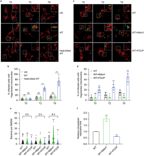
Mdivi1 and FCCP are used as inhibitors and activators of mitochondrial fragmentation.Citation33,Citation34 To determine the impact of mitochondrial morphology on the intracellular replication of S. Tm, BMDMs were pretreated with Mdivi1 or FCCP for 4 h (Fig. S1a), and untreated BMDMs were infected with S. Tm. The proportion of cells with fragmented mitochondria in FCCP-treated BMDMs was 2.53- and 3.10-fold higher than that in untreated BMDMs at 2 and 8 h p.i. (, whereas that with fragmented mitochondria in Mdivi1-treated BMDMs was 1.88- and 2.10-fold lower than that in untreated BMDMs at 2 and 8 h p.i. (. The average number of bacteria in FCCP-treated BMDM decreased by 2.69- and 2.68-fold whereas that in Mdivi1-treated BMDM increased by 1.23- and 2.37-fold at 2 and 8 h p.i. compared with that in untreated BMDM (). These data indicate a negative correlation between the proportion of cells with fragmented mitochondria and S. Tm replication in BMDMs. The effect of mitochondrial morphology on S. Tm replication was confirmed using intracellular replication assays. The relative replication of S. Tm in FCCP-treated BMDMs was 1.62-fold lower, whereas that in Mdivi1-treated BMDMs was 2.03-fold higher than that in untreated BMDMs (). These results indicate that inhibition of mitochondrial fragmentation enhances the intracellular replication of S. Tm within BMDMs.
Together, the above results indicate that S. Tm effectively inhibits LPS-induced host mitochondrial fragmentation in BMDMs, which is critical for intracellular replication of S. Tm.
SipA is required to prevent mitochondrial fragmentation during infection
Next, we investigated the mechanism underlying S. Tm’s inhibition of host mitochondrial fragmentation during infection. Because pathogens can secrete effectors targeting mitochondria to modulate the mitochondrial dynamics of host cells, we evaluated the colocalization of 47 known effectors encoded by SPI1 and SPI2 of S. Tm with mitochondria. First, we constructed eukaryotic expression vectors for 47 effector-GFP fusion proteins and transfected them into RAW264.7 macrophages. Except for 13 effectors that could not be transfected into RAW264.7, we analyzed the colocalization of the other 34 effectors with mitochondria by immunofluorescence. The Pearson’s correlation coefficient for the colocalization of the 11 effectors with mitochondria, including SipB and SopA, which have been previously shown to target mitochondria,Citation28,Citation35 was significantly higher than that observed between GFP expressed by the empty vector and mitochondria (Fig. S2a, b). Among these 11 effector proteins, SipA significantly colocalized with the mitochondria, with a maximum Pearson’s correlation coefficient of 0.64 (), indicating that SipA specifically localizes to the mitochondria. We hypothesized that SipA plays a major role in preventing mitochondrial fragmentation in macrophages.
Figure 2. SipA is required to prevent mitochondrial fragmentation during infection.
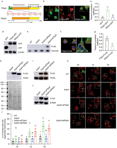
Positively charged mitochondrial targeting sequence (MTS) is a short peptide essential for protein transport to the mitochondria. Analysis using MitoProt (http://ihg.gsf.de/ihg/mitoprot.html) revealed a potential N-terminal MTS in SipA (). SipA likely targets the mitochondria via this N-terminal MTS. To test this hypothesis, MTS-deleted SipA (MSipA) eukaryotic expression vectors were constructed and transfected into RAW264.7 macrophages. Immunofluorescence analysis showed that the deletion of the MTS in SipA resulted in its failure to localize to the mitochondria (). Further colocalization analysis demonstrated that MSipA could not localize to the mitochondria (), indicating that the MTS is crucial for the mitochondrial localization of SipA. Meanwhile, the expression level of full-length SipA (FSipA) or MSipA were evaluated by immunoblotting. The results showed the expression level of MSipA was comparable to that of FSipA ( and S3(a)). To further validate the above conclusion, BMDMs were infected by WT, ΔsipA, ΔsipA complemented with pTr-FSipA-FLAG (ΔsipA+pFSipA), and ΔsipA complemented with pTr-MSipA-FLAG (ΔsipA+pMSipA) at 8 h p.i. The mitochondria in these infected cells were isolated, and the localization of SipA on the mitochondria was detected by immunoblotting. SipA was localized to the mitochondria in ΔsipA+pFSipA-infected BMDMs (), whereas SipA could not be detected in mitochondria from WT-, ΔsipA-, or ΔsipA+pMSipA-infected BMDMs (). Furthermore, SipA localization in the mitochondria of BMDMs infected with ΔsipA+pFSipA or ΔsipA+pMSipA was analyzed by immunofluorescence using confocal laser scanning microscopy. Consistent with these results, FSipA, but not MSipA, was localized to the mitochondria at 8 h p.i. (). Considering that the majority of T3SS effectors possess N-terminal secretion signals which is essential for their translocation, the immunoblotting was performed to determine the secretion and expression of SipA from ΔsipA, ΔsipA+pFSipA and ΔsipA+pMSipA under T3SS inducing conditions for 3 h. The result showed MSipA can still be detected in the bacterial culture supernatants (, S3b) and stably expressed in the bacterial cells ( and S3(c)), indicating the absence of N-terminal MTS doesn’t affect SipA secretion and expression in vitro. Furthermore, the immunoblotting were also performed to detect the secretion of SipA in cell lysate supernatants of BMDMs infected with ΔsipA, ΔsipA+pFSipA and ΔsipA+pMSipA at 8 h p.i. The results showed a comparable protein level for FSipA and MSipA was observed in cell lysate supernatants ( and S3(d)), indicating secretion of SipA does not depend on its MTS. These data indicate that the N-terminal MTS is required for SipA to target the host mitochondria.
We further investigated whether SipA was required to prevent mitochondrial fragmentation during infection. First, BMDMs were infected by WT, ΔsipA, or ΔsipA+pFSipA. The mitochondrial morphology of infected cells was determined by immunofluorescence and observed using confocal laser scanning microscopy. The proportion of cells with fragmented mitochondria in ΔsipA-infected BMDMs was 2.91- and 2.92-fold higher than that in WT-infected BMDMs at 2 and 8 h p.i., respectively (). However, the ΔsipA+pFSipA-infected BMDMs exhibited a similar mitochondrial fragmentation ratio to that of WT-infected BMDMs (). These data indicated that SipA prevents mitochondrial fragmentation during infection. To confirm that targeting mitochondria via the N-terminal MTS is essential for the function of SipA in preventing mitochondrial fragmentation, we infected BMDMs with ΔsipA+pMSipA, and the mitochondrial morphology of the infected cells was detected by immunofluorescence and monitored by confocal laser scanning microscopy. The results showed that the proportion of cells with fragmented mitochondria in ΔsipA+pMSipA-infected BMDMs exhibited no significant difference compared with that in ΔsipA-infected BMDMs but was significantly higher than that in WT-infected BMDMs and ΔsipA+pFSipA-infected BMDMs at 2 and 8 h p.i. (). These data indicate that MTS plays a key role in SipA-mediated inhibition of mitochondrial fragmentation. In conclusion, these data demonstrate that S. Tm secretes SipA during infection to prevent mitochondrial fragmentation by directly targeting mitochondria, which is mediated by the N-terminal MTS of SipA.
Lack of SipA increases mitochondrial dysfunction
Above results showed that SipA targeted mitochondria to prevent mitochondrial fragmentation. As mitochondrial fragmentation leads to mitochondrial dysfunction, we hypothesized that SipA prevents mitochondrial dysfunction during infection. Mitochondrial fragmentation commonly leads to excessive production of mtROS.Citation36 We first investigated whether SipA plays a role in the attenuation of mtROS production. The BMDMs were infected with WT, ΔsipA, or ΔsipA+pFSipA and stained with MitoSOX Red to detect mtROS levels. The immunofluorescence results showed that at 2 and 8 h p.i., the mean fluorescence intensity (MFI) of mtROS in ΔsipA-infected BMDMs was significantly higher than that in WT-infected BMDMs (), whereas the ΔsipA+pFSipA-infected BMDMs exhibited a similar MFI of mtROS to that of WT-infected BMDMs (), indicating that SipA inhibits mtROS production.
Figure 3. Lack of sipA increases mitochondrial dysfunction.
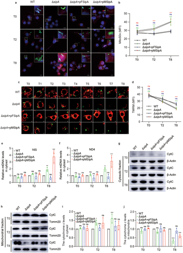
The excessive production of mtROS can further induce the mitochondrial permeability transition,Citation37 resulting in the dissipation of ΔΨm. Thus, we investigated whether SipA prevents the loss of ΔΨm during infection. The BMDMs were infected with WT, ΔsipA, or ΔsipA+pFSipA and stained with a ΔΨm-dependent mitochondrial dye tetramethylrhodamine methyl ester (TMRM). The MFI of ΔΨm was detected by confocal laser scanning microscopy. The time-lapse confocal results revealed a gradual decline of ΔΨm in BMDMs infected with WT, ΔsipA, or ΔsipA+pFSipA over 8 h p.i. (). Furthermore, the MFI analysis results of ΔΨm showed that at 2 and 8 h p.i., the ΔΨm of BMDMs infected with ΔsipA was significantly lower than that in BMDMs infected with WT (), whereas there was no significant difference in MFI of ΔΨm between ΔsipA+pFSipA-infected and WT-infected BMDMs (). These data indicate that SipA can repress the loss of ΔΨm.
The mitochondrial permeability transition leads to the release of mtDNA and cytochrome c from mitochondria into the cytosol. We subsequently assessed the effect of SipA on the translocation of mtDNA from mitochondria into the cytosol. The BMDMs were infected with WT, ΔsipA, or ΔsipA+pFSipA, and the isolated cytosolic mtDNA levels were quantified using qPCR. ΔsipA triggered a significantly higher release of mtDNA from mitochondria than WT at 8 h p.i. (), whereas ΔsipA+pFSipA induced a similar level of mtDNA release compared with WT (). This indicates that SipA can inhibit the release of mtDNA from the mitochondria into the cytosol during S. Tm infection.
To investigate the influence exerted by SipA on the translocation of cytochrome c from mitochondria into the cytosol, the BMDMs were infected with WT, ΔsipA, or ΔsipA+pFSipA and subjected to immunoblotting analysis for cytochrome c level measurements in mitochondria and cytosol. ΔsipA significantly increased cytochrome c levels in the cytosol by 1.36- and 1.61-fold compared with WT at 2 and 8 h p.i. (). Meanwhile, compared with WT-infected BMDMs, cytochrome c levels decreased in the mitochondria of ΔsipA-infected BMDMs (). In contrast, cytochrome c levels in both the cytosol and mitochondria of ΔsipA+pFSipA-infected BMDMs exhibited no significant difference compared with that in WT-infected BMDMs at 2 and 8 h p.i. (). These results demonstrated that SipA efficiently suppressed the release of cytochrome c from the mitochondria into the cytosol during infection.
SipA could prevent mitochondrial fragmentation-induced mitochondrial dysfunction, including the production of mtROS, loss of ΔΨm, and release of mtDNA and cytochrome c from mitochondria. We further investigated whether the MTS of SipA was indispensable for these effects. MtROS production, ΔΨm loss, and mtDNA and cytochrome c release from mitochondria in BMDMs infected with ΔsipA+pMsipA were comparable to those observed in ΔsipA-infected BMDMs (). These findings suggest that N-terminal MTS is essential for mitigating mitochondrial dysfunction by SipA.
SipA enhances S. Tm intracellular replication by targeting mitochondria
As the inhibition of host mitochondrial fragmentation is crucial for S. Tm intracellular replication in BMDMs, we next investigated whether SipA contributes to the intracellular replication of S. Tm by targeting the mitochondria. We quantified the bacterial load in BMDMs infected with WT, ΔsipA, ΔsipA+pFSipA or ΔsipA+pMSipA using confocal laser scanning microscopy. The average bacterial load in ΔsipA- and ΔsipA+pMsipA-infected BMDMs was significantly reduced compared with that in WT-infected BMDMs at 8 h p.i. (), while the average bacterial load showed no significant difference between ΔsipA+pFSipA- and WT-infected BMDMs (). This indicates that SipA-targeted mitochondria are essential for bacterial replication in BMDMs. To further confirm this conclusion, we conducted intracellular replication assays with WT, ΔsipA, ΔsipA+pFSipA or ΔsipA+pMSipA. The relative replication fold of ΔsipA and ΔsipA+pMSipA exhibited 2.07- and 1.58-fold decrease compared with that of WT at 8 h p.i. (). However, ΔsipA+pFSipA restored the replication ability of S. Tm to a level comparable to that of WT (). These data suggest that SipA contributes to the intracellular replication of S. Tm by targeting mitochondria.
Figure 4. SipA enhances S. Tm intracellular replication by targeting mitochondria.
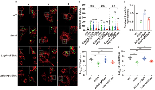
In addition, mice infection assays were performed to confirm this conclusion in vivo. Mice were administered intraperitoneally with WT, ΔsipA, ΔsipA+pFSipA or ΔsipA+pMSipA. After 3 d, the liver and spleen were isolated to quantify the bacterial load. The results demonstrated a significant reduction in bacterial burdens in the liver and spleen of mice infected with ΔsipA compared to those infected with WT (). As expected, the ΔsipA+pFSipA was comparable to the WT in bacterial burdens in the liver and spleen of the infected mice (). However, the bacterial loads in the liver and spleen of ΔsipA+pMSipA-infected mice were only partially restored compared with that in WT-infected mice (), indicating that the interaction between SipA and mitochondria enhances the pathogenicity of S. Tm in animals. Because SipA plays numerous crucial roles during infection, the data suggest that the interaction between SipA and mitochondria is crucial for some of its effects that contribute to successful infection of S. Tm, but not all of them. Finally, we determined the growth curves of WT, ΔsipA, ΔsipA+pFSipA and ΔsipA+pMSipA in Luria – Bertani (LB) and RPMI 1640 medium. There was no growth defect among ΔsipA, ΔsipA+pFSipA and ΔsipA+pMSipA compared with WT in both LB and RPMI 1640 medium (Fig. S4a, b). These findings suggest that SipA enhances in vitro replication capacity and in vivo pathogenicity of S. Tm by selectively targeting mitochondria.
SipA prevents the recruitment of Drp1 to host mitochondria
Mitochondrial fragmentation is closely regulated by Drp1 in eukaryotic cells.Citation38 Thus, we investigated whether SipA inhibited mitochondrial fragmentation by attenuating Drp1 translocation to the mitochondria. The mitochondria of BMDMs infected with WT, ΔsipA, or ΔsipA+pFSipA were isolated, and the Drp1 protein levels recruited to mitochondria were detected by immunoblotting. At 2 and 8 h p.i., BMDMs infected with ΔsipA exhibited a 1.62- and 1.54-fold increase in Drp1 levels in mitochondria compared to WT-infected BMDMs (). Meanwhile, the protein levels of Drp1 in the mitochondria were similar between ΔsipA+pFSipA- and WT-infected BMDMs (). This indicates that a lack of sipA can promote the recruitment of Drp1 to the mitochondria during infection. Furthermore, we also analyzed the levels of Drp1 recruited to mitochondria in ΔsipA+pMSipA-infected BMDMs. The levels of Drp1 in the mitochondria of ΔsipA+pMSipA-infected BMDMs were significantly increased compared to that in WT-infected BMDMs (), indicating that SipA inhibits the recruitment of Drp1 to mitochondria by directly targeting mitochondria.
Figure 5. SipA prevents the recruitment of Drp1 to host mitochondria.
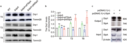
We further investigated whether SipA directly interacts with Drp1 to inhibit its recruitment to mitochondria. The RAW264.7 macrophages were transfected with the SipA-expressing plasmid vector (pcDNA3.1(+)-SipA) or the pcDNA3.1(+) empty vector (control). Both cell lysates were subjected to immunoprecipitation using an anti-Drp1 antibody, followed by immunoblotting. Drp1 and SipA in RAW264.7 macrophages transfected with SipA-expressing plasmid could be detected in input samples. However, SipA was not detected in the corresponding output samples, which was consistent with the control cells, indicating that Drp1 did not coimmunoprecipitate with SipA ( and S5(a)). These results demonstrate that SipA cannot directly bind to Drp1.
Discussion
Pathogens have evolved sophisticated mechanisms to hijack mitochondria and modulate mitochondrial dynamics and function to promote survival and replication.Citation39 However, the mitochondrial dynamics manipulated by S. Tm during infection remain poorly understood. In this study, we demonstrated that S. Tm inhibits mitochondrial fragmentation in macrophages. When engulfed by macrophages, S. Tm secretes the T3SS1 effector, SipA, which directly targets the mitochondria. Subsequently, by interfering with the recruitment of Drp1, SipA alters mitochondrial dynamics to reduce excessive fragmentation while avoiding excessive mtROS production, ΔΨm dissipation, and mtDNA and cytochrome c release from mitochondria. This benefits the intracellular replication of S. Tm and promotes its pathogenicity in the host.
Pathogens employ various strategies to manipulate mitochondrial processes to subvert the fate of the infected host. Numerous effectors secreted by bacteria influence mitochondrial dynamics and function during infection.Citation11 At different infection stages, bacteria can influence mitochondrial dynamics through different effectors and mechanisms. For instance, in the early stages of enteropathogenic Escherichia coli (EPEC) infection, the bacterial effector EspZ interacts with mitochondrial fission protein 1 (FIS1) of host epithelial cells, a mitochondrial outer membrane protein that mediates mitochondrial fragmentation.Citation40 This interaction protects the mitochondrial fusion network and enhances host cell viability, benefitting EPEC colonization. In contrast, at the late stage of infection, EPEC stimulates mitochondrial fragmentation via an EspH-dependent increase in FIS1 levels and causes a loss in ΔΨm,Citation40 leading to host cell death, which likely facilitates pathogen dispersal. In this study, at the early stages of S. Tm infection, SipA targets mitochondria to inhibit mitochondrial fragmentation and cell death, which benefits intracellular survival. The effector SseL of S. Tm induces cell death in macrophages at late time points after invasion.Citation41 After entering macrophages, S. Tm may prevent host mitochondrial fragmentation to promote intracellular replication during the early stages of infection. However, at late time points during infection, it triggers macrophage death,Citation42 which may benefit bacterial dissemination. Multiple effectors are probably engaged in the complex interactions between S. Tm and mitochondria during infection. Additionally, through colocalization analysis by immunofluorescence, we found that in addition to SipA and SipB, other nine effectors could also target the mitochondria of macrophages. These nine effectors likely modulate mitochondrial dynamics in macrophages via different mechanisms. However, the role of these effectors in regulating mitochondrial dynamics requires further investigation.
Multiple bacterial effectors are multitasking proteins that exert different roles during the interaction between host and pathogens. SipB induces the formation of multivesicular structures, including both mitochondrial and endoplasmic reticulum markers.Citation28 In addition to regulating mitochondrial dynamics, SipA performs other functions during infection, such as polymorphonuclear leukocyte recruitment,Citation31 actin cytoskeleton rearrangementCitation29,Citation30 and SCV targeting.Citation24 When SipA is exposed to the cytoplasmic face of the SCV, the N-terminal domain of SipA specifically recruits syntaxin8 on SCVs, which is a soluble N-ethylmaleimide-sensitive factor attachment receptor (SNARE) protein involved in the regulation of phagosome maturation, to promote the fusion of SCV with early endosomes and inhibit the transport of SCV to lysosomes.Citation43 These researches suggest that SipA is a multifunctional protein facilitating various biological processes to enhance S. Tm intracellular replication and pathogenicity. In this study, we revealed that SipA can directly target mitochondria and inhibit mitochondrial fragmentation. Moreover, we also validated that inhibiting mitochondrial fragmentation favors S. Tm intracellular replication, suggesting that SipA targeting mitochondria is one of strategy to promote S. Tm intracellular replication and pathogenicity. Although several mechanisms by which SipA enhances Salmonella intracellular replication and pathogenicity have been demonstrated, the comprehensive understanding of the interplay between these mechanisms in promoting intracellular replication and pathogenicity of S. Tm remains incomplete and warrants further investigation.
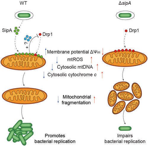
Mitochondrial fragmentation and fusion play critical roles in maintaining functional mitochondria when cells experience metabolic or environmental stress. Mitochondrial fragmentation generates new organelles and facilitates quality control in growing and dividing cells, whereas fusion contributes to the response of mammalian cells to stress and energy demand.Citation44 The Drp1-dependent pathway is the classical mechanism of mitochondrial fragmentation. For example, Legionella pneumophila effector Lpg1137 binds to and cleaves syntaxin17, a SNARE protein that regulates Drp1 activity during infection and induces mitochondrial fragmentation.Citation45 In addition, L. pneumophila effector MitF triggers the activation of the Wiskott – Aldrich syndrome protein (WASP) and actin-related protein 2/3 complex (Arp2/3).Citation13 Actin nucleation by WASP/Arp2/3 progressively facilitates Drp1-mediated mitochondrial fragmentation.Citation13 In this study, S. Tm exploits the T3SS1 effector SipA to inhibit mitochondrial fragmentation in a Drp1-dependent manner (). However, this study failed to identify the precise mechanism underlying the interaction between SipA and Drp1. SipA likely inhibits the recruitment of Drp1 to the mitochondria via unknown intermediates, which can be a subject for future studies ().
Figure 6. Model of S. Tm remodeling mitochondrial dynamics for its efficient replication.
Materials and methods
Mice
Female BALB/c mice (8 weeks old) were purchased from Beijing Vital River Laboratory Animal Technology Co., Ltd. (Beijing, China). All mice were housed in a specific pathogen-free environment and fed a standard mouse chow diet. All animal experiments adhered to the criteria approved by the Institutional Animal Care Committee of Nankai University (Tianjin, China).
Bacterial strains and plasmids
The bacterial strains and plasmids used in this study are listed in Table S1. The primers used in this study are listed in Table S2. Briefly, S. Tm strain ATCC 14028s served as the WT strain throughout this study. All bacterial strains were grown in LB broth containing 1% tryptone, 0.5% yeast extract, and 1% NaCl at 37°C but 30°C for strains carrying the temperature-sensitive plasmid PKD46. When needed, the arabinose and antibiotics were added to the medium at the following final concentrations: 100 mM arabinose, 100 μg/mL ampicillin, 25 μg/mL chloramphenicol, and 50 μg/mL kanamycin.
The mutant strain was generated using the λ-Red recombinase system as described previously.Citation46 Briefly, the chloramphenicol resistance gene of pKD3 was amplified using primers with 38–40 bp of homology to the upstream and downstream regions flanking the mutant gene’s start and stop codons at their 5′ ends. The generated PCR products were electroporated into S. Tm strain ATCC 14028s carrying the temperature-sensitive plasmid PKD46 for homologous recombination. The mutant was selected based on its resistance to chloramphenicol and was further identified by PCR and DNA sequencing.
Complementation strains were generated by cloning the corresponding functional genes carrying the 3×FLAG tag into the pTrc99A plasmid and transforming the plasmid into the mutant strain. Strains carrying GFP were generated by transforming the pETDuet1-GFP plasmid into the corresponding strains. Eukaryotic expression vectors for colocalization by transfection were generated by cloning the corresponding functional genes and native promoters into the pCMV-C-GFP plasmid. A eukaryotic expression vector for immunoprecipitation was established by cloning the corresponding functional genes into the pcDNA3.1(+) plasmid. All resulting clones were verified by DNA sequencing.
BMDM isolation and cell culture
BMDMs were isolated from female BALB/c mice as previously described.Citation47 Briefly, 8-week-old mice were sacrificed, and femur and tibia bone marrow cells were flushed into RPMI 1640 medium (Gibco) supplemented with 10% fetal bovine serum (FBS; Gibco), passed through a 70 μm cell strainer to remove cell clumps, bone, hair, and other cells or tissues followed by centrifuging at 500 × g for 5 min. The collected cells were then suspended in RPMI 1640 medium containing 10% FBS, 1% penicillin/streptomycin, and 10 ng/mL monocyte-colony stimulating factor (M-CSF; MA5–23773; Invitrogen) and left to adhere at 37°C and 5% CO2. The medium was changed every 2 d, and BMDMs were used 7 d post-collection.
The RAW264.7 macrophage cell line was purchased from the Shanghai Institute of Biochemistry and Cell Biology of the Chinese Academy of Sciences (Shanghai, China) and cultured in RPMI 1640 medium supplemented with 10% FBS and incubated at 37°C with 5% CO2 in a humidified atmosphere for 24 h.
Before infection or transfection, BMDMs and RAW264.7 were grown to monolayers with 70–90% coverage in 12-well tissue culture plates with or without glass coverslips, 2-cm glass-bottom cell culture dishes, 10-cm cell culture dishes, or 75 cm2 culture bottles according to experimental requirements.
The infection of BMDMs and intracellular replication assays
BMDMs were infected, as previously described, with slight modifications.Citation48 Briefly, overnight-grown bacteria were inoculated into fresh LB medium at a ratio of 1:100 and further grown to the stationary phase until an OD600 of 1.5. Bacteria were pelleted and opsonized in the RPMI 1640 medium supplemented with 10% FBS for 20 min at 37°C. The bacterial culture was then added to BMDM monolayers at a multiplicity of infection (MOI) of 10. Subsequently, the plates were centrifuged at 1000 × g for 5 min to synchronize infection. After incubation for 40 min at 37°C with 5% CO2, the cell culture supernatant was discarded, and the infected BMDMs were washed thrice with phosphate-buffered saline (PBS) and supplemented with fresh RPMI 1640 medium containing 100 µg/mL gentamicin to kill the extracellular bacteria for 90 min. The infected BMDMs were washed thrice with PBS and incubated with 20 µg/mL gentamicin for the remaining infection time. For intracellular replication assays, BMDMs were seeded in 12-well tissue culture plates for 7 d before infection. At 2 and 8 h p.i., the supernatant was discarded, and the BMDMs were washed thrice with PBS and lysed with 0.1% Triton X-100. Lysates with serial gradient dilutions were plated on LB agar plates to count intracellular bacteria. Additionally, BMDMs were pretreated with the mitochondrial fragmentation inhibitor Mdivi1 (50 µM 475,856; Millipore) and mitochondrial fragmentation-inducing agent FCCP (10 µM, C2920; Sigma) 4 h before infection. BMDMs were then washed thrice with PBS. The intracellular fold replication was expressed as the bacterial burden recovered at 8 h p.i. relative to those at 2 h p.i. Triplicate wells were averaged for each data point, and three independent biological replicates were performed.
Plasmid transfection
Eukaryotic expression vectors were extracted using the EndoFree Plasmid Midi Kit (CW2105S; CWBIO), according to the manufacturer’s instructions. Before transfection, RAW264.7 macrophages were seeded in 2-cm glass-bottom cell culture dishes or 10-cm cell culture dishes for 24 h. Then, RAW264.7 macrophages were transiently transfected with the indicated eukaryotic expression vectors using Lipofectamine LTX DNA Transfection Reagent (15338100; Invitrogen) according to the manufacturer’s instructions. Briefly, endofree plasmid DNA diluted in serum-free medium was mixed with PLUS Reagent. The diluted DNA solution was added to diluted Lipofectamine LTX Reagent and incubated for 5 min at room temperature. The resulting DNA – lipid complex was directly dropped into RAW264.7 and incubated for 24 h at 37°C with 5% CO2.
Immunofluorescence
For the presentation of mitochondrial morphology and intracellular bacteria, BMDMs were seeded in 2-cm glass-bottom cell culture dishes for 7 d, followed by S. Tm infection and staining with 50 nM MitoTracker Deep Red FM (M22426; Invitrogen) according to the manufacturer’s instructions. For the colocalization of the indicated protein with mitochondria after transfection, RAW264.7 macrophages were stained with 50 nM MitoTracker Deep Red FM according to the manufacturer’s instructions. For the indicated protein colocalization with mitochondria after infection, BMDMs attached to glass coverslips were fixed with 4% paraformaldehyde for 10 min at room temperature, permeabilized, and blocked with 0.1% Triton X-100 in 5% bovine serum albumin (BSA) for 1 h at room temperature. Then, BMDMs were incubated with primary antibodies (Tomm20, 1:100, ab56783, Abcam; FLAG, 1:100, F1804, Sigma) overnight at 4°C, washed thrice with PBS, incubated with secondary antibodies (anti-mouse Alexa Fluor 594, 1:200, ab150116, Abcam; anti-rabbit Alexa Fluor 488, 1:200, ab150077, Abcam) for 1 h, and stained with DAPI for 5 min (C0065, Solarbio). After the final wash, the cells were covered with a mounting medium. Following staining, a confocal laser scanning microscope (Zeiss LSM800) was used to observe mitochondrial morphology, intracellular bacteria, the indicated protein colocalization with mitochondria, and image acquisition. For mitochondrial morphology, cells (20/field) of 15 random fields from three independent biological replicates were analyzed to determine the proportion of cells with fragmented mitochondria using ZEN 2.3 (blue edition). For intracellular bacteria, cells (20/field) of five random fields from three independent biological replicates were analyzed to determine the bacteria per BMDM using ZEN 2.3 (blue edition). For colocalization, 30 random fields from three independent biological replicates were analyzed the Pearson’s correlation coefficient with Fiji/ImageJ.
Whole cell lysates after transfection
RAW264.7 macrophages were seeded in 10-cm cell culture dishes for 24 h. After transfection for 24 h, cells were washed thrice and lysed in RIPA buffer (R0010; Solarbio) containing 1 mM PMSF. Then, cell lysates were centrifuged at 15,000 × g for 10 min at 4°C, and the supernatants were collected and analyzed by immunoblotting.
Mitochondrial isolation
Mitochondria were isolated as described previously.Citation49 Briefly, BMDMs were seeded in 75-cmCitation2 culture bottles for 7 d. After infection, BMDMs were collected at 800 × g for 5 min at 4°C, resuspended in ice-cold RSB Hypo buffer (10 mM NaCl, 1.5 mM MgCl2, and 10 mM Tris-HCl (pH7.5)) for 5–10 min, and homogenized to break the cells. Immediately, 2.5 × MS homogenization buffer (525 mM mannitol, 175 mM sucrose, 12.5 mM Tris-HCl (pH7.5), and 2.5 mM EDTA (pH7.5)) was added to the homogenate to acquire a final concentration of 1 × MS homogenization buffer. Then, the homogenate was centrifuged at 1300 × g for 5 min at 4°C, the supernatant was collected, and the centrifugation was repeated twice. The last collected supernatant was centrifuged at 15,000 × g for 15 min at 4°C to isolate the mitochondrial pellet. Finally, the mitochondrial pellet was lysed with 2% CHAPS in TBS buffer containing 1 mM PMSF, and the resulting lysate was quantified using a BCA Protein Assay Kit (C503021–0500; Sangon Biotech) according to the manufacturer’s instructions.
Bacterial secretion assay
The detection of secreted SipA from S. Tm in LB culture supernatants was performed as previously described.Citation50 Briefly, overnight bacterial cultures were subcultured in LB medium supplemented with 0.3 M NaCl under conditions that stimulated expression of the SPI1 T3SS at a ratio of 1:100 for 3 h at 37°C.Citation51 Bacterial culture supernatants were separated by centrifugation at 12,000 × g for 10 min, filtered through a 0.22 µm syringe filter, and concentrated 100-fold with an Ultrafree centrifugal filter device (UFC9030; Millipore) with a 30-kDa cutoff at 4°C followed by quantification through sodium dodecyl sulfate-polyacrylamide gel electrophoresis (SDS-PAGE) and Coomassie blue staining. The separated bacterial cells were resuspended in PBS containing 1 mM PMSF (B111–01; GenStar) and disrupted by sonication at 4°C. Bacterial culture supernatants and bacterial cell lysates were further analyzed by immunoblotting.
Additionally, the detection of secreted SipA in S. Tm-infected BMDMs was conducted as previously described.Citation52 BMDMs were seeded in 10-cm cell culture dishes for 7 d. After 8 h infection, BMDMs were washed thrice with PBS and then treated with 30 µg/mL of proteinase K (AA1907; SparkJade) in PBS for 15 min at 37°C in a CO2 incubator to eliminate cell-surface-associated SipA proteins. Cells detached during the proteinase treatment and were subsequently collected by centrifugation at 600 × g for 5 min and lysed in PBS containing 0.1% Triton X-100 and 1 mM PMSF. The cell lysates were centrifuged at 15,000 × g for 10 min at 4°C and the separated supernatants were filtered through a 0.22 µm syringe filter and proteins were analyzed by immunoblotting.
MtROS determination
MtROS were detected with a mitochondrial ROS detector, MitoSOX Red (M36008; Invitrogen), using the confocal laser scanning microscope. Briefly, BMDMs were seeded in 2-cm glass-bottom cell culture dishes for 7 d. After infection, BMDMs were treated with 5 µM MitoSOX for 10 min at 37°C, washed thrice with PBS, and stained with DAPI for 5 min. After washing thrice with PBS, the confocal laser scanning microscope was used to observe mtROS fluorescence and acquire images. Cells (20/field) of 15 random fields from three independent biological replicates were quantified for mtROS fluorescence intensity using ZEN 2.3 (blue edition).
Measurement of ΔΨm
The ΔΨm was measured with TMRM (I34361; Invitrogen) using the confocal laser scanning microscope. Briefly, BMDMs were seeded in 2-cm glass-bottom cell culture dishes for 7 d. After infection, BMDMs were loaded with 100 nM TMRM for 30 min at 37°C and washed thrice with PBS. Confocal laser scanning microscope was used for ΔΨm fluorescence observation and image acquisition. Cells (20/field) of 15 random fields from three independent biological replicates were quantified for ΔΨm fluorescence intensity using ZEN 2.3 (blue edition).
MtDNA extraction and quantification
Released mtDNA was extracted from the infected BMDMs using the Digitonin method.Citation53 Briefly, BMDMs were seeded in 75-cmCitation2 culture bottles for 7 d. After infection, the BMDMs were collected by centrifugation at 1000 × g for 3 min. Subsequently, the cell pellet was resuspended in digitonin buffer (150 mM NaCl, 50 mM HEPES, and 25 µg/mL digitonin). The cell suspension was rotated for 10 min and centrifugated at 1000 × g for 3 min. The supernatant was centrifuged again at 16,000 × g for 25 min to obtain a new supernatant containing cytosolic mtDNA (cmtDNA) released from the mitochondria. The remaining pellet containing the nuclear genome was prepared for nuclear DNA (nucDNA) acquisition using the SPARKeasy Tissue/Cell DNA Kit (AA1002-B; SparkJade). The purity ratios (A260/280 and A260/230) and DNA yield (nanograms per microliter) of both nucDNA and cmtDNA were determined with the NanoDrop 2000 spectrophotometer (Thermo Fisher Scientific, Waltham, MA, USA).
The mtDNA copy numbers were determined by qPCR performed on ABI 7500 thermocycler sequence detection (Applied Biosystems) using SYBR green fluorescence dye. Two mitochondrial (16S and ND4) and one nuclear (PMP22) genes were amplified.Citation54 The qPCR primers used in this study are listed in Table S2. The PMP22 gene was used as a reference control, and the relative expression level of candidate targets was calculated as fold-change values using the 2−ΔΔCT method. The mtDNA copy numbers acquired by cmtDNA/nucDNA were standardized to the control. The data were collected from three biological replicates.
Cytochrome c release
Cytochrome c release was measured using a cytochrome c Releasing Apoptosis Assay kit (ab65311; Abcam) according to the manufacturer’s manual. Briefly, BMDMs were seeded in 75-cmCitation2 culture bottles for 7 d. After infection, BMDMs were collected and centrifuged at 600 × g for 5 min at 4°C. Subsequently, the cells were homogenized and separated into the cytosolic and mitochondrial fractions by centrifugation. The cytosolic fraction contained the released cytochrome c from the mitochondria, and the mitochondrial fraction contained the remaining cytochrome c. To release the remaining cytochrome c from the mitochondria, the mitochondrial fraction was lysed with 2% CHAPS in TBS buffer containing 1 mM PMSF. Finally, the cytosolic and mitochondrial fractions were quantified using a BCA Protein Assay Kit (C503021–0500; Sangon Biotech) according to the manufacturer’s instructions.
Immunoblotting
Protein samples were boiled in 1 × SDS loading buffer and separated using a 4–12% gradient Bis-Tris Gel (M00653; GenScript) by SDS-PAGE. Subsequently, the separated proteins were transferred onto a 0.45 µm PVDF membrane (IPVH00010; Millipore) and blocked with 5% skimmed milk at room temperature for 2 h. Then, the blots were incubated for another 2 h at room temperature with the following primary antibodies: anti-FLAG (1:1000, F1804; Sigma), anti-GFP (1:1000, K200047M; Solarbio), anti-cytochrome c (1:1000, MA5–11674; Invitrogen), anti-Drp1 (1:1000, ab184247; Abcam), anti-SipA (Shanghai Willget Biotech Co., Ltd.), anti-GroEL (1:1000, ab82592; Abcam), anti-Tomm20 (1:1000, ab56783; Abcam), and anti-β-Actin (1:1000, CW0096; CWBIO). To detect proteins, the blots were incubated with the corresponding horseradish peroxidase (HRP)-conjugated anti-rabbit (EF0002; SparkJade) or anti-mouse (EF0001; SparkJade) secondary antibodies at room temperature for 1 h. Finally, the blots were visualized with an ECL detection kit (D601039; Sangon Biotech) using an AmershamTM Imager 600 system (General Electric). Immunoblotting bands were quantified using the ImageJ software. The data were collected from three biological replicates.
Mice infection
Mouse infection assays were performed as previously described with slight modifications.Citation46 Briefly, the overnight-grown bacteria were subcultured as 1:100 in fresh LB medium and grown to the stationary phase. Subsequently, the bacteria were collected and washed thrice with sterile 0.9% NaCl and serially diluted to 1 × 10Citation7 CFU/mL in 0.9% NaCl for intraperitoneal infection. Female BALB/c mice were injected intraperitoneally with 1 × 10Citation6 CFU in 0.1 mL sterile 0.9% NaCl. The infected mice were euthanized at 3 d p.i. The spleen and liver were harvested, homogenized, serially diluted in PBS, and plated on LB agar plates to count the bacterial CFU. Experiments were performed with three independent biological replicates, where each experiment employed four mice, and the data were pooled (n = 12).
Growth assay
To determine the growth curve of each WT, ΔsipA, ΔsipA+pFSipA, and ΔsipA+pMSipA strains, overnight-grown bacteria were adjusted to an OD600 of 1.5 and diluted 1:1000 in LB and RPMI 1640 medium. Next, a 200 μL aliquot was added to a 96-well microplate and incubated at 37°C with shaking at 180 rpm for 24 h. Absorbance at 600 nm was recorded using a programmed procedure. The experiment was independently performed in triplicate.
Immunoprecipitation
RAW264.7 macrophages were seeded in 10-cm cell culture dishes for 24 h and transfected with the endofree plasmid pcDNA3.1(+) and pcDNA3.1(+)-SipA. After 24 h, the RAW264.7 macrophages were subjected to immunoprecipitation. Briefly, the cells were washed thrice and lysed in 1 mL RIPA buffer containing a complete protease inhibitor cocktail. Lysates were centrifuged at 15,000 × g for 10 min at 4°C, and the supernatants were collected. Subsequently, 50 µL Dynabeads (10001D; Novex) were incubated with anti-Drp1 antibody (ab184247; Abcam) for 30 min at room temperature on a rotation shaker followed by washing with PBST (PBS (pH 7.4) with 0.02% Tween-20). The Dynabeads – antibody complex was incubated with supernatant lysates overnight at 4°C on a rotatory shaker and washed thrice with PBST. Finally, the proteins bound to the beads were eluted with an elution buffer (50 mM glycine, pH 2.8) and boiled in SDS loading buffer. The protein samples were resolved in 4–12% gradient Bis-Tris Gel and analyzed by immunoblotting with anti-Drp1 and anti-SipA antibodies. Experiments were performed with three independent biological replicates.
Statistical analyses
Statistical significance was analyzed with GraphPad Prism 8.0.1 software (GraphPad Inc., San Diego, CA, USA) using the two-way ANOVA, two-tailed unpaired Student’s t-test and Mann–Whitney U-test according to the test requirements (as stated in the figure legends). P-values <0.05, 0.01, and 0.001 were considered statistically significant (*), highly significant (**), and extremely significant (***), respectively, and ns represents no significant difference.
Author contributions
LF, BL, and YTL designed the study. XML, XYZ, XPL, TY, RYL, QW, QSW, DL, and XTC conducted the experiments and data analyses. XML, YTL, BL, and LF wrote and edited the manuscript.
Supplementary material revised clean.docx
Download MS Word (1.2 MB)Disclosure statement
No potential conflict of interest was reported by the author(s).
Data availability statement
The authors confirm that the data supporting the findings of this study are available within the article [and/or] its supplementary materials.
Supplementary material
Supplemental data for this article can be accessed online at https://doi.org/10.1080/19490976.2024.2316932
Additional information
Funding
References
- Cloonan SM, Choi AM. Mitochondria: commanders of innate immunity and disease?. Curr Opin Immunol. 2012;24(1):32–21. PMID:22138315. doi:10.1016/j.coi.2011.11.001
- Chan DC. Fusion and fission: interlinked processes critical for mitochondrial health. Annu Rev Genet. 2012;46:265–287. PMID:22934639. doi:10.1146/annurev-genet-110410-132529.
- Tait SW, Green DR. Mitochondria and cell death: outer membrane permeabilization and beyond. Nat Rev Mol Cell Biol. 2010;11(9):621–32. PMID:20683470. doi:10.1038/nrm2952.
- Chen S, Liao Z, Xu P. Mitochondrial control of innate immune responses. Front Immunol. 2023;14:1166214. PMID:37325622. doi:10.3389/fimmu.2023.1166214.
- Mishra P, Chan DC. Mitochondrial dynamics and inheritance during cell division, development and disease. Nat Rev Mol Cell Biol. 2014;15(10):634–46. PMID:25237825. doi:10.1038/nrm3877.
- Arnoult D, Carneiro L, Tattoli I, Girardin SE. The role of mitochondria in cellular defense against microbial infection. Semin Immunol. 2009;21(4):223–32. PMID:19535268. doi:10.1016/j.smim.2009.05.009.
- Mishra P, Chan DC. Metabolic regulation of mitochondrial dynamics. J Cell Biol. 2016;212(4):379–87. PMID:26858267. doi:10.1083/jcb.201511036.
- Frank S, Gaume B, Bergmann-Leitner ES, Leitner WW, Robert EG, Catez F, Smith CL, Youle RJ. The role of dynamin-related protein 1, a mediator of mitochondrial fission, in apoptosis. Dev Cell. 2001;1(4):515–525. PMID:11703942. doi:10.1016/s1534-5807(01)00055-7.
- Jagasia R, Grote P, Westermann B, Conradt B. DRP-1-mediated mitochondrial fragmentation during EGL-1-induced cell death in C. Elegans Nat. 2005;433(7027):754–760. PMID:15716954. doi:10.1038/nature03316.
- Wasiak S, Zunino R, McBride HM. Bax/Bak promote sumoylation of DRP1 and its stable association with mitochondria during apoptotic cell death. J Cell Biol. 2007;177(3):439–50. PMID:17470634. doi:10.1083/jcb.200610042.
- Cervantes-Silva MP, Cox SL, Curtis AM. Alterations in mitochondrial morphology as a key driver of immunity and host defence. EMBO Rep. 2021;22(9):e53086. PMID:34337844. doi:10.15252/embr.202153086.
- Hicks SW, Galán JE. Exploitation of eukaryotic subcellular targeting mechanisms by bacterial effectors. Nat Rev Microbiol. 2013;11(5):316–26. PMID:23588250. doi:10.1038/nrmicro3009.
- Escoll P, Song OR, Viana F, Steiner B, Lagache T, Olivo-Marin JC, Impens F, Brodin P, Hilbi H, Buchrieser C. Legionella pneumophila modulates mitochondrial dynamics to trigger metabolic repurposing of infected macrophages. Cell Host Microbe. 2017;22(3):302–316.e7. PMID:28867389. doi:10.1016/j.chom.2017.07.020.
- Suzuki M, Danilchanka O, Mekalanos JJ. Vibrio cholerae T3SS effector VopE modulates mitochondrial dynamics and innate immune signaling by targeting Miro GTPases. Cell Host Microbe. 2014;16(5):581–591. PMID:25450857. doi:10.1016/j.chom.2014.09.015.
- Jain P, Luo ZQ, Blanke SR. Helicobacter pylori vacuolating cytotoxin a (VacA) engages the mitochondrial fission machinery to induce host cell death. Proc Natl Acad Sci USA. 2011;108(38):16032–7. PMID:21903925. doi:10.1073/pnas.1105175108.
- Fan T, Lu H, Hu H, Shi L, McClarty GA, Nance DM, Greenberg AH, Zhong G. Inhibition of apoptosis in chlamydia-infected cells: blockade of mitochondrial cytochrome c release and caspase activation. J Exp Med. 1998;187(4):487–496. PMID:9463399. doi:10.1084/jem.187.4.487.
- Kurihara Y, Itoh R, Shimizu A, Walenna NF, Chou B, Ishii K, Soejima T, Fujikane A, Hiromatsu K. Chlamydia trachomatis targets mitochondrial dynamics to promote intracellular survival and proliferation. Cell Microbiol. 2019;21(1):e12962. PMID:30311994. doi:10.1111/cmi.12962.
- Fischer SF, Vier J, Kirschnek S, Klos A, Hess S, Ying S, Häcker G. Chlamydia inhibit host cell apoptosis by degradation of proapoptotic BH3-only proteins. J Exp Med. 2004;200(7):905–916. PMID:15452181. doi:10.1084/jem.20040402.
- Coburn B, Grassl GA, Finlay BB. Salmonella, the host and disease: a brief review. Immunol Cell Biol. 2007;85(2):112–118. PMID:17146467. doi:10.1038/sj.icb.7100007.
- LaRock DL, Chaudhary A, Miller SI. Salmonellae interactions with host processes. Nat Rev Microbiol. 2015;13(4):191–205. PMID:25749450. doi:10.1038/nrmicro3420.
- Haraga A, Ohlson MB, Miller SI. Salmonellae interplay with host cells. Nat Rev Microbiol. 2008;6(1):53–66. PMID:18026123. doi:10.1038/nrmicro1788.
- Giacomodonato MN, Uzzau S, Bacciu D, Caccuri R, Sarnacki SH, Rubino S. SipA, SopA, SopB, SopD and SopE2 effector proteins of Salmonella enterica serovar typhimurium are synthesized at late stages of infection in mice. Microbiology. 2007;153(Pt 4):1221–1228. PMID:17379731. doi:10.1099/mic.0.2006/002758-0.
- Hernandez LD, Hueffer K, Wenk MR, Galán JE. Salmonella modulates vesicular traffic by altering phosphoinositide metabolism. Science. 2004;304(5678):1805–1807. PMID:15205533. doi:10.1126/science.1098188.
- Brawn LC, Hayward RD, Koronakis V. Salmonella SPI1 effector SipA persists after entry and cooperates with a SPI2 effector to regulate phagosome maturation and intracellular replication. Cell Host Microbe. 2007;1(1):63–75. PMID:18005682. doi:10.1016/j.chom.2007.02.001.
- Patel JC, Hueffer K, Lam TT, Galán JE. Diversification of a Salmonella virulence protein function by ubiquitin-dependent differential localization. Cell. 2009;137(2):283–94. PMID:19379694. doi:10.1016/j.cell.2009.01.056.
- Schultz BM, Melo-Gonzalez F, Salazar GA, Porto BN, Riedel CA, Kalergis AM, Bueno SM. New insights on the early interaction between typhoid and Non-typhoid Salmonella Serovars and the Host cells. Front Microbiol. 2021;12:647044. PMID:34276584. doi:10.3389/fmicb.2021.647044.
- Ruan H, Zhang Z, Tian L, Wang S, Hu S, Qiao JJ. The salmonella effector SopB prevents ROS-induced apoptosis of epithelial cells by retarding TRAF6 recruitment to mitochondria. Biochem Biophys Res Commun. 2016;478(2):618–23. PMID:27473656. doi:10.1016/j.bbrc.2016.07.116.
- Hernandez LD, Pypaert M, Flavell RA, Galan JE. A salmonella protein causes macrophage cell death by inducing autophagy. J Cell Biol. 2003;163(5):1123–1131. PMID:14662750. doi:10.1083/jcb.200309161.
- Zhou D, Mooseker MS, Galán JE. An invasion-associated Salmonella protein modulates the actin-bundling activity of plastin. Proc Natl Acad Sci USA. 1999;96(18):10176–81. PMID:10468582. doi:10.1073/pnas.96.18.10176.
- Zhou D, Mooseker MS, Galán JE. Role of the S. typhimurium actin-binding protein SipA in bacterial internalization. Science. 1999;283(5410):2092–2095. PMID:10092234. doi:10.1126/science.283.5410.2092.
- Wall DM, Nadeau WJ, Pazos MA, Shi HN, Galyov EE, McCormick BA. Identification of the Salmonella enterica serotype typhimurium SipA domain responsible for inducing neutrophil recruitment across the intestinal epithelium. Cell Microbiol. 2007;9(9):2299–313. PMID:17697195. doi:10.1111/j.1462-5822.2007.00960.x.
- Deng S, Zhang L, Mo Y, Huang Y, Li W, Peng Q, Huang L, Ai Y. Mdivi-1 attenuates lipopolysaccharide-induced acute lung injury by inhibiting MAPKs, oxidative stress and apoptosis. Pulm Pharmacol Ther. 2020;62:101918. PMID:32251714. doi:10.1016/j.pupt.2020.101918.
- Cassidy-Stone A, Chipuk JE, Ingerman E, Song C, Yoo C, Kuwana T, Kurth MJ, Shaw JT, Hinshaw JE, Green DR. et al. Chemical inhibition of the mitochondrial division dynamin reveals its role in Bax/Bak-dependent mitochondrial outer membrane permeabilization. Dev Cell. 2008;14(2):193–204. PMID:18267088. doi:10.1016/j.devcel.2007.11.019.
- Chen M, Chen Z, Wang Y, Tan Z, Zhu C, Li Y, Han Z, Chen L, Gao R, Liu L. et al. Mitophagy receptor FUNDC1 regulates mitochondrial dynamics and mitophagy. Autophagy. 2016;12(4):689–702. PMID:27050458. doi:10.1080/15548627.2016.1151580.
- Layton AN, Brown PJ, Galyov EE. The Salmonella translocated effector SopA is targeted to the mitochondria of infected cells. J Bacteriol. 2005;187(10):3565–3571. PMID:15866946. doi:10.1128/JB.187.10.3565-3571.2005.
- Willems PH, Rossignol R, Dieteren CE, Murphy MP, Koopman WJ. Redox homeostasis and mitochondrial dynamics. Cell Metab. 2015;22(2):207–218. PMID:26166745. doi:10.1016/j.cmet.2015.06.006.
- Srinivasan S, Guha M, Kashina A, Avadhani NG. Mitochondrial dysfunction and mitochondrial dynamics-the cancer connection. Biochim Biophys Acta Bioenerg. 2017;1858(8):602–14. PMID:28104365. doi:10.1016/j.bbabio.2017.01.004.
- Labbé K, Murley A, Nunnari J. Determinants and functions of mitochondrial behavior. Annual review of cell and developmental biology. Annu Rev Cell Dev Biol. 2014;30:357–391. PMID:25288115. doi:10.1146/annurev-cellbio-101011-155756.
- Escoll P, Mondino S, Rolando M, Buchrieser C. Targeting of host organelles by pathogenic bacteria: a sophisticated subversion strategy. Nat Rev Microbiol. 2016;14(1):5–19. PMID:26594043. doi:10.1038/nrmicro.2015.1.
- Roxas JL, Ramamurthy S, Cocchi K, Rutins I, Harishankar A, Agellon A, Wilbur JS, Sylejmani G, Vedantam G, Viswanathan VK. Enteropathogenic Escherichia coli regulates host-cell mitochondrial morphology. Gut Microbes. 2022;14(1):2143224. PMID:36476073. doi:10.1080/19490976.2022.2143224.
- Jennings E, Thurston TLM, Holden DW. Salmonella SPI-2 type III secretion system effectors: molecular mechanisms and physiological consequences. Cell Host Microbe. 2017;22(2):217–31. PMID:28799907. doi:10.1016/j.chom.2017.07.009.
- Monack DM, Navarre WW, Falkow S. Salmonella-induced macrophage death: the role of caspase-1 in death and inflammation. Microbes Infect. 2001;3(14–15):1201–1212. PMID:11755408. doi:10.1016/s1286-4579(01)01480-0.
- Singh PK, Kapoor A, Lomash RM, Kumar K, Kamerkar SC, Pucadyil TJ, Mukhopadhyay A. Salmonella SipA mimics a cognate SNARE for host Syntaxin8 to promote fusion with early endosomes. J Cell Biol. 2018;217(12):4199–4214. PMID:30309979. doi:10.1083/jcb.201802155.
- Youle RJ, van der Bliek AM. Mitochondrial fission, fusion, and stress. Science. 2012;337(6098):1062–1065. PMID:22936770. doi:10.1126/science.1219855.
- Arasaki K, Mikami Y, Shames SR, Inoue H, Wakana Y, Tagaya M. Legionella effector Lpg1137 shuts down ER-mitochondria communication through cleavage of syntaxin 17. Nat Commun. 2017;8:15406. PMID:28504273. doi:10.1038/ncomms15406.
- Jiang L, Feng L, Yang B, Zhang W, Wang P, Jiang X, Wang L, Slauch JM. Signal transduction pathway mediated by the novel regulator LoiA for low oxygen tension induced salmonella typhimurium invasion. PloS Pathog. 2017;13(6):e1006429. PMID:28575106. doi:10.1371/journal.ppat.1006429.
- Assouvie A, Daley-Bauer LP, Rousselet G. Growing murine bone marrow-derived macrophages. Methods Mol Biol. 2018;1784:29–33. PMID:29761385. 10.1007/978-1-4939-7837-3_3.
- Jiang L, Li W, Hou X, Ma S, Wang X, Yan X, Yang B, Huang D, Liu B, Feng L. Nitric oxide is a host cue for salmonella typhimurium systemic infection in mice. Commun Biol. 2023;6(1):501. PMID:37161082. doi:10.1038/s42003-023-04876-1.
- Clayton DA, Shadel GS. Isolation of mitochondria from tissue culture cells. Cold Spring Harb Protoc. 2014;2014(10):pdb.prot080002. PMID:25275104. doi:10.1101/pdb.prot080002.
- Abreu AG, Abe CM, Nunes KO, Moraes CT, Chavez-Dueñas L, Navarro-Garcia F, Barbosa AS, Piazza RM, Elias WP. The serine protease pic as a virulence factor of atypical enteropathogenic Escherichia coli. Gut Microbes. 2016;7(2):115–125. PMID:26963626. doi:10.1080/19490976.2015.1136775.
- MH K, AJ R, Wilson M, Henderson J, JJ L, DL G, CM K. Type III secretion of the Salmonella effector protein SopE is mediated via an N-terminal amino acid signal and not an mRNA sequence. J Bacteriol. 2005;187(5):1559–1567. PMID:15716426. doi:10.1128/jb.187.5.1559-1567.2005.
- Collazo CM, Galán JE. The invasion-associated type III system of salmonella typhimurium directs the translocation of sip proteins into the host cell. Mol Microbiol. 1997;24(4):747–56. PMID:9194702. doi:10.1046/j.1365-2958.1997.3781740.x.
- Kim J, Gupta R, Blanco LP, Yang S, Shteinfer-Kuzmine A, Wang K, Zhu J, Yoon HE, Wang X, Kerkhofs M. et al. VDAC oligomers form mitochondrial pores to release mtDNA fragments and promote lupus-like disease. Science. 2019;366(6472):1531–6. PMID:31857488. doi:10.1126/science.aav4011.
- Marquis J, Lefebvre G, Kourmpetis YAI, Kassam M, Ronga F, De Marchi U, Wiederkehr A, Descombes P. MitoRS, a method for high throughput, sensitive, and accurate detection of mitochondrial DNA heteroplasmy. BMC Genom. 2017;18(1):326. PMID:28441938. doi:10.1186/s12864-017-3695-5.