
ABSTRACT
Enterohemorrhagic Escherichia coli (EHEC) O157:H7 is a common food-borne pathogen that can cause acute diseases. Lysine acetylation is a post-translational modification (PTM) that occurs in various prokaryotes and is regulated by CobB, the only deacetylase found in bacteria. Here, we demonstrated that CobB plays an important role in the virulence of EHEC O157:H7 and that deletion of cobB significantly decreased the intestinal colonization ability of bacteria. Using acetylation proteomic studies, we systematically identified several proteins that could be regulated by CobB in EHEC O157:H7. Among these CobB substrates, we found that acetylation at the K44 site of CesA, a chaperone for the type-III secretion system (T3SS) translocator protein EspA, weakens its binding to EspA, thereby reducing the stability of this virulence factor; this PTM ultimately attenuating the virulence of EHEC O157:H7. Furthermore, we showed that deacetylation of the K44 site, which is deacetylated by CobB, promotes the interaction between CesA and EspA, thereby increasing bacterial virulence in vitro and in animal experiments. In summary, we showed that acetylation influences the virulence of EHEC O157:H7, and uncovered the mechanism by which CobB contributes to bacterial virulence based on the regulation of CesA deacetylation.
Introduction
Post-translational modifications (PTMs) of proteins are considered important for survival. Approximately half of all proteins in organisms can be modified by small chemical groups or molecular structures, such as methyl (14 Da), acetyl (42 Da), phosphate (80 Da), oligosaccharide structures (2–3 kDa), or even polypeptide chains (up to 10 kDa). The electron-rich and nucleophilic nature of the lysine side chain makes it suitable for undergoing covalent PTM reactions with diverse substrates.Citation1 This residue can potentially be modulated by several PTMs, including acetylation,Citation2 propionylation,Citation3 malonylation,Citation4 crotonylation,Citation5 succinylation,Citation6 dihydroxybutyrylation,Citation7 and the newly discovered lactylation.Citation8 The difference between them is that the side-chain molecules that are added are different, and these subtle differences often affect the chemical properties of the modified residues and/or adjacent polypeptide regions. Moreover, acylation can affect the net charge, conformation, and binding properties of proteins, ultimately affecting their function.
Among these modifications, lysine acetylation, which we will refer to as acetylation, is the third most common form of PTM and is well known to regulate protein‒DNA and protein‒protein interactions.Citation9 Acetylation can be catalyzed by lysine acetyltransferases (KATs) and lysine deacetylases (KDACs).Citation10 In addition to the non-enzymatic mechanism dependent on the concentration of acetyl-coenzyme A, lysine acetylation occurs by the transfer of the acetyl group (-CO-CH3) from acetyl-coenzyme A to amino acid residues by KAT and can be reversed by KDAC. As an example, YfiQ is a Gcn5-like acetyltransferase, and CobB is an NAD+-dependent (Sir2-like) lysine deacetylase in E. coli.Citation11–15 Many studies have indicated that reversible lysine acetylation is involved in the regulation of bacterial virulence. For example, the acetylation of PhoP K201 inhibits the transcription of PhoP-regulated genes to attenuate the magnesium and acid tolerance response, thereby regulating Salmonella virulence.Citation16 Acetylation of glucosyltransferases GtfB and GtfC, which are enzymatically modified by ActG, can lead to a decrease in their activities, thereby further regulating the virulence and pathogenicity of Streptococcus.Citation17 Acetylation of the transcription factor RcsB prevents DNA binding, activates flagellar biosynthesis and motility, and increases acid stress susceptibility to regulate the virulence of E. coli.Citation11
Enterohemorrhagic Escherichia coli (EHEC) O157:H7 is an important foodborne pathogen that can cause various diseases, including bloody diarrhea, hemorrhagic colitis, and life-threatening systemic hemolytic uremic syndrome.Citation18 As an important EHEC serotype, O157:H7 has a low infectious dose of approximately 50 colony-forming units (CFUs) and is considered the most common cause of pathogenic gastrointestinal diseases in developed countries.Citation19,Citation20 This pathogen encodes a T3SS that is required for tight adherence to the intestinal surface and formation of attaching and effacing (A/E) lesions by actin rearrangement of the host cell during infection.Citation21 The T3SS of EHEC is encoded by a 35-kilobase locus of enterocyte effacement (LEE) pathogenicity island, which includes many important virulence factors, such as EspA, EspB, and EspD.Citation22 In particular, as one of the major EHEC translocator proteins, EspA forms a filamentous conduit along which secreted proteins travel before arriving at the translocation pore in the plasma membrane of the host cell.Citation23 EspA has a high tendency to self-oligomerize and is thus maintained in the bacterial cytoplasm by association with a specific chaperone, CesA.Citation24,Citation25 In addition, rabbits infected with the espA mutant strain showed fewer intestinal A/E lesions than rabbits infected with the wild-type (WT) strain, indicating that the absence of espA can significantly reduce bacterial virulence.Citation26 The virulence of EHEC is mainly regulated by environmental signals in the gut, such as ammonium concentration, biotin, magnesium ions, and serotonin.Citation27–30 However, little research has been conducted on the regulation of EHEC O157:H7 virulence by PTMs. In this study, we used a combination of antibody affinity enrichment and liquid chromatography with tandem mass spectrometry (LC‒MS/MS) to analyze the acetylation profile of EHEC O157:H7 in the presence or absence of CobB and found that the chaperone CesA, one of the substrates of CobB, contributes to EHEC virulence by adjusting its acetylation level.
Results
CobB is involved in regulating the virulence of EHEC O157:H7
CobB has been reported to exhibit deacetylation activity in a variety of prokaryotes.Citation31,Citation32 To detect the acetylation levels of EHEC O157:H7 in vivo and examine whether CobB is a lysine deacetylase in O157:H7, we conducted a Western blot analysis of the wild-type (WT), cobB mutant (ΔcobB), and cobB-overexpressing (cobB++) strains using a pan-anti-acetyllysine antibody. The pan-anti-acetyllysine antibody was generated using a synthetic random acetyl-lysine peptide library, which consists of a pool of antibodies that recognize acetylated lysine residues flanked by various sequences, as the antigen, and this analysis revealed the overall abundance of acetylated proteins.Citation33,Citation34 At first, we performed SDS‒PAGE and staining with Coomassie Brilliant Blue to ensure equivalent loading of proteins in each lane. The actual blot () showed that there was a high abundance of acetylated proteins in EHEC O157:H7, and the overall abundance of acetylated intracellular proteins increased when cobB was deleted. However, when we overexpressed cobB in EHEC O157:H7, the overall abundance of acetylated proteins decreased slightly, especially the acetylation of proteins with a molecular weight near 35 kDa. Our results confirmed that CobB is a deacetylase in EHEC O157:H7. Because the homologous gene cobB in E. coli strain K12 encodes an NAD+-dependent lysine deacetylase,Citation35 CobB may also function as a lysine deacetylase in EHEC O157:H7.
Figure 1. Assays investigation of deacetylation of CobB and its role in virulence regulation.
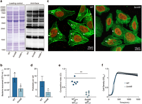
CobB is an important deacetylase that plays an important role in a wide range of prokaryotes. Thus, we investigated whether CobB affects the virulence of EHEC O157:H7 through an in vitro cell adhesion assay. HeLa cells were infected with the EHEC O157:H7 WT and ΔcobB strains, and the adherence levels were quantified by evaluating the number of bacteria that adhered to the HeLa cells. The cell adhesion assays results showed that deletion of cobB decreased the number of bacteria that adhered to HeLa cells by 2.3-fold compared to that of WT (). Fluorescent actin staining (FAS) assays were performed to evaluate A/E lesions and pedestal numbers – the most important features associated with EHEC adhesion to host cells,Citation21 in cells infected with WT or ΔcobB strains. The results showed that the pedestal number per cell found for cells infected with ΔcobB was significantly reduced () compared with that obtained for WT-infected cells. To verify the contribution of cobB to virulence in vivo, groups of infant rabbits were orally infected with the WT or ΔcobB strains, and intestinal colonization was assessed based on the bacterial load on the colon at 3 days post infection. Competitive infection assays showed that the colonization ability of the ΔcobB strain in rabbit colons was reduced by approximately 8-fold compared with that of the WT (). Importantly, the generation of growth curves in DMEM revealed that the WT and ΔcobB strains grew at similar rates (), indicating that the decrease in the gut colonization of ΔcobB was not due to different growth rate. The above results indicate that the presence of CobB contributes to the virulence of EHEC O157:H7.
Identification of the substrates of CobB using quantitative acetylome comparison
To better understand the landscape of acetylation in EHEC O157:H7 and how it regulates protein deacetylation to influence bacterial virulence, comparative acetylation proteomics were used to identify the protein modification sites regulated by CobB. First, WT and ΔcobB cells were lysed in urea-containing buffer and then digested with trypsin. Immune affinity enrichment of the acetylated peptides was conducted using a pan-anti-acetyllysine antibody conjugated to protein A agarose beads. The enriched peptides were profiled via LC‒MS/MS. The acquired mass spectrometric data were processed and analyzed by PEAKS software to identify acetylated peptides and quantify their relative abundance in the two groups of cells. We considered a peptide to be acetylated if the false recovery rate (FDR) was less than or equal to 1%. Finally, we identified 8978 acetylated peptides and 2128 acetylated proteins (Supplementary Datasets), which accounted for 39% of all proteins in EHEC O157:H7. Among the 2128 acetylated proteins, there were several heavily acetylated proteins, such as RNA polymerase beta prime subunit (40 sites), enolase (ENO) (20 sites), glyceraldehyde-3-phosphate dehydrogenase A (GapA) (16 sites), the HTH-type transcriptional regulator MalT (8 sites), and ribosomal proteins acetylated at multiple sites (Supplementary Datasets).
Since CobB has been shown to be a deacetylase enzyme in EHEC O157:H7, we focused on proteins with an increased abundance of acetylation in response to cobB deletion. At the peptide level, 8540 acetylated sites overlapped, 120 acetylated sites were only present in WT, and 318 acetylated sites were only present in ΔcobB ().
Figure 2. Identification of the substrates of CobB by using quantitative acetylome comparison of ΔcobB and WT.
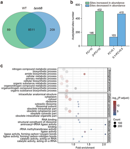
Among all acetylated peptides present in both WT and ΔcobB, we compared those that had a fold change greater than two. As shown in , the abundance of 581 acetylated peptides increased significantly (fold change ≥2.0, p < 0.05) in ΔcobB; these peptides belonged to 426 proteins. GO analysis showed that these proteins were mainly enriched in metabolism, biosynthesis, and diversity-binding activities mainly occurring in the ribosome and cytosol (). Among these substrates with increased abundance in ΔcobB, 11 sites increased more than 100-fold in 11 proteins (Supplementary Datasets), such as K251 of Serine hydroxymethyltransferase (998-fold), K195 of ENO (331-fold) and K138 of GapA (300-fold). In particular, acetylation of the K44 site on the LEE chaperone CesA in the T3SS was increased nearly 705-fold in ΔcobB, suggesting that the acetylation of K44 on the CesA protein may be regulated by CobB. Overall, our data revealed that lysine acetylation in EHEC O157:H7 is prevalent and suggests that acetylation-mediated signaling is involved in a broad range of cellular functions.
Deacetylation of the K44 site of the CesA protein enhanced EHEC O157:H7 adhesion to HeLa cells and bacterial colonization in the colon
A previous study showed that CesA is a chaperone for the translocator protein EspA, and is essential for maintaining the stability of EspA within bacterial cells prior to secretion.Citation25 EspA is an important virulence factor that is required to form A/E lesions in vivo. Therefore, we speculated that reversible acetylation of the K44 site on CesA may regulate the virulence of EHEC O157:H7 by altering the stability of EspA.
To test this hypothesis, we performed cell adhesion assays and infant rabbit colonization experiments by generating deletion mutants of cesA (ΔcesA) to study the role of CesA in regulating the virulence of EHEC O157:H7. As shown in , the ability of ΔcesA to adhere to HeLa cells was reduced by 3.8-fold compared to that of the WT strain; however, the level of adherence was restored to that of WT by complementation with cesA (ΔcesA+). FAS assays further showed that both the percentage of HeLa cells that formed pedestals and the number of pedestals formed per infected cell were significantly reduced by cesA deletion (). Infant rabbit colonization experiments showed that colonization by the ΔcesA strain decreased by approximately 7.8-fold compared with colonization by the WT strain, whereas in vivo colonization by the ΔcesA+ strain was restored to the level of the WT strain.
Figure 3. Assays of bacterial adhesion to HeLa cells and bacterial colonization of rabbits.
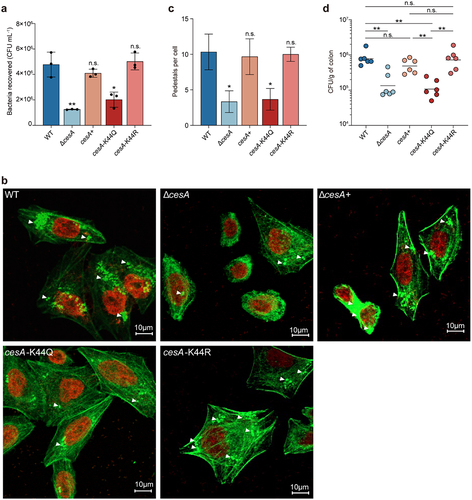
To further study the role of the acetylated K44 site in CesA, we constructed two plasmids (pBlue-cesA-K44Q and pBlue-cesA-K44R) harboring cesA with K44 mutated to Q or R. The K-to-Q (replacement of lysine by glutamine) substitution mimics constitutive acetylation by changing the positive charge to a neutral charge, whereas the K-to-R (replacement of lysine by arginine) substitution mimics the deacetylated state. We then introduced the pBlue-cesA-K44Q and pBlue-cesA-K44R plasmids into the ΔcesA strain, generating the strains cesA-K44Q and cesA-K44R, respectively. As shown in , cell adhesion and infant rabbit colonization assays were performed to examine the role of acetylated K44 in the CesA-mediated regulation of EHEC O157:H7 virulence. The adherence of strain cesA-K44Q to HeLa cells was reduced 2.37-fold compared to that of the WT (), and there were fewer A/E lesions in cells infected with cesA-K44Q than in cells infected with cesA-K44R or the complemented strain ΔcesA (+) (). Furthermore, the rabbit colonization results showed that the intestinal colonization ability of strain cesA-K44Q was significantly decreased compared to that of WT and ΔcesA (+), but the intestinal colonization ability of cesA-K44R was similar to that of WT and ΔcesA (+) (). Overall, the above results indicate that deacetylation of the K44 site on the CesA protein increases the virulence of EHEC O157:H7.
Deacetylation of the K44 site on CesA improves the stability of EspA
CesA is critical for maintaining the stability of EspA.Citation25 To investigate whether the stability of EspA was influenced by acetylation of the K44 site of CesA, we performed a Western blot analysis of EspA in strains cesA-K44Q and cesA-K44R for in vivo degradation assays. After the cells had grown in DMEM to the exponential phase, protein translation was blocked with 200 mg/mL chloramphenicol, a sample was collected every 20 min, and the EspA protein level was detected using an anti-EspA antibody. As shown in , the amount of EspA protein in the cesA-K44Q strain gradually decreased with time, whereas that in the cesA-K44R strain remained stable.
Figure 4. Assays of EspA stability and interaction with CesA.
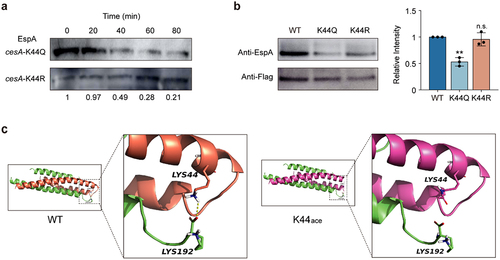
Previous studies have shown that CesA binds directly to EspA.Citation36 To further verify the acetylation of K44 directly affects the binding affinity between CesA and EspA and ultimately reduces the stability of EspA. We constructed three plasmids expressing CesA-FLAG, CesA-K44Q-FLAG, and CesA-K44R-FLAG and introduced these plasmids into ΔcesA, generating the strains cesA-Flag, cesA-K44Q-Flag, and cesA-K44R-Flag, respectively. We then immunoprecipitated CesA-FLAG, CesA-K44Q-FLAG, and CesA-K44R-FLAG from ΔcesA using an anti-Flag antibody for Western blot analysis. The amount of EspA that interacted with CesA was detected with an antibody against EspA. The results indicated that the K44Q mutation in CesA caused a marked decrease in EspA binding to CesA, whereas K44R had no such effect ().
The crystal structure of the CesA-EspA complex has been previously determined.Citation36 To further examine the possible effects of acetylation on the complex, we performed molecular dynamics simulations of the structures of wild-type EspA and CesA with K44 acetylation. The docking results demonstrated a stable interaction between wild-type EspA and CesA through hydrogen bonds. However, when acetylation was present at the K44 site, the hydrogen bond between the two disappeared (). These results showed that deacetylation of the K44 site in CesA promoted the stability of EspA, which contributed to the virulence of EHEC O157:H7.
CobB directly interacts with CesA
CobB has been identified as a NAD+-dependent lysine deacetylase.Citation31 To investigate whether deacetylation of the CesA protein was directly mediated by CobB in EHEC O157:H7, we immunoprecipitated CesA-FLAG from ΔcesA, double-mutant ΔcobBΔcesA and cobB complemented strain ΔcobB (+) ΔcesA, and then analyzed the acetylation level of CesA-FLAG by Western blot using a pan-anti-acetyllysine antibody. The results showed that the acetylation level of the CesA-FLAG protein in ΔcobBΔcesA was greater than that in ΔcesA (). To further test whether CobB could regulate the acetylation level of CesA in vitro, we purified the proteins CesA and CobB and incubated CesA with CobB in the presence or absence of NAD+ or NAM in vitro. Western blot analysis showed that the acetylation of CesA decreased significantly in the presence NAD+, but no significant change in the acetylation of CesA was detected in the presence of NAD+ and NAM ().
Figure 5. Assays of CesA interactions with CobB.
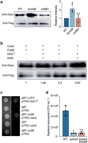
Furthermore, to confirm the interaction between CesA and CobB, a bacterial two-hybrid assay was conducted. Two plasmids were constructed: pBT-cobB, which expresses the bait protein (λcI-CobB fusion protein), and pTRG-cesA, which expresses the target protein (RNAPα-CesA fusion protein). If bait and target proteins that are cotransformed into the reporter strain interact, RNA polymerase is recruited to the promoter of the HIS3 reporter gene, where it activates transcription, which allows the growth of the reporter strain in the presence of 3-amino-1,2,4-triazole (3-AT).Citation37–39 As shown in , cell growth was observed in the presence of 5 mM 3-AT when both pBT-cobB and pTRG-cesA were cotransformed into the E. coli selection host. No cell growth was detected in the negative controls. The results confirmed that CobB interacts with CesA.
Additionally, we investigated whether CobB influences bacterial virulence via CesA. We used WT, ΔcesA and ΔcobBΔcesA to perform cell adhesion assays with HeLa cells. As expected, the adherence of ΔcesA was significantly lower than that of the WT. However, there was no further decrease in cell adherence in ΔcobBΔcesA compared to that in ΔcesA (). These data indicate that CobB regulates the virulence of EHEC O157:H7 by deacetylating CesA.
Discussion
CobB, a sirtuin-like deacetylase, plays an important role in the physiological functions of many bacteria. For example, CobB deacetylates acetyl-CoA synthase (Acs) to alter cellular energy metabolism processes.Citation14 Additionally, CobB regulates chemotaxis by deacetylating the chemotaxis response regulator protein, CheY.Citation40 In addition, CobB affects the OAT activity of NhoA, an N-hydroxyarylamine O-acetyltransferase, and can help maintain the catalytic activity of TopA, a topoisomerase I, to reduce DNA supercoiling.Citation41 Furthermore, CobB-mediated deacetylation attenuates the virulence of Yersinia pestis and reduces the virulence of V. cholerae in Drosophila.Citation32,Citation42–44 However, whether CobB is involved in regulating the virulence of EHEC O157:H7 has not yet been reported. In the present study, we systematically identified many proteins that could be regulated by CobB in EHEC O157:H7 using acetylation proteomics and discovered that CobB promotes the virulence of O157:H7 by reversing the acetylation at the K44 site on CesA, a chaperone for EspA. Given that CobB is a widely conserved protein in bacteriaCitation31 and that CesA and EspA are present in many other EPEC and EHEC serotypes, as revealed by genome sequence analysis, acetylation-mediated control by CobB may also contribute to the virulence of other EHEC and EPEC strains.
In addition to CesA, many of the central metabolic pathway enzymes identified here were lysine acetylated, suggesting that acetylation targets a broad range of fundamental cellular processes, ranging from the control of binding to metabolic pathways. Proteins such as enolase (ENO), glyceraldehyde-3-phosphate dehydrogenase A (GapA), and 2-isopropylmalate synthase (IPMSs) are key enzymes in the glycolytic or biosynthetic pathwayCitation45–47 and are hyperacetylated in ΔcobB. ENO is a glycolytic enzyme that catalyzes the dehydration of 2-phospho-d-glycerate to form phosphoenolpyruvate and the reverse of gluconeogenesis. Previous studies have shown that acetylation of ENO at K326 is regulated by CobB, thereby influencing cell growth.Citation48 The acetylation of K195 on ENO was significantly higher in ΔcobB, suggesting a markedly stronger regulation of cell growth by the acetylation of K195. MalT is an HTH-type transcriptional activator that controls the expression of the maltose regulon in E. coli,Citation49 and its activity is strictly dependent on the presence of ATP.Citation50 We observed that acetylation at K25 occurs in the ATPase domain of MalT, which is important for ATP binding.Citation49 This finding indicates that acetylation likely modulates the ability of MalT to bind ATP, which may influence maltose uptake and utilization.Citation49 Moreover, ribosomal proteins acetylated at multiple sites probably affect the ribosome assembly and activity.Citation51 Collectively, these results suggest that lysine acetylation of proteins may play an important role in a variety of bacterial physiological processes. Further studies are warranted to fully understand the role of lysine acetylation in the regulation of EHEC virulence.
Materials and methods
Ethics statement
All animal experiments were performed in accordance with the standards set forth in the Guide for the Care and Use of Laboratory Animals. The experimental protocols were approved by the Institutional Animal Care Committee of Nankai University.
Strains, plasmids, primers, and media
All wild-type strains and their derivatives, as well as the plasmids, and primers used in this study, are described in Supplementary Table S1 and Supplementary Table S2. Mutant strains were generated using the λ-red recombinase system. Briefly, the pKD46 plasmid was introduced into the WT strain to express three proteins (Exo, Beta, and Gam) required for homologous recombination. Kanamycin- and chloramphenicol-resistant fragments were amplified using the pKD3 or pKD4 plasmid as a template. All the strains were cultured overnight in LB or DMEM at 37°C.
Bacterial adhesion to HeLa cells
Adherence assays were performed as described previously.Citation30 Briefly, bacteria were added to HeLa cells at a multiplicity of infection (MOI) of 10. At 3 h post-infection, the HeLa cells were washed three times with PBS to remove non-adherent bacteria and were treated with 1 mL of 0.1% Triton X-100 for 5 min. The gradient-diluted cell lysates were spread on agar plates and the bacterial colonies were counted. The adhesion efficiency of different strains was compared by calculating the number of bacteria per mL.
Growth curve
The overnight strains cultured in LB broth at 37°C and 180 rpm were transferred to 50 mL of LB broth. Subsequently, a 200 μl aliquot was added to a 96-well plate. The absorbance at OD600 nm was measured and recorded every 30 min for 24 h at 37°C and 180 rpm automatically using a multifunctional microplate tester (TECAN Spark, Shanghai, China). Three independent experiments were performed, and the results were analyzed.
Intestinal colonization assay
Three-day-old female New Zealand white rabbits were housed with their mother. Infant rabbits were infected as described previously.Citation52 Overnight growing bacteria were centrifuged at 5500 × g and resuspended in PBS at a 10-fold concentration. Each rabbit was gavaged with 100 µL of bacterial solution containing 109 CFUs of bacteria at the logarithmic stage of growth. After 3 days of infection, length of 1–3 cm of colon tissue was separated and weighed, after which the homogenates were diluted and plated on LB agar. The attachment efficiency in vivo was determined by counting the number of CFUs per gram of the colon.
For the in vivo competition assay, WTnadi, WTkana, ΔcobB and WTnadi strains were grown overnight at 37°C in LB broth. Approximately 105 CFUs of the WTnadi strain were mixed with the WTkana at a ratio of 1:1. Similarly, 105 CFUs of the ΔcobB strain were mixed with the WTnadi at a ratio of 1:1, and the mixtures were gavaged into six rabbits. The competitive index (CI) was determined as the output ratio divided by the input ratio.
Mass spectrometry-based label-free quantitative acetylated proteomics
Mass spectrometry analysis was performed blindly using the Applied Protein Technology (Shanghai, China). Briefly, urea (8 M urea, 100 mM Tris/HCl, pH 8.5) buffer was used for sample lysis and protein extraction. Trypsin was then added to the samples for overnight digestion. Acetylated peptides were enriched using a PTMScan Acetyl-Lysine Motif Kit (Cell Signalling Technology, #13416). LC-MS/MS analysis was performed on a timsTOF Pro mass spectrometer (Bruker) coupled to a nanoelute (Bruker Daltonics) for 60 min. The raw MS data for each sample were combined and searched using PEAKS software with the NCBI_EDL933_17828_20210510.fasta protein database for identification and quantification.
Immunoprecipitation and Western blot
The cells were pelleted by centrifugation at 4°C, washed three times, resuspended in PBS, and subsequently disrupted by sonication. The cell lysates were mixed by rotation with pretreated anti-Flag magnetic beads (MCE, # HY-K0207), and proteins with FLAG tags were obtained according to the manufacturer’s protocol. Purified CesA-Flag protein was analyzed by Western blot using an anti-Flag antibody (Meck, #F1804) and pan-anti-acetyllysine antibody (PTM Bio, #PTM-101).
For Western blot, purified proteins were loaded onto a 4%-12% SDS-polyacrylamide gel electrophoresis (SDS-PAGE) gel and transferred to a polyvinylidene fluoride (PVDF) membrane (Millipore, #IPVH00010). The membranes were blocked with 5% (w/v) skimmed milk powder dissolved in Tris-buffered saline (TBS) containing 0.05% (v/v) Tween 20, incubated for 1 h at room temperature and then incubated overnight at 4°C with anti-Flag and pan-anti-acetyllysine antibody as the primary antibodies at a dilution of 1:10000. Next, goat anti-mouse (Thermo Scientific, #31430) or goat anti-rabbit (Thermo Scientific, #31460) IgG conjugated with horseradish peroxidase (HRP) was added to the blot at a dilution of 1:10000 and incubated. Finally, the signal was detected using an ECL system (Thermo Scientific, USA) and images were acquired using an Amersham Imager 600 system (General Electric).
Bioinformatic analysis
The GO annotations of the proteins were divided into three broad categories: biological process, cellular component, and molecular function. Enrichment analysis was performed using Fisher’s exact test with all quantified proteins as the background dataset. The Benjamini–‒Hochberg correction for multiple comparisons was further applied to adjust the p values. Only functional categories and pathways with p values under the threshold of 0.05 were considered significant.
Molecular docking
Crystal structures were obtained from PDB (PDB:1XOU). The Protein Preparation Wizard module in Maestro v.12.8 was employed for processing the protein structure. The overall structural charges and protonation states were prepared as necessary. The orientation of the water molecules and other functional groups, such as amides and hydroxyls, was checked. The acetylated structure was obtained using AutoDock Vina software for covalent docking. The results were analyzed and visualized using PyMOL software.
Degradation of EspA in vivo
The stability of EspA was determined using in vivo degradation experiments. cesA-K44Q or cesA-K44Q strains grown overnight were inoculated at 1:100 in DMEM at 37°C. When the OD600 nm reached 0.7, translation was blocked with 200 mg/mL chloramphenicol, and 2 mL of the bacterial solution was collected every 20 min and centrifuged at 4°C. The cell pellets were resuspended in radioimmunoprecipitation assay (RIPA) lysis buffer (Solarbio, #R0020) for 10 min. The supernatant was then separated for Western blot analysis.
In vitro deacetylation assay
The purified CobB and CesA-Flag proteins were incubated in reaction buffer [50 mM Tris-HCl, 137 mM NaCl, 2.7 mM KCl, 1 mM MgCl2, and 1 mM DTT (pH 8)] with or without 0.5 mM NAD+ and in the presence or absence of 10 mM NAM. The mixture was incubated at 25°C for 10 h. The reaction products were separated by SDS‒PAGE and analyzed by Western blot using a pan-anti-acetyllysine antibody.
Fluorescent actin staining
Fluorescent actin staining was performed as previously described.Citation53 Briefly, overnight bacteria grown at 37°C and 180 rpm were subcultured at 1:100 in DMEM. Cultures in the exponential phase were added to HeLa cells grown overnight on sterile coverslips in 6-well plates at a ratio of 1:100 and incubated for 3 h. The coverslips were then washed with PBS and fixed with 4% formaldehyde. After permeabilization with 0.2% Triton-X, the actin filaments of the cells were stained and visualized with fluorescein isothiocyanate-labeled phalloidin, and the cell nuclei of both were observed with propidium iodide. A minimum of 50 HeLa cells were counted to determine the number of A/E lesions formed by each bacterial strain.
Bacterial two-hybrid assay
The protein–protein interaction between CobB and CesA was detected using the BacterioMatch II Two-Hybrid System Vector (Agilent Technologies; #240065) according to a previously reported procedure.Citation16 The cobB and cesA genes were cloned and inserted into the pBT and pTRG plasmids, respectively. The reporter strain was cotransformed with the recombinant vectors pBT-cesA and pTRG-cobB and subsequently spotted onto screening media supplemented with 5 mM 3-amino-1,2,4-triazole (3-AT), 12.5 mg/ml tetracycline, 12.5 mg/ml streptomycin, and 25 mg/ml chloramphenicol. A cotransformant containing pBT-LGF2 and pTRG-Gal11P was used as a positive control for growth on the screening medium. A cotransformant containing the empty vector pBT and pTRG was used as a negative control
Author contributions
B. L. and M. W. designed the research; L. L., B. Y., J. W., Y. W., B. X., Y. L., W. L., P. W., Y. W., M. L., X. Z., R. L., G. M., and T. F. performed the experiments; L. L., B. Y., Y. W., J. Q., B. X. analyzed the data; and B. L., L. L., B. Y., J. W., and Y. W. wrote the manuscript.
0311_Revised_Supplementary Information.docx
Download MS Word (25.6 KB)0311_Revised_Supplementary Datasets.xlsx
Download MS Excel (1.7 MB)Disclosure statement
No potential conflict of interest was reported by the author(s).
Data availability statement
The relevant data are provided in the manuscript (Supplementary Information). MS proteomics data were deposited in the ProteomeXchange Consortium (http://proteomecentral.proteomexchange.org) via the iProX partner repository with the dataset identifier PXD045950.
Supplementary material
Supplemental data for this article can be accessed online at https://doi.org/10.1080/19490976.2024.2331435
Additional information
Funding
References
- Chen Y, Sprung R, Tang Y, Ball H, Sangras B, Kim SC, Falck JR, Peng J, Gu W, Zhao Y. et al. Lysine propionylation and butyrylation are novel post-translational modifications in histones. Mol Cell Proteomics: MCP. 2007 May;6(5):812–14. doi:10.1074/mcp.M700021-MCP200.
- Hu LI, Lima BP, Wolfe AJ. Bacterial protein acetylation: the dawning of a new age. Mol Microbiol. 2010 Jul 1;77(1):15–21. doi:10.1111/j.1365-2958.2010.07204.x.
- Sun M, Xu J, Wu Z, Zhai L, Liu C, Cheng Z, Xu G, Tao S, Ye B-C, Zhao Y. et al. Characterization of Protein Lysine Propionylation in Escherichia coli: Global Profiling, Dynamic Change, and Enzymatic Regulation. J Proteome Res. 2016 Dec 2;15(12):4696–4708. doi:10.1021/acs.jproteome.6b00798.
- Peng C, Lu Z, Xie Z, Cheng Z, Chen Y, Tan M, Luo H, Zhang Y, He W, Yang K. et al. The first identification of lysine malonylation substrates and its regulatory enzyme. Mol Cell Proteomics: MCP. 2011 Dec;10(12):M111 012658. doi:10.1074/mcp.M111.012658.
- Tan M, Luo H, Lee S, Jin F, Yang J, Montellier E, Buchou T, Cheng Z, Rousseaux S, Rajagopal N. et al. Identification of 67 histone marks and histone lysine crotonylation as a new type of histone modification. Cell. 2011 Sep 16;146(6):1016–28. doi:10.1016/j.cell.2011.08.008.
- Zhang Z, Tan M, Xie Z, Dai L, Chen Y, Zhao Y. Identification of lysine succinylation as a new post-translational modification. Nat Chem Biol. 2011 Jan;7(1):58–63. doi:10.1038/nchembio.495.
- Hong G, Su X, Xu K, Liu B, Wang G, Li J, Wang R, Zhu M, Li G. Salt stress downregulates 2-hydroxybutyrylation in Arabidopsis siliques. J Proteomics. 2022 Jan 6;250:104383. doi:10.1016/j.jprot.2021.104383.
- Zhang D, Tang Z, Huang H, Zhou G, Cui C, Weng Y, Liu W, Kim S, Lee S, Perez-Neut M. et al. Metabolic regulation of gene expression by histone lactylation. Nature. 2019 Oct;574(7779):575–580. doi:10.1038/s41586-019-1678-1.
- Okada AK, Teranishi K, Ambroso MR, Isas JM, Vazquez-Sarandeses E, Lee J-Y, Melo AA, Pandey P, Merken D, Berndt L. et al. Lysine acetylation regulates the interaction between proteins and membranes. Nat Commun. 2021 Nov 9;12(1):6466. doi:10.1038/s41467-021-26657-2.
- Narita T, Weinert BT, Choudhary C. Functions and mechanisms of non-histone protein acetylation. Nat Rev Mol Cell Biol. 2019 Mar;20(3):156–174. doi:10.1038/s41580-018-0081-3.
- Castano-Cerezo S, Bernal V, Post H, Fuhrer T, Cappadona S, Sánchez‐Díaz NC, Sauer U, Heck AJ, Altelaar AM, Cánovas M. et al. Protein acetylation affects acetate metabolism, motility and acid stress response in Escherichia coli. Mol Syst Biol. 2014 Nov 27;10(11):762. doi:10.15252/msb.20145227.
- Wolfe AJ. Bacterial protein acetylation: new discoveries unanswered questions. Curr Genet. 2016 May;62(2):335–41. doi:10.1007/s00294-015-0552-4.
- Dong H, Zhang J, Zhang H, Han Y, Lu C, Chen C, Tan X, Wang S, Bai X, Zhai G. et al. YiaC and CobB regulate lysine lactylation in Escherichia coli. Nat Commun. 2022 Nov 4;13(1):6628. doi:10.1038/s41467-022-34399-y.
- Starai VJ, Celic I, Cole RN, Boeke JD, Escalante-Semerena JC. Sir2-dependent activation of acetyl-CoA synthetase by deacetylation of active lysine. Science. 2002 Dec 20;298(5602):2390–2392. doi:10.1126/science.1077650.
- AbouElfetouh A, Kuhn ML, Hu LI, Scholle MD, Sorensen DJ, Sahu AK, Becher D, Antelmann H, Mrksich M, Anderson WF. et al. The E. coli sirtuin CobB shows no preference for enzymatic and nonenzymatic lysine acetylation substrate sites. Microbiologyopen. 2015 Feb;4(1):66–83. doi:10.1002/mbo3.223.
- Ren J, Sang Y, Tan Y, Tao J, Ni J, Liu S, Fan X, Zhao W, Lu J, Wu W. et al. Acetylation of lysine 201 inhibits the DNA-Binding ability of PhoP to regulate salmonella virulence. PloS Pathog. 2016 Mar;12(3):e1005458. doi:10.1371/journal.ppat.1005458.
- Ma Q, Pan Y, Chen Y, Yu S, Huang J, Liu Y, Gong T, Zou J, Li Y, Li Y. Acetylation of glucosyltransferases regulates Streptococcus mutans biofilm formation and virulence. PloS Pathog. 2021 Dec;17(12):e1010134. doi:10.1371/journal.ppat.1010134.
- Liu B, Liu Y, Yang B, Wang Q, Liu X, Qin J, Zhao K, Li F, Feng X, Li L. et al. Escherichia coli O157: H7 senses microbiota-produced riboflavin to increase its virulence in the gut. Proc Natl Acad Sci U S A. 2022 Nov 29;119(48):e2212436119. doi:10.1073/pnas.2212436119.
- Kaper JB, Nataro JP, Mobley HL. Pathogenic Escherichia coli. Nat Rev Microbiol. 2004 Feb;2(2):123–40. doi:10.1038/nrmicro818.
- Karmali MA, Gannon V, Sargeant JM. Verocytotoxin-producing Escherichia coli (VTEC). Vet Microbiol. 2010 Jan 27;140(3–4):360–70. doi:10.1016/j.vetmic.2009.04.011.
- Jarvis KG, Giron JA, Jerse AE, McDaniel TK, Donnenberg MS, Kaper JB. Enteropathogenic Escherichia coli contains a putative type III secretion system necessary for the export of proteins involved in attaching and effacing lesion formation. Proc Natl Acad Sci U S A. 1995 Aug 15;92(17):7996–8000. doi:10.1073/pnas.92.17.7996.
- Fernandez-Brando RJ, Yamaguchi N, Tahoun A, McAteer SP, Gillespie T, Wang D, Argyle SA, Palermo MS, Gally DL. Type III Secretion-Dependent Sensitivity of Escherichia coli O157 to Specific Ketolides. Antimicrob Agents Chemother. 2016 Jan;60(1):459–470. doi:10.1128/AAC.02085-15.
- Crepin VF, Shaw R, Abe CM, Knutton S, Frankel G. Polarity of enteropathogenic Escherichia coli EspA filament assembly and protein secretion. J Bacteriol. 2005 Apr;187(8):2881–2889. doi:10.1128/JB.187.8.2881-2889.2005.
- Chen L, Ai X, Portaliou AG, Minetti CSA, Remeta D, Economou A, Kalodimos C. Substrate-activated conformational switch on chaperones encodes a targeting signal in type III secretion. Cell Rep. 2013 Mar 28;3(3):709–715. doi:10.1016/j.celrep.2013.02.025.
- Creasey EA, Friedberg D, Shaw RK, Umanski T, Knutton S, Rosenshine I, Frankel G. CesAB is an enteropathogenic Escherichia coli chaperone for the type-III translocator proteins EspA and EspB. Microbiol. 2003 Dec;149(12):3639–3647. doi:10.1099/mic.0.26735-0.
- Abe A, Heczko U, Hegele RG, Brett Finlay B. Two enteropathogenic Escherichia coli type III secreted proteins, EspA and EspB, are virulence factors. J Exp Med. 1998 Nov 16;188(10):1907–1916. doi:10.1084/jem.188.10.1907.
- Jia T, Liu B, Mu H, Qian C, Wang L, Li L, Lu G, Zhu W, Guo X, Yang B. et al. A novel small RNA promotes motility and virulence of enterohemorrhagic Escherichia coli O157: H7 in response to ammonium. mBio. 2021 Mar 9;12(2):10–128. doi:10.1128/mBio.03605-20.
- Kumar A, Russell RM, Pifer R, Menezes-Garcia Z, Cuesta S, Narayanan S, MacMillan JB, Sperandio V. The Serotonin Neurotransmitter Modulates Virulence of Enteric Pathogens. Cell Host Microbe. 2020 Jul 8;28(1):41–53.e8. doi:10.1016/j.chom.2020.05.004.
- Liu Y, Han R, Wang J, Yang P, Wang F, Yang B. Magnesium Sensing Regulates Intestinal Colonization of Enterohemorrhagic Escherichia coli O157:H7. mBio. 2020 Nov 10;11(6):10–128. doi:10.1128/mBio.02470-20.
- Yang B, Feng L, Wang F, Wang L. Enterohemorrhagic Escherichia coli senses low biotin status in the large intestine for colonization and infection. Nat Commun. 2015 Mar 20;6(1):6592. doi:10.1038/ncomms7592.
- Liu CX, Wu FL, Jiang HW, He X, Guo, SJ, Tao, SC. Global identification of CobB interactors by an Escherichia coli proteome microarray. Acta Biochim Biophys Sin (Shanghai). 2014 Jul;46(7):548–55. doi:10.1093/abbs/gmu038.
- Sang Y, Ren J, Ni J, Tao J, Lu J, Yao Y-F. Protein Acetylation Is Involved in Salmonella enterica Serovar Typhimurium Virulence. J Infect Dis. 2016 Jun 1;213(11):1836–1845. doi:10.1093/infdis/jiw028.
- Kim SY, Sim CK, Zhang Q, Tang H, Brunmeir R, Pan H, Karnani N, Han W, Zhang K, Xu F. et al. An alternative strategy for pan-acetyl-lysine antibody generation. PloS One. 2016;11(9):e0162528. doi:10.1371/journal.pone.0162528.
- Xu W, Zhao S. Generation and characterization of pan-specific anti-acetyllysine antibody. Methods Mol Biol. 2013;981:137–150.
- Zhao K, Chai X, Marmorstein R. Structure and substrate binding properties of cobB, a Sir2 homolog protein deacetylase from Escherichia coli. J Mol Biol. 2004 Mar 26;337(3):731–41. doi:10.1016/j.jmb.2004.01.060.
- Yip CK, Finlay BB, Strynadka NC. Structural characterization of a type III secretion system filament protein in complex with its chaperone. Nat Struct Mol Biol. 2005 Jan;12(1):75–81. doi:10.1038/nsmb879.
- Dove SL, Hochschild A. Conversion of the omega subunit of Escherichia coli RNA polymerase into a transcriptional activator or an activation target. Genes Dev. 1998 Mar 1;12(5):745–54. doi:10.1101/gad.12.5.745.
- Dove SL, Joung JK, Hochschild A. Activation of prokaryotic transcription through arbitrary protein-protein contacts. Nature. 1997 Apr 10;386(6625):627–630. doi:10.1038/386627a0.
- Joung JK, Ramm EI, Pabo CO. A bacterial two-hybrid selection system for studying protein-DNA and protein-protein interactions. Proc Natl Acad Sci U S A. 2000 Jun 20;97(13):7382–7387. doi:10.1073/pnas.110149297.
- Li R, Gu J, Chen YY, Xiao C-L, Wang L-W, Zhang Z-P, Bi L-J, Wei H-P, Wang X-D, Deng J-Y. et al. CobB regulates Escherichia coli chemotaxis by deacetylating the response regulator CheY. Mol Microbiol. 2010 Jun 1;76(5):1162–74. doi:10.1111/j.1365-2958.2010.07125.x.
- Zhou Q, Zhou YN, Jin DJ, Tse-Dinh Y-C. Deacetylation of topoisomerase I is an important physiological function of E. coli CobB. Nucleic Acids Res. 2017 May 19;45(9):5349–5358. doi:10.1093/nar/gkx250.
- Liimatta K, Flaherty E, Ro G, Nguyen DK, Prado C, Purdy AE. A Putative Acetylation System in Vibrio cholerae Modulates Virulence in Arthropod Hosts. Appl Environ Microbiol. 2018 Nov 1;84(21):e01113–18. doi:10.1128/AEM.01113-18.
- Liu W, Tan Y, Cao S, Zhao H, Fang H, Yang X, Wang T, Zhou Y, Yan Y, Han Y. et al. Protein acetylation mediated by YfiQ and CobB is involved in the virulence and stress response of Yersinia pestis. Infect Immun. 2018 Jun;86(6):10–128. doi:10.1128/IAI.00224-18.
- Xu Z, Wang L, Wang X, Wan M, Tang M, Ding Y. Characterizing the effect of the lysine deacetylation modification on enzyme activity of pyruvate kinase I and pathogenicity of vibrio alginolyticus. Front Vet Sci. 2022;9:877067. doi:10.3389/fvets.2022.877067.
- Antikainen J, Kuparinen V, Lahteenmaki K, Korhonen TK. Enolases from Gram-positive bacterial pathogens and commensal lactobacilli share functional similarity in virulence-associated traits. FEMS Immunol Med Microbiol. 2007 Dec;51(3):526–534. doi:10.1111/j.1574-695X.2007.00330.x.
- Toyoda K, Teramoto H, Inui M, Yukawa H. Expression of the gapA gene encoding glyceraldehyde-3-phosphate dehydrogenase of Corynebacterium glutamicum is regulated by the global regulator SugR. Appl Microbiol Biotechnol. 2008 Nov;81(2):291–301. doi:10.1007/s00253-008-1682-0.
- Yoshida A, Kosono S, Nishiyama M. Characterization of two 2-isopropylmalate synthase homologs from thermus thermophilus HB27. Biochem Biophys Res Commun. 2018 Jun 22;501(2):465–470. doi:10.1016/j.bbrc.2018.05.013.
- Dong H, Zhai G, Chen C, Bai X, Tian S, Hu D, Fan E, Zhang K. Protein lysine de-2-hydroxyisobutyrylation by CobB in prokaryotes. Sci Adv. 2019 Jul;5(7):eaaw6703. doi:10.1126/sciadv.aaw6703.
- Dardonville B, Raibaud O. Characterization of malT mutants that constitutively activate the maltose regulon of Escherichia coli. J Bacteriol. 1990 Apr;172(4):1846–52. doi:10.1128/jb.172.4.1846-1852.1990.
- Richet E, Raibaud RO. MalT, the regulatory protein of the Escherichia coli maltose system, is an ATP-dependent transcriptional activator. EMBO J. 1989 Mar;8(3):981–7. doi:10.1002/j.1460-2075.1989.tb03461.x.
- Hansen AM, Chaerkady R, Sharma J, Díaz-Mejía JJ, Tyagi N, Renuse S, Jacob HKC, Pinto SM, Sahasrabuddhe NA, Kim M-S. et al. The Escherichia coli phosphotyrosine proteome relates to core pathways and virulence. PloS Pathog. 2013;9(6):e1003403. doi:10.1371/journal.ppat.1003403.
- Ritchie JM, Thorpe CM, Rogers AB, Waldor MK. Critical roles for stx2, eae, and tir in enterohemorrhagic Escherichia coli-induced diarrhea and intestinal inflammation in infant rabbits. Infect Immun. 2003 Dec;71(12):7129–7139. doi:10.1128/IAI.71.12.7129-7139.2003.
- Feng L, Yang B, Xu Y, Xiong Y, Wang F, Liu B, Yang W, Yao T, Wang L. Elucidation of a complete mechanical signaling and virulence activation pathway in enterohemorrhagic Escherichia coli. Cell Rep. 2022 Apr 5;39(1):110614. doi:10.1016/j.celrep.2022.110614.