
ABSTRACT
Obesity, insulin resistance (IR), and the gut microbiome intricately interplay in Metabolic-associated Steatotic Liver Disease (MASLD), previously known as Non-Alcoholic Fatty Liver Disease (NAFLD), a growing health concern. The complex progression of MASLD extends beyond the liver, driven by “gut-liver axis,” where diet, genetics, and gut-liver interactions influence disease development. The pathophysiology of MASLD involves excessive liver fat accumulation, hepatocyte dysfunction, inflammation, and fibrosis, with subsequent risk of hepatocellular carcinoma (HCC). The gut, a tripartite barrier, with mechanical, immune, and microbial components, engages in a constant communication with the liver. Recent evidence links dysbiosis and disrupted barriers to systemic inflammation and disease progression. Toll-like receptors (TLRs) mediate immunological crosstalk between the gut and liver, recognizing microbial structures and triggering immune responses. The “multiple hit model” of MASLD development involves factors like fat accumulation, insulin resistance, gut dysbiosis, and genetics/environmental elements disrupting the gut-liver axis, leading to impaired intestinal barrier function and increased gut permeability. Clinical management strategies encompass dietary interventions, physical exercise, pharmacotherapy targeting bile acid (BA) metabolism, and microbiome modulation approaches through prebiotics, probiotics, symbiotics, and fecal microbiota transplantation (FMT). This review underscores the complex interactions between diet, metabolism, microbiome, and their impact on MASLD pathophysiology and therapeutic prospects.
Introduction
By the beginning of the third millennium, the prevalence of obesity and metabolic diseases dramatically increased, and became a real burden for health systems in the Western world.Citation1
Metabolic dysfunction-associated steatotic liver disease (MASLD), previously known as Non-Alcoholic Fatty Liver disease (NAFLD), is a global disease that affects about 25% of the world population.Citation2 Nowadays, the overall prevalence of MASLD is growing in parallel with the worldwide epidemic of obesity. The sedentary lifestyle, lack of physical exercise, an hypercaloric diet mainly composed by fat, refined sugars and carbohydrates are the main reasons and risk factors for the development of MASLD.Citation3 However other risk factors, as gender, genetic predisposition, age, smoking, or drug and alcohol consumption can influence on MASLD progression and make its pathophysiology more heterogeneous and complex.Citation4
Although the liver was initially assessed as the main organ involved in the development of MASLD, nowadays it became clear that a complex crosstalk between several organs involved in the progression of MASLD. In fact, MASLD is rather a systemic disease where obesity, metabolic syndrome (MS), white adipose tissue (WAT) inflammation and fatty liver (FL) tightly interact and push each other to further pathophysiological stages of such as hepatic inflammation, fibrosis, cirrhosis and hepatocellular carcinoma (HCC).Citation5
Recently, a growing body of experimental and clinical evidence indicated that central aspects of liver function also strongly depend on the coordinated action of gut. In fact, anatomical cellular and molecular paths connect both organs and synchronize their work. The intimate connection and the strict mutual cooperation between the gut and the liver realizes a functional entity called “gut-liver axis”.Citation6
This connection involves reciprocal interactions at cellular and molecular levels between the gut, its microbiota, and the liver, which are influenced by various factors such as the diet, genetics, and the environment.Citation7
MASLD is associated with changes in intestinal microbiota as well as intestinal barrier integrity suggesting an important role of the gut-liver axis in the development of disease.Citation8 The effect of obesity, insulin resistance (IR), the interaction and metabolic crosstalk between the gut and the gut microbiome on MASLD will be discussed in this review.
Hepatic features of MASLD
MASLD is characterized by an excessive fat accumulation (>5%) in the liver.Citation9,Citation10
The increased uptake of free fatty acids (FFA) and lipogenesis, defects in FFA oxidation, and decreased lipids export all contribute to the impaired hepatic lipid metabolism.Citation11 Extensive fat accumulation in the hepatic parenchyma led to excessive mitochondrial reactive oxygen species (ROS) production. Oxidative stress can induce mitochondrial dysfunction, and hepatocyte cytotoxicity, ending in cell death.Citation12 Extensive hepatocyte cell death and liver damage induce the recruitment of immune cells and the activation of proinflammatory pathways that further increase liver damage and induce the over presence of proinflammatory cytokines (TNFα, TGFβ, IL6, IL1β) that consequently activate the hepatic stellate cells (HSCs).Citation13 These events trigger the production of extracellular matrix (ECM) inducing collagen deposition and leading in 1–2% of the cases to advanced stages of fibrosis and cirrhosis. Advanced liver fibrosis and cirrhosis are the major risk factors for HCC.Citation14,Citation15
Gut-liver axis
The gut is one of the most extensive mucosal surfaces in the human body and serves as a barrier that safeguards against pathogenic microorganisms and toxic substances. The gastrointestinal (GI) system is also responsible for the digestion and absorption of food.Citation16 The gut and the liver are in constant crosstalk due to anatomical and functional interactions. The gut is the initial organ that receives the nutrients from the food. Consequently, the liver is exposed to substances that come directly from the gut via portal vein supplementation.Citation16,Citation17
The gut is divided into two parts, the small intestine that is also segmented into the duodenum, jejunum and ileum and the large intestine that is formed mainly by the colon and the rectum.Citation6 The structure of the small intestine is characterized by the presence of villi and crypts that increase the surface of absorption. Its internal structure is complex, and substances can be absorbed by active or passive transportCitation18 ().
Figure 1. A. Epithelium organization of the small intestine. Graphical representation of villi, crypts and different type of intestinal cells. B. Colon epithelium. Graphical representation of crypts and different type of colonic cells. Figure created with BioRender.com.
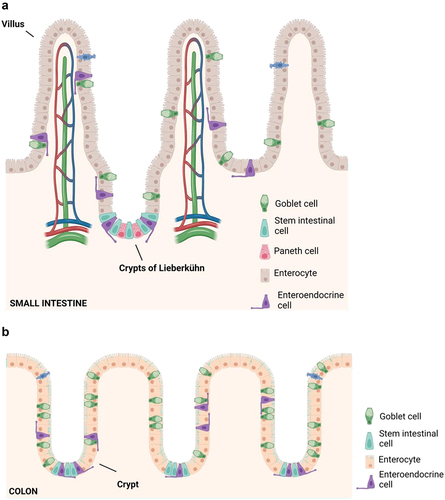
The colon lacks villi and displays a flatter surface. Epithelial cells are continuously renewed from invaginations known as the crypts of Lieberkühn, where multipotent stem cells give rise to the different cell types of intestinal epithelium: columnar absorptive cells or enterocytes, mucous secreting goblet cells, enteroendocrine cells and Paneth cellsCitation6,Citation19,Citation20 ().
The gut and the liver are anatomically linked through portal circulation which is the physical connection of the gut-liver axis. However, the presence of the intestinal barrier restricts the extent to which the intestine and liver are connected.Citation6
The gut is semi-permeable, it allows the passage of some substance while others remain in the GI tract.Citation21 Hence, GI tract is not only responsible for digestion and absorption of nutrients and other substances essential for life, but it also constitutes the primary defense against pathogens and hazardous substances preventing them to reach the blood, liver, spleen, and other organs.Citation22
However, some environmental factors, changes in the gut microbiome, toxins and substances like the alcohol or drugs, excess of fat and intestinal inflammation itself can induce changes in the enterocytes and in the mucosa, and consequently increase the intestinal permeability also called – leaky gut.Citation16
The gut barriers
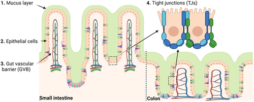
The gut barrier is comprised of three major lines of defense: 1. The mechanical barrier; 2. The immune barrier; and 3. The biological barrier. These barriers can interact with each other to maintain gut homeostasisCitation23 ().
Figure 2. Gut mechanical barrier components. 1. Mucus layer is the outer mechanical barrier composed by mucus. 2. Epithelial cells: enterocytes, goblet cells, enteroendocrine cells, Paneth cells and microfold cells. 3. Tight junctions (TJs) are composed of proteins that control the paracellular pathway, as well as adherent junctions, desmosomes, and gap junctions. 4. Gut vascular barrier (GVB) constitutes the inner layer of defense, and it is composed by endothelial cells linked by TJs among others and different proteins that play a fundamental role regulating blood vessel permeability. Figure created with BioRender.com.
The mechanical barrier
One of the components of the gut barrier is the mechanical layer composed by: a) The intestinal epithelial cells (IEC); b) Goblet cells; c). Paneth cells, d) Glial cells and e) Mucus layer.Citation22,Citation24,Citation25 All layers offer protection against mechanical, chemical, and biological agents.Citation26 All cells of the gut barrier are replenished by a group of stem cells located in the intestinal crypts.Citation23
The mucus layer
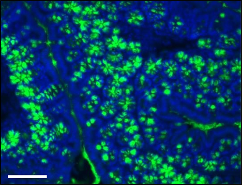
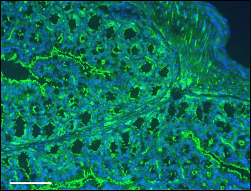
The mucosal surface of the GI tract is covered by mucus a substance secreted by goblet cells. The major building blocks and critical structural component of the mucosal barrier are mucins, which are large, highly glycosylated proteins giving the mucus its gel-like properties. The mucus produced by the Goblet cells is the classical gel-forming mucins (MUC), MUC2 (), MUC5AC, MUC6 and MUC5B which are secreted by the intestine, stomach surface, stomach glands and salivary glands, respectively.Citation27
Figure 3. Photomicrograph of immunofluorescence (IF) preparation of colon stained with mucin-2 antibody. Nuclei are stained in blue. Staining was performed in 5 μm sections from paraffin embedded colon from a C57Bl/6J mouse 20 weeks age fed with chow diet. Scale = 100 μm.
The main function of the mucus layer is to shield the intestinal cells from external agents and to facilitate nutrient absorption.Citation27 The stomach and the colon have double layer of mucus, while the small intestine has only a single layer.Citation28
n the stomach and first part of the duodenum the mucus is thicker as it serves as a stable, non-stirred layer that supports surface neutralization of acid and maintains a pH gradient from acidic to nearly neutral at the mucosal surface. Furthermore, the mucus layer acts as a physical barrier, preventing luminal pepsin from reaching the underlying mucosal surface.Citation29
In the colon there are two layers of mucus. The inner layer is densely packed, firmly attached to the epithelium and is impenetrable to bacteria. When the inner layer is penetrable to bacteria, they reach the epithelial cells and trigger inflammation. The outer layer is movable, has an expanded volume and is the natural habitat for the commensal bacteria.Citation28 Due to proteolytic cleavages of MUC2 and increased pore sizes bacteria into the mucin net-like structure and gain access to the plentiful mucin-bound carbohydrates that can be utilized by the bacteria as an energy source. In turn, the commensal bacteria produce a variety of metabolites, some of which are useful to the host.Citation27
The epithelial cells
The gut epithelium is not permeable to hydrophilic solutes, which means that molecules and nutrients can only pass through it via specific transporters. There are two primary pathways for transport: the transcellular route, which includes aqueous pores, active carrier-mediated absorption for nutrients and endocytosis, and the paracellular route, which allows ions and hydrophilic molecules to pass through.Citation30
Protein junctional complexes
The paracellular pathway is controlled by a group of proteins known as junctional complexes that include tight junctions (TJs), adherent junctions, desmosomes and gap junctions. They are located at the apical ends of the lateral membranes of IECs. They are composed by transmembrane proteins, occludins, claudins, junctional adhesion molecules and tricellulins,Citation31,Citation32 the Marvel domain-containing proteins and immunoglobulin superfamily, which interact with the cytoskeletal actomyosin ring.Citation33 The cytosolic scaffold proteins, such as zona occludens (ZO) and claudin proteins interact among them and anchor the transmembrane proteins to the actin cytoskeleton. This interaction is vital to maintain TJ barrier integrity and minimize the gut permeabilityCitation34 . Representative picture of ZO-1 in colon is shown in .
Figure 4. Schematic representation of TJ in gut. The paracellular pathway is controlled by a group of proteins known as junctional complexes that include, adherent junctions, gap junctions and tight junctions (composed by transmembrane proteins as claudins, occludin and junctional adhesion molecules (JAM)) which interact with the zona occludens (ZO) family of scaffolding proteins and the cytoskeletal actomyosin ring. Figure created with BioRender.com.
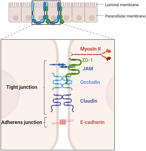
Figure 5. Photomicrograph of immunofluorescence (IF) preparation of colon stained with ZO-1. Nuclei are stained in blue. Staining was performed in 5 μm sections from paraffin embedded colon from a C57Bl/6J mouse 20 weeks age fed with chow diet. mounting. Scale = 100 μm.
Studies on gut permeability reveal that TJs selectively enable the passage of substances across the gut barrier. This selectivity is achieved through the existence of two distinct classes of trans-TJ flux pathways, each with varying size and charge selectivity. These pathways can also be differentiated based on their capacity. The pore pathway has the ability to transport significant amounts of small, uncharged solutes and specific ions, whereas the leak pathway permits only small quantities of larger molecules and ions regardless of charge to pass.Citation35 As a result, TJs provide a mechanical boundary between the luminal space and other components of the intestinal barrier.Citation36
The gut vascular barrier
The gut vascular barrier (GVB) represents the inner layer of defense in the multi-layered intestinal barrier system that finely regulates the translocation of substances from the intestinal lumen to the systemic circulation.Citation37
The GVB is mainly composed by gut endothelial cells and pericytes, which are linked by adherent junctions, TJs, catenin and cadherin proteins, and play a vital role in regulating the permeability of blood vessels.Citation38 These cells are fenestrated. The small pores are delimited by a fenestrae diaphragm regulated by plasmalemma vesicle-associated protein-1 (PV-1), which is essential for maintaining endothelial homeostasis and permeability.Citation39 Moreover, the defective GVB has been associated to an increased expression of PV-1.Citation38,Citation40,Citation41
In addition to their role in regulating vascular permeability, endothelial cells also play a role in mucosal immunology, expressing Toll-like receptors (TLRs) and adhesion molecules such as E-selectin, vascular cell adhesion molecule-2 (VCAM-1), and intercellular adhesion molecule-1 (ICAM-1). They form a layer with pericytes and enteric glial cells underneath the IEC called the GVB. Enteric glial cells help to maintain the integrity of the intestinal barrier by communicating with enteric neurons and releasing soluble factors such as S-nitroso glutathione, which controls paracellular permeability by increasing TJ protein expression.Citation39,Citation42
The immune barrier
The immune barrier is composed by a diverse range of immune cells and other types of cells that are part from the intestinal epithelium that exert immune functions.Citation43
The epithelium layer is the first line of defense against pathogens. IECs have fundamental immune-regulatory functions. They promote the development and differentiation of immune-regulatory CD8αα intraepithelial lymphocytes through the trans-presentation of IL15. Moreover, the overexpression of IL15 is crucial for facilitation the movement and localization of protective γδ-intraepithelial lymphocytes within the small intestine’s epithelial lining.Citation44
Furthermore, IECs exhibit the expression of anti-inflammatory cytokines like IL10, which likely contributes to fostering tolerance toward commensal bacteria maintaining the integrity of the epithelium.Citation45
IECs recognize pathogenic molecule patterns where MyD88 is a central adaptor molecule involved. Additionally, IECs are also involved in the adaptive immune regulation of gut homeostasis mediated by secretory immunoglobulin A (SIgA).Citation46
Specialized IECs such as Goblet and Paneth cells, play a crucial role in strengthening the barrier function. They achieve this by secreting cytokines and antimicrobial peptides such as defensins that control the overgrowth of commensal and pathogenic bacteria.Citation43,Citation46
The goblet cells have recently been shown to have a novel gate-keeping role for the presentation of oral antigens to the immune system. Goblet cells deliver small intestinal luminal material to the lamina propria dendritic cells of the tolerogenic CD103+-type.Citation27
Another type of specialized IECs is concentrated in the follicle-associated epithelium which overlays the luminal surface of lymphoid structures. These microfold cells or M cells are specialized epithelial cells that participate in inflammatory responses by capturing antigens in the luminal surface of the intestinal mucosa and transporting them to PPs through transcytosis for antigen presentation.Citation47 These cells mainly recognize bacteria-derived molecules, through TLRs and NOD-like receptors (NLRs), which activate defense mechanisms through the secretion of cytokines and chemokines that signal to the underlying immune cells and induction of IgA-secreting plasma.Citation43
Intestinal epithelial cells maintain a strong and intricate interaction with the intestinal immune cells. Interactions between IECs and macrophages and other immune cell types promotes a harmonious crosstalk mechanism that sustains a healthy environment.Citation43
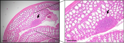
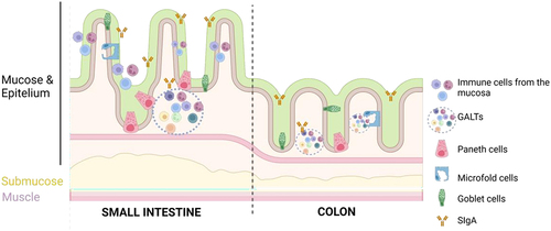
A network of intestinal innate and adaptive immune agents (dendritic cells (DCs), macrophages and lymphocytes, among others) reside within the epithelium and gut lamina propria. Moreover, lymphoid cells that include DCs, T cells, Treg cells and B cells can accumulate into gut-associated lymphoid tissue (GALT) that comprises the Peyer Patches (PPs) and Isolated Lymphoid Follicles (ILFs) depending on its location and composition.Citation43,Citation46,Citation48,Citation49 PPs are structured and highly organized lymphoid structures that are mainly located in the small intestine, whereas ILFs are more diffuse structures found in the small and large intestine.Citation46 Photomicrograph of GALT structures in colon is represented in . These sites serve as hubs for promoting tolerance to food antigens, regulating the balance of intestinal microflora, and warding off potential pathogens. Consequently, they serve as pivotal sites for triggering the adaptive immune response, capable of generating SIgA, which functions as a primary defense mechanism in the intestinal tissue.Citation50,Citation51
Figure 6. Representative photomicrograph of hematoxylin and eosin (H&E) staining in distal colon. Staining was performed in 5 μm sections from paraffin embedded colon from a C57Bl/6J mouse 20 weeks age fed with chow diet. GALT structure is marked with an arrow. Scale = 100 μm.
SIgA is mainly secreted by plasma cells located within mucosal membranes lining the gastrointestinal tract. It is produced in response to microbial- and food-derived antigens and plays different roles in intestinal mucosal secretions. It acts as first line of defense against pathogens and facilitates mucus surface colonization by commensal microbiota and regulates immune homeostasis.Citation52
This combination of physical and biochemical defenses acts as a barrier against both commensal and pathogenic microorganisms ().
Figure 7. Immune barrier. Immune cells are mainly located in the gut mucose where secretory immunoglobulin a (SIgA) can be found. Gut barrier include macrophages, lymphocytes, paneth cells (more abundant in the small intestine) and microfold cells (M). Moreover, the gut-associated lymphoid tissue (GALT) is located in the lamina propria and can be classified into Peyer’s patches (PP) or isolated lymphoid follicles. Figure created with BioRender.com.
The microbial barrier
The microbial gut barrier is the third component of the gut barrier. The intestinal microbiota consists of highly diverse communities of prokaryotic and eukaryotic microorganisms as well as viruses. After birth the gut becomes colonized with comparably few different microorganisms and the ecosystem is relatively unstable. Thereafter, diversity of the microbiota increases and eventually results in the formation of a complex microbial ecosystem.Citation53
Human adult microbiota is composed of 100 trillion microorganisms that include commensal bacteria, pro- and anti-inflammatory, pathogenic, and nonpathogenic bacteria, fungi, and viruses that maintain gut homeostasis.Citation54,Citation55 The intestinal microbiota increases in density from the small intestine to the colon, where it reaches approximately 10 trillion cells per gram of colonic content.Citation53
Gut microbiome is a complex and dynamic community that keeps a symbiotic relationship with the host.Citation56–58 During the life of an individual the microbiome changes in percentage and population. Under healthy and physiological conditions, it maintains host immune homeostasis.Citation56–58 Some phyla are usually present in the gut microbiome: Firmicutes, Bacteroidetes, Proteobacteria, Verrucomicrobia, Actinobacteria and Fusobacteria.Citation59
It provides efficient protection from infection by enteric pathogens (“colonization resistance”), stimulates maturation of the immune system and facilitates the conversion of non-digestible complex carbohydrates.Citation60,Citation61
Most often the microbiome takes part in metabolic processes including the fermentation of polysaccharides and the regulation of bile acid (BA) production. Besides, the contribute to the choline metabolism and the process of energy harvest, providing protection against pathogens or even stimulating the endogenous ethanol production.Citation62–64
However, changes in the diet, drug toxicity, a proinflammatory environment and the presence of other microbiota members have been defined as the main microbiome modifiers and dysbiosis inductors.Citation65
Hence, the excessive proliferation of some bacterial species, or the loss of some commensal bacteria, as well as variations in the total number of bacteria, is named dysbiosis, that is frequently associated to the pathogenesis of several inflammatory diseases and potential infections.Citation66,Citation67
Gut barriers and microbiome crosstalk
The interactions among microbiome and mechanical and immunological components that constitute the gut barrier are essential for intestinal and systemic homeostasis.
Microbiota and mechanical gut barrier interaction
Mucus metabolism is influenced by several factors, being one of them the microbiota that can affect the structure and function of the outer mucus layer. It has been described that germ-free animals have thinner mucus layer and fewer goblet cells.Citation68 Meanwhile, some bacterial subproducts such as lipopolysaccharide (LPS) a cell wall component from gram-negative bacteria and peptidoglycan, cell wall component from gram-positive bacteria can stimulate mucus secretion. Conversely, some resident bacteria like Akkermansia municiphila, can break down the mucus to obtain energy for themselves and other commensal bacteria in a balanced stage.Citation69,Citation70 This process is more active when the diet is low in fiber, as fiber serves as energy source for the microbiota.Citation71 Additionally, immune cells play a role in regulating mucus metabolism through the secretion of cytokines.Citation26
One of the primary roles of gut microbiota is to obtain nutrients for the intestinal cells and break down undigested dietary products, such as fiber and protein.Citation61 Specifically, by anaerobically fermenting complex carbohydrates that have not been digested, the gut microbiota can create short-chain fatty acids (SCFAs) like butyric, propionic and acetic acid. These SCFAs serve as an energy source for IECs, they can influence on cell proliferation and reduce cytokine production by neutrophils and macrophages, leading to an immunotolerogenic phenotype.Citation72 Moreover, SCFAs can enhance the production of the mucus layer by modifying the transcription of mucin genes in goblet cells and can also encourage the reassembly of TJs, thereby strengthening the intestinal epithelial barrier.Citation73,Citation74
Microbiota and immune system interaction
The gut microbiota also contributes to the development of the host’s immune system by producing metabolites, microorganism-associated molecular patterns (MAMPs), including pathogen-associated molecular patterns (PAMPs), and antigens.Citation75 Bacterial translocation refers to the process by which pathogens or their products move from the intestinal lumen to mesenteric lymph nodes (MNL).Citation76 LPS, which is present in gram-negative bacteria cell wall, is one example of a MAMP recognized by receptors on cells from the innate immune system, including TLRs. These receptors are pattern recognition receptors (PRRs) that are typically found on the surface of immune and intestinal cells and transmembrane proteins, and are capable of identifying bacterial, viral, or parasitic ligands.Citation77 When activated by pathogens or commensal bacteria, TLRs can trigger an immune response through the induction of the nuclear factor- κB (NF-κB), a group of transcription factors involved in the production of inflammatory cytokines. This symbiotic interaction between commensal bacteria and the host immune system plays a protective role in maintaining intestinal homeostasis.Citation78,Citation79
Gut and liver crosstalk
The concept of “gut-liver axis” had initially been proposed to describe the presence of antibodies directed against intestinal microorganisms and food antigens in the circulation of patients with liver cirrhosis,Citation80 the common end stage of chronic liver disease (CLD).
Both gut and liver diseases are frequently associated with a perturbed regulation of gut-liver communication and dysbiosis. This fact, together with the disruption of mechanical gut barrier, altered immune homeostasis and imbalanced bile salts pool, triggers enhanced gut permeability “leaky gut” and systemic inflammation. Alterations of the structure and functions of the gut microbiota have major effects on the gut and liver. In fact, studies show that the microbiota has an ethiopathogenic role in gut and liver diseases and that, in turn, gut and liver disease alter the enteric microbiota composition.Citation81,Citation82
Yet, the clinical relevance of the gut-liver axis is not limited to aspects concerning microbiota and bacterial dissemination. Gut- and liver-derived mediators including cytokines, hormones, bile salts and other factors, directly link the function of both organs.
Moreover, liver disease disrupts gut homeostasis and leads to changes in gut microbiota composition and intestinal permeability, which correlates with the severity of liver dysfunction. Portal hypertension causes alterations in intestinal barrier function, allowing normally restricted substances to enter the bloodstream. The translocation of bacterial products or fragments triggers the immune system activation and inflammation. This process not only exacerbates liver dysfunction, but also initiates a series of reactions throughout the body, leading to a systemic inflammatory condition characteristic of advanced liver cirrhosis.Citation83
Metabolic crosstalk between the gut and the liver
BA are amphipathic molecules that are synthetized in the hepatocytes as primary BA, cholic acid (CA) and chenodeoxycholic acid (CDCA), that are transported from the canalicular side of the hepatocyte by the bile salt export pump (BSEP) along with phospholipids and cholesterol. Conjugated BA are actively transported into the ileum. After reabsorption, BA induce the synthesis of fibroblast growth factor 15/19 (FGF-15/19). They exit the basolateral side of ileocyte via the heterodimeric organic solute transporter (Ost-a/b). BA and FGF-15/19 are transported back to the liver via the portal blood. Conjugated BA are actively transported into the hepatocyte primarily by the Na+/taurocholate co-transporting polypeptide (NTCP). FGF-15/19 binds to and activates hepatic fibroblast growth factor receptor 4 (FGFR4), which in turn activates JNK signaling pathway. Activation of JNK downregulates the gene encoding cholesterol 7α-hydroxylase (CYP7A1), inhibiting BA synthesisCitation84 ().
Figure 8. Enterohepatic BA cycle. Bile synthesis is performed in the liver by Cyp7a1 and Cyp27a11 enzymes. When synthesis is completed, BA are released in the bile canaliculi through the BSEP pump and stored in the gallbladder. In the ileum, bile salts are absorbed by ASBT and efluxed by OST-α/β to the circulation. Back to the liver they are uptaken by NTCP, a transporter located in the basolateral membrane of the hepatocytes. ABST, apical sodium-bile acid transporter; BSEP, bile salt export pump; FXR, farnesoid X receptor; NTCP, sodium taurocholate co-transporting polypeptide; OST, organic solute transporter.
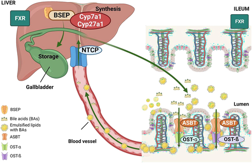
During bile salt enterohepatic circulation, several hundred milligrams of BA escape this cycle and enter in the colon where they are metabolized by gut microbiota, generating secondary BAs. These substances and derived metabolites are passively absorbed from the colon and returned to the liver via the portal vein, where they will be re-conjugated to either glycine or taurine and join primary bile salt cycle.Citation84
Briefly, one of the functions of BA is to emulate fats and bring them near the intestinal brush border membrane which results in fat absorption in the gut. The ratio of taurine to glycine BA depends on the diet in human, but not in rodents.Citation84 In diet- and obesity-induced MASLD, high fat diet (HFD) can modify BA composition due to liver damage, which potentially has an impact on the gut microbiome.Citation85–88
Moreover, BA have other metabolic actions and have been recognized as signaling molecules in the body through Farnesoid X Receptor (FXR) and TGR5, playing a key role in the control of hepatic de novo lipogenesis, very low density lipoprotein (VLDL) and plasma TG turnover. FXR is strongly expressed in the liver and the intestine, where is a regulator of BA enterohepatic circulation but can interact with fat metabolism. Some studies demonstrate that gut microbiota can also modify BA secretion though FXR, fostering lipid peroxidation and hepatic steatosis.Citation17,Citation89,Citation90
It has been recently reported that FXR deficient mice are protected against diet-induced obesity. FXR suppression is alleviated by microbial metabolism of TβMCA and βMCA. Higher levels of TβMCA are related to lower levels of FXR activity.Citation91,Citation92 It has been described that conventionally raised microbiome mice under a HFD feeding increase lipid accumulation in the liver due to higher expression in Cd36, ApoC2 and Vldlr genes when compared to germ-free (GF) mice.Citation91 These data would suggest that, in part, gut microbiota can interact to FXR and partially induce hepatic steatosis.
It has been also described that FXR activation inhibits Srebp1-c expression, and increases insulin sensitivity, reducing obesity and supressing inflammation. Moreover, the activated TGR-5 would bind to secondary BAs, stimulating GLP-1 and playing an important role in feeding signals and glucose homeostasis.Citation93
Immunological crosstalk between the gut and the liver
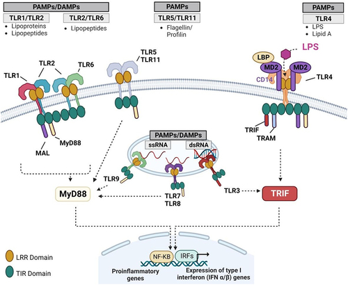
Gut microbiome is a particularly important mediator of the gut-liver axis.Citation94 Microbiome serves as the primary mechanism of interaction with the liver via TLRs. Rodents express 13 TLRs, whereas humans do have only 10. The presence of multiple widely expressed TLRs allows for the recognition of various microorganisms, triggering the appropriate immune response by the innate immune system.
PAMPs consist of microbial molecular structures like LPS from Gram-negative bacteria, lipoteichoic acid and peptidoglycan (PGN) from Gram-positive bacteria, lipoglycans, lipopeptides, and lipomannans from mycobacteria, zymosan from yeast, as well as DNA from viruses and bacteria. Damage-associated molecular patterns (DAMPs) include components of the extracellular matrix and plasma membrane, nuclear and cytosolic proteins, and elements from damaged organelles.Citation95
Each TLR possesses the ability to recognize specific molecular patterns. TLR1, TLR2, TLR4, TLR5, and TLR6 bind to molecules associated with bacterial membranes such as LPS, lipoprotein, and PGN. On the other hand, TLR3, TLR7, TLR8, and TLR9 detect viral, bacterial, or endogenous nucleic acids. TLR4, in conjunction with TLR2, can identify antigens from bacteria, fungi, parasites, viruses, and DAMPs. LPS, which is a constituent of the cell wall in gram-negative bacteria and interacts with TLR4, is one of the extensively studied mediators of host-microbe interactionsCitation95 ().
Figure 9. TLR types. TLR1, 2, 4, 5 and 6 bind to molecules associated with bacterial membranes. Concretely, TLR4, one of the most studied TLRs, binds to the LPS from the Gram-negative bacteria cell wall. TLR3, 7, 8 and 9 recognize viral, bacterial, or endogenous nucleic acids. Immune response initiation is mediated by TLR activation. DAMP, damage associated molecular patterns; LPS, lipopolysaccharide; PAMP, pathogen associated molecular patterns; TLR, toll-like receptor.
MASLD and Gut-barrier disruption
MASLD development is intricate and involves multiple factors. A more comprehensive “multiple hit model” has been recently proposed to explain disease’s onset and has gained acceptance. According to this model, the initial hit leads to increased liver fat levels, followed by the influence of various factors such as IR, gut microbiota, and genetic/environmental elements. These factors collectively affect the inflammatory environment within hepatocytes.Citation96
Fat accumulation, hepatocyte injury, and particularly, intestinal barrier damage are crucial elements in the pathophysiology of MASLD. The intestinal barrier plays a vital role in absorbing essential nutrients and preventing the intrusion of microorganisms from the gut lumen. When alterations occur in the intestinal barrier function, it leads to increased intestinal permeability, which significantly contributes to the initiation and progression of intra and extrahepatic damage in MASLD.Citation96,Citation97 MASLD is linked to gut barrier disruption, changes in TJs, rise in intestinal permeability and dysbiosis.
Mechanical barrier disruption in MASLD
Loss of mucus and chemical substances in the gut barrier can lead to bacterial overgrowth in the GI tract, disrupting gut homeostasis and permeability.Citation96
Recent studies have demonstrated that heightened inflammation in the intestinal mucosa and damage to the intestinal epithelial barrier increase the possibility of microbial translocation contributing to MASLD. Concretely, disruption and alterations in the TJs has been described in Metabolic dysfunction Associated Steatohepatitis (MASH) patients and rodents’ model.Citation98,Citation99 Under conditions of hypoxia and inflammatory stimulation, TJ proteins exhibit contractions and shift to the cytoplasm. Consequently, the cell pores expand significantly, leading to increased permeability of the intestinal mucosa. This, in turn, allows for the translocation of intestinal bacteria and the release of bacterial byproducts (e.g., LPS) into the bloodstream and liver through the portal system. As a result, liver Kupfer cells (KCs) are stimulated leading to the release of inflammatory factors.Citation96
The disruption of the GVB plays a crucial role in facilitating the entry of gut bacteria and bacterial products into the bloodstream.Citation42 PV-1 expression, which serves as a marker of GVB permeability, is elevated during pathogenic events like the systemic dissemination of bacteria, including MASH among others. Studies have revealed that GVB disruption is noticeable during the early stages of MASH. Enteric pathogens have been found to breach the GVB by interfering with the WNT/β-catenin pathway in endothelial cells.Citation37,Citation38
Immunological barrier disruption in MASLD
The primary constituents of the immune barrier are lymphocyte- and plasma cell-secreted IgA. IgA exhibits a specific affinity for Gram-negative bacteria present in the GI tract. However, when the intestinal mucosa is impaired, the functionality of IgA is hindered, which, in turn, facilitated bacterial translocation within the intestine and contributes to inflammation.Citation100,Citation101
The gut-liver axis plays a pivotal role in the development of MASLD. Sterile inflammation triggered by DAMPs and MAMPs is recognized a significant factor in causing liver damage.Citation102
Recent research has revealed elevated levels of serum IgA in MASLD patients, which is produced by plasma cells in secondary lymphoid organs. Long-term inflammation and fibrosis in both human and mouse models of MASLD were linked to liver-resident IgA producing cells expressing PDL1, hindering body’s ability to effectively avoid cancer.Citation103,Citation104 In a mouse model of MASH, B cells within the liver stimulated by microbial factors from the hut, contributed to liver inflammation and fibrosis.Citation105
Moreover, individuals with MASLD exhibit a decrease in FOXP3-expressing regulatory T cells (Tregs), alongside an increase in Th1 and CD8+T cells within the lamina propria of the gut.Citation106 Mast cells (MCs) which are immune cells located in the intestinal barrier, play a role in regulating both innate and adaptive immunity. These MCs release cytokines, histamine and proteases, which can affect the integrity of the intestinal barrier. The primary proteases, trypsin and chymases, are responsible for ZO-1 cleavage, reducing the expression of JAM-A, and increasing the permeability of the gut epithelium.Citation107 Recent research suggests that histamine can elevate the permeability of the intestinal epithelium and the translocation of gut bacteria in murine models.Citation108
SIgA produced by lymphocytes and plasma cells plays a crucial role in immune function at the gut barrier. There is evidence indicating that serum IgA levels are notably higher in individuals with severe NASH compared to those in the early stages of the disease, and this elevation is linked to advanced fibrosis.Citation103,Citation109
Microbial barrier disruption in MASLD
High- fat, cholesterol, and refined carbohydrate diet, currently known as western diet (WD) can induce microbiome changes and gut dysbiosis, decreasing populations of commensal bacteria that keep gut barrier integrity and increasing other bacterial populations, such as gram-negative bacteria known to have a proinflammatory effect and to induce a harmful environment.Citation8,Citation110,Citation111 Hence, these facts could induce an increase in gut permeability, triggering the activation of TLRs family and its consequent inflammation by LPS and endotoxin among other bacterial subproducts.Citation54,Citation65
Consistently, some studies have identified that obesity, high-kcal, fat, and carbohydrate diet can lead to gut dysbiosis, gut damage and metabolic disarrangements.Citation112,Citation113 Others have described that obese an overweight adults have less microbial gene count.Citation114–116 Moreover, lower Firmicutes/Bacteroidetes ratio has been correlated with lean humans when they were compared to obese individuals.Citation116,Citation117
Clinical data from the past two decades has convinced scientists that MASLD patients exhibit a reduced gut microbiome signature, as indicated by increased percentage of Proteobacteria, Enterobacteria, Escherichia, and Bacteroides species and decreased the percentage of Firmicutes species in the gut microbiome profile.Citation118
As a result, gut permeability could be directly affected by high fat contents but also by dysbiosis. Nonetheless increased gut inflammation can also induce by itself gut damage, dysbiosis and an increase in gut permeability. Despite dysbiosis has been pointed out as one of the primary mechanisms by which the altered microbiome induces gut inflammation, and consequently alter the gut permeability, it remains unclear the beginning of this feedback loop.
Diet, metabolism and microbiome, feedback loop in the development of obesity and MASLD
Lately, the role of SCFAs have been described as a key in the development of obesity and gut dysbiosis. Overproduction of SCFAs can be stimulated by WD consumption and could increase the energy intake (kcal) coming from the food.
SCFAs origin is the fermentation of some carbohydrates that have not been digested. Propionate, acetate, and butyrate are the most common SCFAs. While butyrate constitutes an energy source for colon epithelial cells and exhibits immunomodulatory and anti-inflammatory properties that contribute to the homeostasis of the gut barrier, acetate has been described as an obesogenic SCFA. Overall, colonic derived SCFAs account for 10% of harvested energy from the diet, with acetate being the main source of energy. Hence, the metabolic capacity of the microbiome can increase the energy extraction from the diet through the fermentation of complex polysaccharides present in the WD to SCFAs.Citation119,Citation120
Other studies show that obese profiles might also exhibit low levels of SCFAs. In this situation high content of hydrogen sulfide (H2S) has been detected. H2S can be produced either by the host or the microbiome itself. High level of this component can be explained by the direct impact of specific fat sources in BA production. Despite of its effect in metabolism it is not clear, high saturated fats diets have been related to increased levels of taurine-conjugated bile salts and obesity.Citation121
Several recent studies have demonstrated that the specific combination of metabolic cofactors composed of L-carnitine (an enhancer of FFA uptake across the mitochondrial membrane), nicotinamide riboside (NAD+ precursor), n-acetyl cysteine, and betaine (glutathione precursors and betaine a methyl donor) is a promising treatment against MASLD.Citation122 Such multi-ingredient supplementation improves pathological MASLD features in the liver, reducing inflammation, steatosis, and IR.Citation123,Citation124 Moreover, multi-ingredient supplementation administrated to diet-induced MASLD mice is able to ameliorate gut morphological changes, increase epithelial cell proliferation and the number of goblet cells, restore TJ barrier integrity, and reduce intestinal inflammation by improving intestinal microbiota composition diversity, as well as by modulating short-chain fatty acids (SCFAs) concentrations in feces. In addition, supplementation with metabolic cofactors induces the reduction of gut microbiota-derived propionate levels linked to decreased levels of Firmicutes contributing to the prevention of propionate-induced lipid accumulation in the liver.Citation8
The amino acid histidine is another key energy source for the microbiota, scavenging it from the host. Different metabolic medical conditions, such as obesity, heart failure, and hepatic steatosis are connected with decreased histidine levels.Citation125,Citation126 Importantly, plasma histidine levels are negatively associated with several bacterial families that are also increased in MASLD, in particular from the phylum Proteobacteria. Patients with a higher degree of liver steatosis have higher clr-transformed levels of hutH, hutU, and hutI, suggesting a higher catabolism of histidine by the gut microbiota, associated with lower histidine plasma levels. Notably, histidine supplementation improved MASLD in different animal models (diet-induced MASLD in mouse and flies, ob/ob mouse, and ovariectomized rats) and reduced de novo lipogenesis.Citation127
Not only subproducts of microbes but also the gut microbes by themselves have an impact in host metabolism. It is known that certain profile of gut microbiota could raise the activity of lipoprotein lipase (LPL), a fat uptake enzyme. Moreover, AMP-activated protein kinase (AMPK), an energy metabolite in the liver and skeletal muscle, can be also disrupted by gut microbiota, and its decrease would reduce the lipid oxidation.Citation120 Therefore, the gut microbiota environment together with a specific diet can have an impact on host metabolism.
As previously mentioned, one of the inflammatory pathways in MASLD is mediated by KCs that can be activated by TLRs. Host inflammation can be increased by gut microbes. Previous work shows that the loss of specific TLRs or use if innate immune adaptors as MyD88 exhibit a protection against HFD-induced obesity. Moreover, it has been revealed that TLR4 activation is sensitive to saturated fatty acid metabolism and hepatic and serum lipid profile induced by HFD.Citation128
Moreover, it has been previously described that ethanol endogenous production boosted by dysbiosis directly contributes to MASLD. Microbiota fermentation of undigestible carbohydrates from the diet can induce alcohol endogenous production in the intestinal lumen. Endogenous ethyl alcohol reaches the liver by the portal vein which contributes to induce liver damage that aggravates MASLD pathology. Liver metabolizes the ethanol, product of fermentation by ADH and cytochrome P450 isozymes, contributing to mitochondrial dysfunction and being considered a causative factor for the development of MASLD.Citation129–131
Clinical management of MASLD
Despite extensive research on understanding the pathophysiology of MASLD, no targeted therapies are yet available.Citation132
Dietary intervention for MASLD management
Epidemiological research has demonstrated the link between eating habits and liver disease. It is now widely acknowledged that diet plays a crucial role in the development of CLD and is also a fundamental aspect of its management. Recent investigations have suggested that a diet high in sugar, saturated fats, and cholesterol contributes to the progression and emergence of MASLD. Conversely a diet abundant in fruits, protein, polyunsaturated fats and vegetables is associated with a reduced risk of MASLD.Citation133 Some studies proposing dietary interventions advocate a regimen in which carbohydrates primarily come from cereals, fruits, and vegetables, protein accounts for approximately 12% of the total daily energy intake, and fat is minimized and derived from vegetables. Besides, these interventions emphasize the avoidance of alcohol and smoking. Changing dietary habits highlight the significant role of diet in the treatment of liver disease.Citation133,Citation134
The interplay between the gut microbiota and dietary habits is a dynamic process where the gut microbiota influences the host’s response to diet, and, in turn, the host can impact the gut microbiota through change in dietary patterns.Citation135
Diet plays a role in shaping the composition, diversity, and richness of the gut microbiota over time. Human studies have demonstrated noticeable shifts in the gut microbiota just 24 h after transitioning from a high-fat/low fiber to a low-fat, high-fiber diet.Citation136 Balanced and healthy diet enhances the integrity of the gut barrier, increases mucus production, lowers luminal pH, and reduces the leakage of microbes into the bloodstream. This leads to improved insulin sensitivity and increase in anti-inflammatory markers.Citation113,Citation137
Dietary fiber plays a crucial role in creating an optimal gut environment that supports the flourishing of beneficial bacteria (eubiosis), resulting in beneficial physiological effects such as reduced plasma cholesterol and glucose levels. Moreover, it enhances the presence of SCFAs- producing species as Akkermansia, Bifidobacterium, Lactobacillus, Ruminococcus, etc..Citation138 Furthermore, these substances serve as an energy source for enterocytes, increase mucus production, contribute to maintain immune homeostasis, and act as important signaling molecules systemically.Citation139 Clinical trials of dietary interventions mentioned above have been summarized in Suppl. Table. S1.
Physical exercise for MASLD management
Previous discussions have highlighted the association between CLD and its worsening in the presence of obesity and MS features. Moreover, individuals diagnosed with both conditions are at heightened risk of developing cardiovascular diseases, such as myocardial infarction and stroke. Due to the increased susceptibility of these patients to severe liver damage and cardiovascular issues, it is of utmost importance to prioritize weight loss and lifestyle changes in their overall care.Citation140
Several studies indicate that physical exercise can directly benefit the liver and indirectly affect it through non-hepatic pathways.Citation141–143 Advantages of physical activity, including structured exercises extend beyond and serves as a fundamental treatment for patients with MASLD. Both aerobic and resistance training have been shown to effectively reduce hepatic steatosis and alleviate the cardiovascular risk associated with MASLD.Citation142,Citation143
Recent studies demonstrated that athletes had a higher diversity of gut microorganisms; however, the mechanisms remain unclear.Citation144 In line with these data, cardiorespiratory fitness has been reported to improve gut barrier integrity in patients undergoing coronary artery disease.Citation145 Some animal studies in rats and mice demonstrated that exercise effectively counteracted HFD-induced microbial imbalance, leading to intestinal barrier preservation, which in turn prevented deregulation of gut liver axis and improved BA homeostasis.Citation141,Citation146,Citation147 Specifically, some reports indicated that both short- and long-term exercise programs, spanning a range of intensities, can enhance the body’s ability to counter oxidative stress, stimulate the turnover of lymphocytes, and boost the expression of anti-inflammatory cytokines within the intestinal lining.Citation148 It is proposed that exercise promotes intestinal motility, potentially leading to the shedding of loosely attached microbes from the GI epithelium.
Physical exercise fosters the proliferation of other beneficial microorganisms that play a role in maintaining healthy mucosal immune system and gut barrier balance.Citation149
Furthermore, animal research indicates that engaging in aerobic exercise improved intestinal mucosal morphology and resulted in the upregulation of claudin 1 and occludin. This was accompanied by a decrease in endotoxemia, suggesting that exercise training has the potential to partially rehabilitate the function of the intestinal barrier.Citation147,Citation150 The studies and interventions citated above have been summarized in Suppl. Table. S2.
Pharmacotherapy. FXR modulators
The lipotoxicity, the inflammation and the fibrosis are the most described mechanisms that contribute to MASH development. One of the target pathways is the regulation of BA synthesis, specifically the modulation of FXR. As it has mentioned above, FXR us a central molecule in the BA metabolism, and it manages post-prandial stage signals, limits the lipogenesis and the gluconeogenesis.Citation17,Citation89
Several studies with FXR agonists have been published. In 2021 Clifford et al. demonstrated in murine that the use of GSK2324 a FXR agonist reduced lipid uptake as well as decreased lipogenesis, as a result hepatic steatosis was significantly reduced.Citation151 Moreover, other FXR agonists have been used in randomized control trials as cicloflexor (GS-9674) in patients with MASH, decreasing hepatic steatosis, the transaminases in serum and the circulating BAs. However, patients suffered from pruritus as side effect.Citation152 The use of MET-409, a structurally novel molecule that agonists FXR has also reported beneficial effects in lipid accumulation in the liver and lower levels of hepatic transaminases, however, the use of high doses reports high expression of FGF-19, a potential indicator of drug accumulation that could also explain the increase of LDL-c cholesterol and pruritus. However, the use of lower doses of MET-409 could balance its adverse or beneficial effects.Citation153
Nevertheless, it is not clear whether FXR should be agonized or antagonized. Several studies demonstrated that FXR antagonists can be used also as a potential drug for MASLD. Concomitantly, TGR5 agonists, antagonizing FXR can stimulate adipose tissue thermogenesis, boost energy metabolism, and reduce inflammation.Citation154 Moreover, they have been reported to alleviate obesity and MS in mouse models.Citation155
All in all, FXR agonists can be effective in the reduction of the accused MASH-associated lipotoxicity, however more translational studies are needed due to the differences between animal models and humans, side effects should be reduced, and more translational markers included. Furthermore, it should be elucidated whether its agonism results more interesting than its antagonism. Studies and clinical interventions mentioned above have been summarized in Suppl. Table. S3.
Microbiome modulation in MASLD
Close connection between the gut and the liver (gut-liver axis) and dysbiosis as one of highlighted features in CLD, sheds light on another putative therapy for the management of the disease: the restauration of intestinal microbial diversity.
Preclinical and clinical studies suggest that the microbiome could be used as a novel target to alleviate the pathophysiology of MASLD. One of the most common effects that microbiota changes have on the host, is the release of bacterial metabolites that play important roles in energy homeostasis. After food ingest the body releases compounds named BA for digestion. BA are known to control bacterial overgrowth and maintain intestinal barrier function.Citation156 Moreover, bacteria also influence in BA metabolism transforming the primary BA into secondary BA in the colon, modulating FXR and TGR5 expression, having a direct effect on glucose tolerance and homeostasis, insulin sensitivity, lipid metabolism, triglycerides (TG) and cholesterol levels and energy expenditure by the host.Citation84
Prebiotics, probiotics and symbiotics
Probiotics are a group of beneficial microorganisms that actively colonize the human gut and reproductive system, aiming to improve the imbalanced microbiota of the host. Studies have shown that supplementing with probiotics can decrease the presence of pathogenic bacteria by absorbing endotoxins, enhance the balance of microecology, and reduce the production and entry of harmful substances into the liver. As a result, probiotics play a preventive and alleviating role in the pathological process of MASLD.Citation157
On the other hand, prebiotics are indigestible food ingredients that can effectively improve host health by selectively stimulating the growth and activity of specific bacterial colonies. They work by influencing the activity of probiotics and have a positive impact on the human body.Citation158 When probiotics and prebiotics are combined, the are named symbiotics and their collective effects are mutually beneficial.Citation158
Preclinical and clinical studies suggest that the use of probiotics could alleviate the pathophysiology of MASLD. Some animal studies use combination of Bifidobacterium and Lactobacillus. The cholesterol and TG content in the liver was significantly reduced together with the hepatic transaminases in serum.Citation159
Other studies suggest the use of probiotics in combination with metronidazole, boosting the effect on lipid profile, liver function, oxidative stress, and inflammatory markers in rats with MASLD.Citation160
The administration of Lactobacillus and Bifidobacterium as a probiotic in pilot patients despite no significant effect was demonstrated on MASLD, the mucosal immune function was stabilized.Citation161 Other clinical trials reported minor differences with the use of probiotics to treat obesity a MASLD. Despite the single use of probiotics to treat MASLD pathophysiology reports no significant differences, it is suggested its combined use with other treatments.Citation7
Furthermore, the beneficial effect may be a result of a combination of actions, which may be related to the enzymes or metabolites produced by specific strains.Citation162 Regarding intestinal barrier function, increasing evidence shows that probiotics stimulate immune function against enteric pathogens and at the same time regulates intestinal inflammation by PPARγ expression.Citation162,Citation163 Besides, probiotics contribute to the improvement of intestinal barrier function by not only suppressing the host’s inflammatory response but also by altering the characteristics and secretion of intestinal mucus. The composition of intestinal microorganisms, leading to changes in the nature of mucus and increased secretion.Citation162 For instance, Limosilactobacillus reuteri, present in probiotic formulations has been shown to enhance the intestinal barrier by increasing mucus thickness in a mouse model of colitis.Citation164 Additionally, probiotics promote the expression and localization of TJ proteins and genes related to mucin production.Citation162 All the studies cited above have been summarized in Suppl. Table. S4.
Faecal Microbiota Transplant (FMT)
Gut microbiota is associated to metabolic diseases, including obesity, IR, and MASLD as demonstrated by correlative studies that transplant microbiota from obese humans or mice into mice, inducing greater symptoms of MASLD in the recipient animals.Citation165,Citation166
In this line, FMT has been proposed as a treatment for MASLD. Animal studies report that autologous or orthologous FMT from lean healthy donors potentiate loss of body weight and adiposity, and beneficial effects on MS.Citation167,Citation168
Regarding the application of FMT as a treatment, single or combined, for MAFLD, few animal studies show that the total FMT from healthy donors to HFD animals resulted into an improvement of gut permeability and decreased steatosis and inflammation in the liver of recipients.Citation169
In the clinical practice, FMT is only approved to treat recurrent Clostridium difficile infection.Citation170,Citation171 However, some randomized clinical trials that propose FMT application to improve obesity, MS and MASLD have been developed.Citation172–174 Currently, these studies have some limitations as the reduced sample size and the heterogeneity of donors and recipients. Despite significant changes in bacterial populations, it is hard to find remarkable improvements in MASLD pathophysiology when FMT is used as single treatment. Most of the clinical trials show a tendency of body weight and adiposity reduction in long term FMT application. Concerning the effects of FMT in liver, fat accumulation in the liver was decreased.Citation174
Some studies report that metabolic changes of FMT include enhanced linoleic acid metabolism contributing to the improvement of gut barrier integrity. Recent results show that effectively repaired microbiome through FMT applied to E. coli K88 infection, would increase Lactobacillus populations that exerts properties of maintenance of the intestinal integrity barrier upregulating mucosal MUC expression levels, butyrate production and ZO-1 expressionCitation175 as well as in other studies with affected gut barrier.Citation176 All the studies mentioned above have been summarized in Suppl. Table. S5.
Future perspectives
MASLD stands out as the most prevalent CLD, having reached epidemic levels globally among both adults and children.Citation2 Despite its widespread impact, there is currently no approved pharmacological treatment for MASLD.Citation132
MASLD management is a complex condition associated with factors like obesity, arterial hypertension, IR, and abnormal lipid profiles, that typically requires an intricate intervention once diagnosed.Citation177
Predicting the onset and progression of MASLD is still challenging, yet genetic assessment emerges as a promising avenue in the pursuit of precision and individual medicine. Advances in understanding the human genome, coupled with next-generation sequencing technologies, pave the way for fully integrated genomic medicine. This approach would enable the classification of patients into high or low-risk categories for disease onset or severity, facilitating therapeutic interventions.Citation177
Recent studies redefine MASLD as a systemic disease with impacts extending beyond the liver. Inflammation and disturbances in hepatic metabolism are linked to alterations in the gut, activation of the gut-liver axis, and dysbiosis.Citation5
The concept of gut liver axis constitutes other unexplored via of treatment, suggesting that a healthier gut would enhance the hepatic function and vice versa. Some innovative treatments in research already incorporate this notion, particularly the microbiome modulation therapies. Strategies such as probiotics or FMT have being considered, viewing bugs as potential therapeutic agents. However, FMT for this purpose remains clinically unapproved, necessitating further studies to standardize procedures and ascertain its actual beneficial effects on the disease, alone or in combination with other treatments or recommendations.Citation178 Additional approaches involve the use of drugs targeting the microbiome, such as specific antibiotics of phages. These aim to promote the growth of bacteria with favorable metabolic effects for the host, and to avoid the presence of harmful bacteria.Citation179,Citation180 These particular strategies also seek to provide individualized treatments, considering the significant variations in the microbiome among individuals.
Importantly, another aspect of consideration is the recent shift in the definition of fatty liver disease, incorporating the term MAFLD alongside NAFLD, having implications for diagnosis and prevalence. MASLD prevalence surpasses that of NAFLD, carrying a higher risk of overall mortality and increasing the variable factors among patients.Citation181 Thus, diagnosis challenges go beyond the requirement to differentiate pure MASLD from alcoholic liver disease (ALD), as clinicians frequently observe a combination, referred as Metabolic associated liver disease in combination with increased alcohol intake (MetALD), that constitutes a big gray area in the hepatology field.Citation4,Citation182,Citation183
Conclusions
The development of MASLD is a complex process influenced by various factors, not limited to the liver. The integrity of the intestinal barrier is crucial for nutrient absorption and defense against microorganism intrusion from the gut. Disruptions in the intestinal barrier result in increased permeability, contributing significantly to both intrahepatic and extrahepatic damage in MASLD. This condition is associated with gut barrier dysfunction, changes in tight junctions, elevated intestinal permeability, and dysbiosis. Impairment of the intestinal mucosa is linked to compromised immune barriers, leading to heightened inflammation and intestinal damage, ultimately increasing gut permeability.
The interplay between the immune and mechanical gut barriers is closely intertwined with the gut microbiota. The intricate relationship between the adult human microbiota and the host is essential for maintaining gut homeostasis and immune function. A healthy microbiota consists of a balanced array of beneficial and harmful microorganisms that interact dynamically with their environment. The gut microbiota contributes not only to the maturation of the immune system and protection against pathogens but also influences the mechanical and immunological aspects of the gut barrier. It affects mucus composition, generates essential metabolites, and participates in antigen recognition and immune modulation, all of which are vital for barrier function. Dysbiosis, the disruption of this delicate equilibrium, has emerged as a significant factor in the pathogenesis of various inflammatory disorders and potential infections.
Current clinical management of MASLD primarily relies on dietary interventions and physical exercise. These strategies have demonstrated favorable effects on liver and gut physiology, as well as the restoration of a balanced microbiome. Recent research suggests the microbiome as a potential target that can complement existing clinical therapies to enhance their benefits. Nevertheless, the exact benefits of FMT and the intricate microbiota-host interactions remain unclear. In-depth research in this area is warranted to assess the potential of microbiota modulation in clinical pathways and to optimize existing procedures.
List of Abbreviations
| ALD | = | Alcohol-associated liver disease |
| AMPK | = | AMP-activated protein kinase |
| BA | = | Bile acid |
| BSEP | = | Bile salt export pump |
| CA | = | Cholic acid |
| CDCA | = | Chenodeoxycholic acid |
| CLD | = | Chronic liver disease |
| DAMP | = | Damage-associated molecular pattern |
| ECM | = | Extracellular matrix |
| FGF | = | Fibroblast growth factor |
| FGFR | = | Fibroblast growth factor receptor |
| FL | = | Fatty liver |
| FMT | = | Fecal microbiota transplant |
| FXR | = | Farnesoid X receptor |
| GALT | = | Gut associated lymphoid tissue |
| GF | = | Germ free |
| GI | = | Gastrointestinal |
| GVB | = | Gut vascular barrier |
| HCC | = | Hepatocellular carcinoma |
| H&E | = | Hematoxylin and eosin staining |
| HFD | = | High fat diet |
| HSC | = | Hepatic stellate cell |
| ICAM −1 | = | Intercellular adhesion molecule-1 |
| IEC | = | Intestinal epithelial cell |
| IgA | = | Immunoglobulin A\ |
| ILF | = | Isolated lymphoid follicles |
| IR | = | Insulin resistance |
| KC | = | Kupfer cell |
| LPL | = | Lipoprotein lipase |
| LPS | = | Lipopolysaccharide |
| MAMP | = | Microorganism-associated molecular patterns |
| MASH | = | Metabolic dysfunction associated steatohepatitis |
| MASLD | = | Metabolic dysfunction associated steatotic liver disease |
| MetALD | = | Metabolic associated liver disease and increased alcohol intake |
| MLN | = | Mesenteric lymph nodes |
| MNL | = | Mesenteric lymph nodes lymphocytes |
| MS | = | Metabolic syndrome |
| MUC2 | = | Mucin-2 |
| NAFLD | = | Non-alcoholic fatty liver disease |
| NASH | = | Non-alcoholic steatohepatitis |
| NF-κB | = | Nuclear factor- κB |
| NLR | = | NOD-like receptors |
| NTCP | = | Na+/taurocholate cotransporting polypeptide |
| OST | = | Organic solute transporter |
| PAMP | = | Pathogen-associated molecular patterns |
| PGN | = | Peptidoglycan |
| PPs | = | Peyer Patches |
| PRR | = | Pattern recognition receptors |
| PV-1 | = | Plasmalemma vesicle-associated protein-1 |
| ROS | = | Reactive oxygen species |
| SCFA | = | Short chain fatty acid |
| sIgA | = | Secretory Immunoglobulin A |
| TG | = | Triglyceride |
| TJ | = | Tight junction |
| TLR | = | Toll-like receptor |
| VCAM | = | Vascular cell adhesion molecule |
| VLDL | = | Very low density lipoprotein |
| WAT | = | White adipose tissue |
| WD | = | Western diet |
| ZO | = | Zona occludens. |
Breaking the barriers SUPPL.docx
Download MS Word (29.2 KB)Disclosure statement
No potential conflict of interest was reported by the author(s).
Data availability statement
The authors confirm that the data supporting the findings of this study are available within the article.
Supplementary material
Supplemental data for this article can be accessed online at https://doi.org/10.1080/19490976.2024.2331460
Additional information
Funding
References
- Bluher M. Obesity: global epidemiology and pathogenesis. Nat Rev Endocrinol. 2019;15(5):288–27. doi:10.1038/s41574-019-0176-8.
- Younossi Z, Anstee QM, Marietti M, Hardy T, Henry L, Eslam M, George J, Bugianesi E. Global burden of NAFLD and NASH: trends, predictions, risk factors and prevention. Nat Rev Gastroenterol Hepatol. 2018;15(1):11–20. doi:10.1038/nrgastro.2017.109.
- Vancells Lujan P, Vinas Esmel E, Sacanella Meseguer E. Overview of Non-Alcoholic Fatty Liver Disease (NAFLD) and the role of sugary food consumption and other dietary components in its development. Nutrients. 2021;13(5):1442. doi:10.3390/nu13051442.
- Aberg F, Byrne CD, Pirola CJ, Mannisto V, Sookoian S. Alcohol consumption and metabolic syndrome: clinical and epidemiological impact on liver disease. J Hepatol. 2023;78(1):191–206. doi:10.1016/j.jhep.2022.08.030.
- Zarghamravanbakhsh P, Frenkel M, Poretsky L. Metabolic causes and consequences of nonalcoholic fatty liver disease (NAFLD). Metabol Open. 2021;12:100149. doi:10.1016/j.metop.2021.100149.
- Proszkowiec-Weglarz M. Gastrointestinal anatomy and physiology. In: Colin G, Scanes S, editors. Sturkie’s Avian Physiology. Seventh ed. London, UK: Academic Press;2022. p. 485–527.
- Saez-Lara MJ, Robles-Sanchez C, Ruiz-Ojeda FJ, Plaza-Diaz J, Gil A. Effects of probiotics and synbiotics on obesity, insulin resistance syndrome, type 2 diabetes and non-alcoholic fatty liver disease: a review of human clinical trials. Int J Mol Sci. 2016;17(6):928. doi:10.3390/ijms17060928.
- Quesada-Vazquez S, Bone C, Saha S, Triguero I, Colom-Pellicer M, Aragones G, Hildebrand F, Del Bas JM, Caimari A, Beraza N. et al. Microbiota dysbiosis and gut barrier dysfunction associated with non-alcoholic fatty liver disease are modulated by a specific metabolic cofactors’ combination. Int J Mol Sci. 2022;23(22):23. doi:10.3390/ijms232213675.
- Gluchowski NL, Becuwe M, Walther TC, Farese RV Jr. Lipid droplets and liver disease: from basic biology to clinical implications. Nat Rev Gastroenterol Hepatol. 2017;14(6):343–55. doi:10.1038/nrgastro.2017.32.
- Kawano Y, Cohen DE. Mechanisms of hepatic triglyceride accumulation in non-alcoholic fatty liver disease. J Gastroenterol. 2013;48(4):434–41. doi:10.1007/s00535-013-0758-5.
- Hong T, Chen Y, Li X, Lu Y. The role and mechanism of oxidative stress and nuclear receptors in the development of NAFLD. Oxid Med Cell Longev. 2021;2021:6889533. doi:10.1155/2021/6889533.
- Spahis S, Delvin E, Borys JM, Levy E. Oxidative stress as a critical factor in nonalcoholic fatty liver disease pathogenesis. Antioxid Redox Signal. 2017;26(10):519–41. doi:10.1089/ars.2016.6776.
- Higashi T, Friedman SL, Hoshida Y. Hepatic stellate cells as key target in liver fibrosis. Adv Drug Deliv Rev. 2017;121:27–42. doi:10.1016/j.addr.2017.05.007.
- Roehlen N, Crouchet E, Baumert TF. Liver fibrosis: mechanistic concepts and therapeutic perspectives. Cells. 2020;9(4):875. doi:10.3390/cells9040875.
- Dhar D, Baglieri J, Kisseleva T, Brenner DA. Mechanisms of liver fibrosis and its role in liver cancer. Exp Biol Med (Maywood). 2020;245(2):96–108. doi:10.1177/1535370219898141.
- Leigh SJ, Morris MJ. Diet, inflammation and the gut microbiome: mechanisms for obesity-associated cognitive impairment. Biochim Biophys Acta Mol Basis Dis. 2020;1866(6):165767. doi:10.1016/j.bbadis.2020.165767.
- Quesada-Vazquez S, Aragones G, Del Bas JM, Escoté X. Diet, gut microbiota and non-alcoholic fatty liver disease: three parts of the same axis. Cells. 2020;9(1):176. doi:10.3390/cells9010176.
- Kiela PR, Ghishan FK. Physiology of intestinal absorption and secretion. Best Pract Res Clin Gastroenterol. 2016;30(2):145–59. doi:10.1016/j.bpg.2016.02.007.
- Mowat AM, Agace WW. Regional specialization within the intestinal immune system. Nat Rev Immunol. 2014;14(10):667–85. doi:10.1038/nri3738.
- Shaker A, Rubin DC. Intestinal stem cells and epithelial–mesenchymal interactions in the crypt and stem cell niche. Transl Res. 2010;156(3):180–187. doi:10.1016/j.trsl.2010.06.003.
- Farre R, Fiorani M, Abdu Rahiman S, Matteoli G. Intestinal permeability, inflammation and the role of nutrients. Nutrients. 2020;12(4):12. doi:10.3390/nu12041185.
- Liu S, Song P, Sun F, Ai S, Hu Q, Guan W, Wang M. The concept revolution of gut barrier: from epithelium to endothelium. Int Rev Immunol. 2021;40(6):401–408. doi:10.1080/08830185.2020.1841185.
- Vancamelbeke M, Vermeire S. The intestinal barrier: a fundamental role in health and disease. Expert Rev Gastroenterol Hepatol. 2017;11(9):821–34. doi:10.1080/17474124.2017.1343143.
- Hansson GC. Mucins and the microbiome. Annu Rev Biochem. 2020;89(1):769–93. doi:10.1146/annurev-biochem-011520-105053.
- Bansil R, Turner BS. The biology of mucus: composition, synthesis and organization. Adv Drug Deliv Rev. 2018;124:3–15. doi:10.1016/j.addr.2017.09.023.
- Cornick S, Tawiah A, Chadee K. Roles and regulation of the mucus barrier in the gut. Tissue Barriers. 2015;3(1–2):e982426. doi:10.4161/21688370.2014.982426.
- Pelaseyed T, Bergstrom JH, Gustafsson JK, Ermund A, Birchenough GM, Schutte A, van der Post S, Svensson F, Rodríguez‐Piñeiro AM, Nyström EEL. et al. The mucus and mucins of the goblet cells and enterocytes provide the first defense line of the gastrointestinal tract and interact with the immune system. Immunol Rev. 2014;260(1):8–20. doi:10.1111/imr.12182.
- Johansson ME, Phillipson M, Petersson J, Velcich A, Holm L, Hansson GC. The inner of the two Muc2 mucin-dependent mucus layers in colon is devoid of bacteria. Proc Natl Acad Sci U S A. 2008;105(39):15064–9. doi:10.1073/pnas.0803124105.
- Atuma C, Strugala V, Allen A, Holm L. The adherent gastrointestinal mucus gel layer: thickness and physical state in vivo. Am J Physiol Gastrointest Liver Physiol. 2001;280(5):G922–9. doi:10.1152/ajpgi.2001.280.5.G922.
- Vanuytsel T, Tack J, Farre R. The role of intestinal permeability in gastrointestinal disorders and Current methods of evaluation. Front Nutr. 2021;8:717925. doi:10.3389/fnut.2021.717925.
- Furuse M, Hirase T, Itoh M, Nagafuchi A, Yonemura S, Tsukita S, Tsukita S. Occludin: a novel integral membrane protein localizing at tight junctions. J Cell Biol. 1993;123(6):1777–1788. doi:10.1083/jcb.123.6.1777.
- Powell K. Sticking it out with tight junctions. J Cell Biol. 2005;171(6):916–7. doi:10.1083/jcb1716fta1.
- Chelakkot C, Ghim J, Ryu SH. Mechanisms regulating intestinal barrier integrity and its pathological implications. Experimental & Molecular Medicine. 2018;50(8):1–9. doi:10.1038/s12276-018-0126-x.
- Gonzalez-Mariscal L, Betanzos A, Nava P, Jaramillo BE. Tight junction proteins. Prog Biophys Mol Biol. 2003;81(1):1–44. doi:10.1016/S0079-6107(02)00037-8.
- Shen L, Weber CR, Raleigh DR, Yu D, Turner JR. Tight junction pore and leak pathways: a dynamic duo. Annu Rev Physiol. 2011;73(1):283–309. doi:10.1146/annurev-physiol-012110-142150.
- Otani T, Furuse M. Tight junction structure and function revisited. Trends Cell Biol. 2020;30(10):805–17. doi:10.1016/j.tcb.2020.08.004.
- Spadoni I, Zagato E, Bertocchi A, Paolinelli R, Hot E, Di Sabatino A, Caprioli F, Bottiglieri L, Oldani A, Viale G. et al. A gut-vascular barrier controls the systemic dissemination of bacteria. Science. 2015;350(6262):830–4. doi:10.1126/science.aad0135.
- Mouries J, Brescia P, Silvestri A, Spadoni I, Sorribas M, Wiest R, Mileti E, Galbiati M, Invernizzi P, Adorini L. et al. Microbiota-driven gut vascular barrier disruption is a prerequisite for non-alcoholic steatohepatitis development. J Hepatol. 2019;71(6):1216–28. doi:10.1016/j.jhep.2019.08.005.
- Di Tommaso N, Santopaolo F, Gasbarrini A, Ponziani FR. The gut–vascular barrier as a new protagonist in intestinal and extraintestinal diseases. Int J Mol Sci. 2023;24(2):24. doi:10.3390/ijms24021470.
- Herrnberger L, Ebner K, Junglas B, Tamm ER. The role of plasmalemma vesicle-associated protein (PLVAP) in endothelial cells of Schlemm’s canal and ocular capillaries. Exp Eye Res. 2012;105:27–33. doi:10.1016/j.exer.2012.09.011.
- Bosma EK, van Noorden CJF, Schlingemann RO, Klaassen I. The role of plasmalemma vesicle-associated protein in pathological breakdown of blood–brain and blood–retinal barriers: potential novel therapeutic target for cerebral edema and diabetic macular edema. Fluids Barriers CNS. 2018;15(1):24. doi:10.1186/s12987-018-0109-2.
- Brescia P, Rescigno M. The gut vascular barrier: a new player in the gut–liver–brain axis. Trends Mol Med. 2021;27(9):844–855. doi:10.1016/j.molmed.2021.06.007.
- Takiishi T, Fenero CIM, Camara NOS. Intestinal barrier and gut microbiota: shaping our immune responses throughout life. Tissue Barriers. 2017;5(4):e1373208. doi:10.1080/21688370.2017.1373208.
- Hu MD, Ethridge AD, Lipstein R, Kumar S, Wang Y, Jabri B, Turner JR, Edelblum KL. Epithelial IL-15 is a critical regulator of γδ intraepithelial lymphocyte motility within the intestinal mucosa. J Immunol. 2018;201(2):747–756. doi:10.4049/jimmunol.1701603.
- Hyun J, Romero L, Riveron R, Flores C, Kanagavelu S, Chung KD, Alonso A, Sotolongo J, Ruiz J, Manukyan A. et al. Human intestinal epithelial cells express interleukin-10 through toll-like receptor 4-mediated epithelial-macrophage crosstalk. J Innate Immun. 2015;7(1):87–101. doi:10.1159/000365417.
- Yap YA, Marino E. An insight into the intestinal web of mucosal immunity, microbiota, and diet in inflammation. Front Immunol. 2018;9:2617. doi:10.3389/fimmu.2018.02617.
- Buckley A, Turner JR. Cell biology of tight junction barrier regulation and mucosal disease. Cold Spring Harb Perspect Biol. 2018;10(1):10. doi:10.1101/cshperspect.a029314.
- Riedel S, Pheiffer C, Johnson R, Louw J, Muller CJF. Intestinal barrier function and immune homeostasis are missing links in obesity and type 2 diabetes development. Front Endocrinol (Lausanne). 2021;12:833544. doi:10.3389/fendo.2021.833544.
- Fan H, Wang A, Wang Y, Sun Y, Han J, Chen W, Wang S, Wu Y, Lu Y. Innate lymphoid cells: regulators of gut barrier function and immune homeostasis. J Immunol Res. 2019;2019:1–15. doi:10.1155/2019/2525984.
- Layhadi JA, Shamji MH. Uncovering the immunological properties of isolated lymphoid follicles. Allergy. 2021;76(4):1292–3. doi:10.1111/all.14598.
- Knoop KA, Newberry RD. Isolated lymphoid follicles are dynamic reservoirs for the induction of intestinal IgA. Front Immunol. 2012;3:84. doi:10.3389/fimmu.2012.00084.
- Pietrzak B, Tomela K, Olejnik-Schmidt A, Mackiewicz A, Schmidt M. Secretory IgA in intestinal mucosal secretions as an adaptive barrier against microbial cells. Int J Mol Sci. 2020;21(23):21. doi:10.3390/ijms21239254.
- Yatsunenko T, Rey FE, Manary MJ, Trehan I, Dominguez-Bello MG, Contreras M, Magris M, Hidalgo G, Baldassano RN, Anokhin AP. et al. Human gut microbiome viewed across age and geography. Nature. 2012;486(7402):222–7. doi:10.1038/nature11053.
- Ghosh S, Whitley CS, Haribabu B, Jala VR. Regulation of intestinal barrier function by microbial metabolites. Cell Mol Gastroenterol Hepatol. 2021;11(5):1463–82. doi:10.1016/j.jcmgh.2021.02.007.
- Sender R, Fuchs S, Milo R. Revised estimates for the number of human and bacteria cells in the body. PLoS Biol. 2016;14(8):e1002533. doi:10.1371/journal.pbio.1002533.
- Thursby E, Juge N. Introduction to the human gut microbiota. Biochem J. 2017;474(11):1823–36. doi:10.1042/BCJ20160510.
- Clemente JC, Ursell LK, Parfrey LW, Knight R. The impact of the gut microbiota on human health: an integrative view. Cell. 2012;148(6):1258–70. doi:10.1016/j.cell.2012.01.035.
- Zheng D, Liwinski T, Elinav E. Interaction between microbiota and immunity in health and disease. Cell Res. 2020;30(6):492–506. doi:10.1038/s41422-020-0332-7.
- Rinninella E, Raoul P, Cintoni M, Franceschi F, Miggiano GAD, Gasbarrini A, Mele M. What is the healthy gut microbiota composition? A changing ecosystem across age, environment, diet, and diseases. Microorganisms. 2019;7(1):14. doi:10.3390/microorganisms7010014.
- Caballero-Flores G, Pickard JM, Nunez G. Microbiota-mediated colonization resistance: mechanisms and regulation. Nat Rev Microbiol. 2022;21(6):347–360. doi:10.1038/s41579-022-00833-7.
- Adak A, Khan MR. An insight into gut microbiota and its functionalities. Cell Mol Life Sci. 2019;76(3):473–93. doi:10.1007/s00018-018-2943-4.
- Rowland I, Gibson G, Heinken A, Scott K, Swann J, Thiele I, Tuohy K. Gut microbiota functions: metabolism of nutrients and other food components. Eur J Nutr. 2018;57(1):1–24. doi:10.1007/s00394-017-1445-8.
- Rinninella E, Cintoni M, Raoul P, Lopetuso LR, Scaldaferri F, Pulcini G, Miggiano GA, Gasbarrini A, Mele MC. Food components and dietary habits: keys for a healthy gut microbiota composition. Nutrients. 2019;11(10):2393. doi:10.3390/nu11102393.
- Oliphant K, Allen-Vercoe E. Macronutrient metabolism by the human gut microbiome: major fermentation by-products and their impact on host health. Microbiome. 2019;7(1):91. doi:10.1186/s40168-019-0704-8.
- Brown K, DeCoffe D, Molcan E, Gibson DL. Diet-induced dysbiosis of the intestinal microbiota and the effects on immunity and disease. Nutrients. 2012;4(8):1095–119. doi:10.3390/nu4081095.
- Hou K, Wu ZX, Chen XY, Wang JQ, Zhang D, Xiao C, Zhu D, Koya JB, Wei L, Li J. et al. Microbiota in health and diseases. Signal Transduct Target Ther. 2022;7(1):135. doi:10.1038/s41392-022-00974-4.
- Vijay A, Valdes AM. Role of the gut microbiome in chronic diseases: a narrative review. Eur J Clin Nutr. 2022;76(4):489–501. doi:10.1038/s41430-021-00991-6.
- Sharma R, Schumacher U, Ronaasen V, Coates M. Rat intestinal mucosal responses to a microbial flora and different diets. Gut. 1995;36(2):209–14. doi:10.1136/gut.36.2.209.
- Petersson J, Schreiber O, Hansson GC, Gendler SJ, Velcich A, Lundberg JO, Roos S, Holm L, Phillipson M. Importance and regulation of the colonic mucus barrier in a mouse model of colitis. Am J Physiol Gastrointest Liver Physiol. 2011;300(2):G327–33. doi:10.1152/ajpgi.00422.2010.
- Herath M, Hosie S, Bornstein JC, Franks AE, Hill-Yardin EL. The role of the gastrointestinal mucus system in intestinal homeostasis: implications for neurological disorders. Front Cell Infect Microbiol. 2020;10:248. doi:10.3389/fcimb.2020.00248.
- Makki K, Deehan EC, Walter J, Backhed F. The impact of dietary fiber on gut microbiota in host health and disease. Cell Host Microbe. 2018;23(6):705–15. doi:10.1016/j.chom.2018.05.012.
- Vinolo MA, Rodrigues HG, Hatanaka E, Sato FT, Sampaio SC, Curi R. Suppressive effect of short-chain fatty acids on production of proinflammatory mediators by neutrophils. J Nutr Biochem. 2011;22(9):849–55. doi:10.1016/j.jnutbio.2010.07.009.
- Miao W, Wu X, Wang K, Wang W, Wang Y, Li Z, Liu J, Li L, Peng L. Sodium butyrate promotes reassembly of tight junctions in caco-2 monolayers involving inhibition of MLCK/MLC2 pathway and phosphorylation of PKCβ2. Int J Mol Sci. 2016;17(10):1696. doi:10.3390/ijms17101696.
- Cunningham KE, Turner JR. Myosin light chain kinase: pulling the strings of epithelial tight junction function. Ann N Y Acad Sci. 2012;1258(1):34–42. doi:10.1111/j.1749-6632.2012.06526.x.
- Ruff WE, Greiling TM, Kriegel MA. Host–microbiota interactions in immune-mediated diseases. Nat Rev Microbiol. 2020;18(9):521–538. doi:10.1038/s41579-020-0367-2.
- Wiest R, Lawson M, Geuking M. Pathological bacterial translocation in liver cirrhosis. J Hepatol. 2014;60(1):197–209. doi:10.1016/j.jhep.2013.07.044.
- Ray A, Cot M, Puzo G, Gilleron M, Nigou J. Bacterial cell wall macroamphiphiles: pathogen-/microbe-associated molecular patterns detected by mammalian innate immune system. Biochimie. 2013;95(1):33–42. doi:10.1016/j.biochi.2012.06.007.
- Kawai T, Akira S. The roles of TLRs, RLRs and NLRs in pathogen recognition. Int Immunol. 2009;21(4):317–37. doi:10.1093/intimm/dxp017.
- Kawai T, Akira S. Signaling to NF-κB by toll-like receptors. Trends Mol Med. 2007;13(11):460–469. doi:10.1016/j.molmed.2007.09.002.
- Volta U, Bonazzi C, Bianchi FB, Baldoni AM, Zoli M, Pisi E. IgA antibodies to dietary antigens in liver cirrhosis. Ric Clin Lab. 1987;17(3):235–42. doi:10.1007/BF02912537.
- Schnabl B, Brenner DA. Interactions between the intestinal microbiome and liver diseases. Gastroenterology. 2014;146(6):1513–24. doi:10.1053/j.gastro.2014.01.020.
- Tilg H, Cani PD, Mayer EA. Gut microbiome and liver diseases. Gut. 2016;65(12):2035–44. doi:10.1136/gutjnl-2016-312729.
- Nicoletti A, Ponziani FR, Biolato M, Valenza V, Marrone G, Sganga G, Gasbarrini A, Miele L, Grieco A. Intestinal permeability in the pathogenesis of liver damage: from non-alcoholic fatty liver disease to liver transplantation. World J Gastroenterol. 2019;25(33):4814–4834. doi:10.3748/wjg.v25.i33.4814.
- Hylemon PB, Zhou H, Pandak WM, Ren S, Gil G, Dent P. Bile acids as regulatory molecules. J Lipid Res. 2009;50(8):1509–20. doi:10.1194/jlr.R900007-JLR200.
- Nakade Y, Kitano R, Sakamoto K, Kimoto S, Yamauchi T, Inoue T, Kobayashi Y, Ohashi T, Sumida Y, Ito K. et al. Characteristics of bile acid composition in high fat diet-induced nonalcoholic fatty liver disease in obese diabetic rats. PloS One. 2021;16(2):e0247303. doi:10.1371/journal.pone.0247303.
- Aranha MM, Cortez-Pinto H, Costa A, da Silva IB, Camilo ME, de Moura MC, Rodrigues CMP. Bile acid levels are increased in the liver of patients with steatohepatitis. Eur J Gastroenterol Hepatol. 2008;20(6):519–525. doi:10.1097/MEG.0b013e3282f4710a.
- Kodama M, Kanno K, Kishikawa N, Takei H, Nittono H, Tazuma S. Decrease in major secondary bile acid, hyodeoxycholic acid, was the main alteration in hepatic bile acid compositions in a hypertensive nonalcoholic fatty liver disease model. J Hepato-Biliary-Pancreat Sci. 2019;26:557–567. doi:10.1002/jhbp.678.
- Lin H, An Y, Tang H, Wang Y. Alterations of bile acids and gut microbiota in obesity induced by high fat diet in rat model. J Agric Food Chem. 2019;67(13):3624–32. doi:10.1021/acs.jafc.9b00249.
- Fuchs M. Non-alcoholic fatty liver disease: the bile acid-activated farnesoid x receptor as an emerging treatment target. J Lipids. 2012;2012:934396. doi:10.1155/2012/934396.
- Swann JR, Want EJ, Geier FM, Spagou K, Wilson ID, Sidaway JE, Nicholson JK, Holmes E. Systemic gut microbial modulation of bile acid metabolism in host tissue compartments. Proc Natl Acad Sci U S A. 2011;108(Suppl 1):4523–4530. doi:10.1073/pnas.1006734107.
- Parseus A, Sommer N, Sommer F, Caesar R, Molinaro A, Stahlman M, Greiner TU, Perkins R, Bäckhed F. Microbiota-induced obesity requires farnesoid X receptor. Gut. 2017;66(3):429–437. doi:10.1136/gutjnl-2015-310283.
- Sayin SI, Wahlstrom A, Felin J, Jantti S, Marschall HU, Bamberg K, Angelin B, Hyötyläinen T, Orešič M, Bäckhed F. et al. Gut microbiota regulates bile acid metabolism by reducing the levels of tauro-beta-muricholic acid, a naturally occurring FXR antagonist. Cell Metab. 2013;17(2):225–35. doi:10.1016/j.cmet.2013.01.003.
- Jiao Y, Lu Y, Li XY. Farnesoid X receptor: a master regulator of hepatic triglyceride and glucose homeostasis. Acta Pharmacol Sin. 2015;36(1):44–50. doi:10.1038/aps.2014.116.
- Tripathi A, Debelius J, Brenner DA, Karin M, Loomba R, Schnabl B, Knight R. The gut–liver axis and the intersection with the microbiome. Nat Rev Gastroenterol Hepatol. 2018;15(7):397–411. doi:10.1038/s41575-018-0011-z.
- Kiziltas S. Toll-like receptors in pathophysiology of liver diseases. World J Hepatol. 2016;8(32):1354–69. doi:10.4254/wjh.v8.i32.1354.
- Liu L, Yin M, Gao J, Yu C, Lin J, Wu A, Zhu J, Xu C, Liu X. Intestinal barrier function in the pathogenesis of nonalcoholic fatty liver disease. J Clin Transl Hepatol. 2023;11(2):452–458. doi:10.14218/JCTH.2022.00089.
- Farre R, Vicario M. Abnormal Barrier Function in Gastrointestinal Disorders. Handb Exp Pharmacol. 2017;239:193–217.
- Xin D, Zong-Shun L, Bang-Mao W, Lu Z. Expression of intestinal tight junction proteins in patients with non-alcoholic fatty liver disease. Hepatogastroenterology. 2014;61:136–140.
- Rahman K, Desai C, Iyer SS, Thorn NE, Kumar P, Liu Y, Smith T, Neish AS, Li H, Tan S. et al. Loss of junctional adhesion molecule a promotes severe steatohepatitis in mice on a diet high in saturated fat, fructose, and cholesterol. Gastroenterology. 2016;151(4):733–46 e12. doi:10.1053/j.gastro.2016.06.022.
- Cui Y, Wang Q, Chang R, Zhou X, Xu C. Intestinal barrier function–non-alcoholic fatty liver disease interactions and possible role of gut microbiota. J Agric Food Chem. 2019;67(10):2754–2762. doi:10.1021/acs.jafc.9b00080.
- Pellicciotta M, Rigoni R, Falcone EL, Holland SM, Villa A, Cassani B. The microbiome and immunodeficiencies: lessons from rare diseases. J Autoimmun. 2019;98:132–48. doi:10.1016/j.jaut.2019.01.008.
- Kubes P, Mehal WZ. Sterile inflammation in the liver. Gastroenterology. 2012;143(5):1158–72. doi:10.1053/j.gastro.2012.09.008.
- McPherson S, Henderson E, Burt AD, Day CP, Anstee QM. Serum immunoglobulin levels predict fibrosis in patients with non-alcoholic fatty liver disease. J Hepatol. 2014;60(5):1055–62. doi:10.1016/j.jhep.2014.01.010.
- Shalapour S, Lin XJ, Bastian IN, Brain J, Burt AD, Aksenov AA, Vrbanac AF, Li W, Perkins A, Matsutani T. et al. Inflammation-induced IgA+ cells dismantle anti-liver cancer immunity. Nature. 2017;551(7680):340–5. doi:10.1038/nature24302.
- Karl M, Hasselwander S, Zhou Y, Reifenberg G, Kim YO, Park KS, Ridder DA, Wang X, Seidel E, Hövelmeyer N. et al. Dual roles of B lymphocytes in mouse models of diet-induced nonalcoholic fatty liver disease. Hepatology. 2022;76(4):1135–49. doi:10.1002/hep.32428.
- Albillos A, de Gottardi A, Rescigno M. The gut-liver axis in liver disease: pathophysiological basis for therapy. J Hepatol. 2020;72(3):558–77. doi:10.1016/j.jhep.2019.10.003.
- Wilcz-Villega EM, McClean S, O’Sullivan MA. Mast cell tryptase reduces junctional adhesion molecule-A (JAM-A) expression in intestinal epithelial cells: implications for the mechanisms of barrier dysfunction in irritable bowel syndrome. Am J Gastroenterol. 2013;108(7):1140–51. doi:10.1038/ajg.2013.92.
- Potts RA, Tiffany CM, Pakpour N, Lokken KL, Tiffany CR, Cheung K, Tsolis RM, Luckhart S. Mast cells and histamine alter intestinal permeability during malaria parasite infection. Immunobiology. 2016;221(3):468–474. doi:10.1016/j.imbio.2015.11.003.
- Tomita K, Teratani T, Yokoyama H, Suzuki T, Irie R, Ebinuma H, Saito H, Hokari R, Miura S, Hibi T. et al. Serum immunoglobulin a concentration is an independent predictor of liver fibrosis in nonalcoholic steatohepatitis before the cirrhotic stage. Dig Dis Sci. 2011;56(12):3648–54. doi:10.1007/s10620-011-1771-2.
- Fang J, Yu CH, Li XJ, Yao JM, Fang ZY, Yoon SH, Yu WY. Gut dysbiosis in nonalcoholic fatty liver disease: pathogenesis, diagnosis, and therapeutic implications. Front Cell Infect Microbiol. 2022;12:997018. doi:10.3389/fcimb.2022.997018.
- Tokuhara D. Role of the gut microbiota in regulating non-alcoholic fatty liver disease in children and adolescents. Front Nutr. 2021;8:700058. doi:10.3389/fnut.2021.700058.
- Khan MJ, Gerasimidis K, Edwards CA, Shaikh MG. Role of gut microbiota in the aetiology of obesity: proposed mechanisms and review of the literature. J Obes. 2016;2016:7353642. doi:10.1155/2016/7353642.
- Chakaroun RM, Massier L, Kovacs P. Gut microbiome, intestinal permeability, and tissue bacteria in metabolic disease: perpetrators or bystanders? Nutrients. 2020;12(4):12. doi:10.3390/nu12041082.
- Peters BA, Shapiro JA, Church TR, Miller G, Trinh-Shevrin C, Yuen E, Friedlander C, Hayes RB, Ahn J. A taxonomic signature of obesity in a large study of American adults. Sci Rep. 2018;8(1):9749. doi:10.1038/s41598-018-28126-1.
- Palmas V, Pisanu S, Madau V, Casula E, Deledda A, Cusano R, Uva P, Vascellari S, Loviselli A, Manzin A. et al. Gut microbiota markers associated with obesity and overweight in Italian adults. Sci Rep. 2021;11(1):5532. doi:10.1038/s41598-021-84928-w.
- Kim MH, Yun KE, Kim J, Park E, Chang Y, Ryu S, Kim H-L, Kim H-N. Gut microbiota and metabolic health among overweight and obese individuals. Sci Rep. 2020;10(1):19417. doi:10.1038/s41598-020-76474-8.
- Magne F, Gotteland M, Gauthier L, Zazueta A, Pesoa S, Navarrete P, Balamurugan R. The firmicutes/bacteroidetes ratio: a relevant marker of gut dysbiosis in obese patients? Nutrients. 2020;12(5):1474. doi:10.3390/nu12051474.
- Duarte SMB, Stefano JT, Oliveira CP. Microbiota and nonalcoholic fatty liver disease/nonalcoholic steatohepatitis (NAFLD/NASH). Ann Hepatol. 2019;18(3):416–21. doi:10.1016/j.aohep.2019.04.006.
- Ley RE, Turnbaugh PJ, Klein S, Gordon JI. Microbial ecology: human gut microbes associated with obesity. Nature. 2006;444(7122):1022–3. doi:10.1038/4441022a.
- Backhed F, Ding H, Wang T, Hooper LV, Koh GY, Nagy A, Semenkovich CF, Gordon JI. The gut microbiota as an environmental factor that regulates fat storage. Proc Natl Acad Sci U S A. 2004;101(44):15718–15723. doi:10.1073/pnas.0407076101.
- Devkota S, Wang Y, Musch MW, Leone V, Fehlner-Peach H, Nadimpalli A, Antonopoulos DA, Jabri B, Chang EB. Dietary-fat-induced taurocholic acid promotes pathobiont expansion and colitis in Il10−/− mice. Nature. 2012;487(7405):104–108. doi:10.1038/nature11225.
- Quesada-Vazquez S, Colom-Pellicer M, Navarro-Masip E, Aragones G, Del Bas JM, Caimari A, Escoté X. Supplementation with a Specific Combination of Metabolic Cofactors Ameliorates Non-Alcoholic Fatty Liver Disease, Hepatic Fibrosis, and Insulin Resistance in Mice. Nutrients. 2021;13(10):3532. doi:10.3390/nu13103532.
- Zhang C, Bjornson E, Arif M, Tebani A, Lovric A, Benfeitas R, Ozcan M, Juszczak K, Kim W, Kim JT. et al. The acute effect of metabolic cofactor supplementation: a potential therapeutic strategy against non-alcoholic fatty liver disease. Mol Syst Biol. 2020;16(4):e9495. doi:10.15252/msb.209495.
- Mardinoglu A, Ural D, Zeybel M, Yuksel HH, Uhlen M, Boren J. The potential use of metabolic cofactors in treatment of NAFLD. Nutrients. 2019;11(7):11. doi:10.3390/nu11071578.
- Hayashi T, Yamashita T, Takahashi T, Tabata T, Watanabe H, Gotoh Y, Shinohara M, Kami K, Tanaka H, Matsumoto K. et al. Uncovering the role of gut microbiota in amino acid metabolic disturbances in heart failure through metagenomic analysis. Front Cardiovasc Med. 2021;8:789325. doi:10.3389/fcvm.2021.789325.
- Niu YC, Feng RN, Hou Y, Li K, Kang Z, Wang J, Sun C-H, Li Y. Histidine and arginine are associated with inflammation and oxidative stress in obese women. Br J Nutr. 2012;108(1):57–61. doi:10.1017/S0007114511005289.
- Quesada-Vazquez S, Castells-Nobau A, Latorre J, Oliveras-Canellas N, Puig-Parnau I, Tejera N, Tobajas Y, Baudin J, Hildebrand F, Beraza N. et al. Potential therapeutic implications of histidine catabolism by the gut microbiota in NAFLD patients with morbid obesity. Cell Rep Med. 2023;4(12):101341. doi:10.1016/j.xcrm.2023.101341.
- Ferreira DF, Fiamoncini J, Prist IH, Ariga SK, de Souza HP, de Lima TM. Novel role of TLR4 in NAFLD development: modulation of metabolic enzymes expression. Biochim Biophys Acta. 2015;1851(10):1353–1359. doi:10.1016/j.bbalip.2015.07.002.
- Zhu L, Baker SS, Gill C, Liu W, Alkhouri R, Baker RD, Gill SR. Characterization of gut microbiomes in nonalcoholic steatohepatitis (NASH) patients: a connection between endogenous alcohol and NASH. Hepatology. 2013;57(2):601–609. doi:10.1002/hep.26093.
- Hyun J, Han J, Lee C, Yoon M, Jung Y. Pathophysiological aspects of alcohol metabolism in the liver. Int J Mol Sci. 2021;22(11):22. doi:10.3390/ijms22115717.
- Chen X, Zhang Z, Li H, Zhao J, Wei X, Lin W, Zhao X, Jiang A, Yuan J. Endogenous ethanol produced by intestinal bacteria induces mitochondrial dysfunction in non-alcoholic fatty liver disease. J Gastroenterol Hepatol. 2020;35(11):2009–2019. doi:10.1111/jgh.15027.
- Singh S, Osna NA, Kharbanda KK. Treatment options for alcoholic and non-alcoholic fatty liver disease: a review. World J Gastroenterol. 2017;23(36):6549–70. doi:10.3748/wjg.v23.i36.6549.
- Guo X, Yin X, Liu Z, Wang J. Non-Alcoholic Fatty Liver Disease (NAFLD) pathogenesis and natural products for prevention and treatment. Int J Mol Sci. 2022;23(24):23. doi:10.3390/ijms232415489.
- Hajdarevic B, Vehabovic I, Catic T, Masic I. The role of diet therapy in the treatment of liver disease. Mater Sociomed. 2020;32(3):200–206. doi:10.5455/msm.2020.32.200-206.
- Wegh CAM, Schoterman MHC, Vaughan EE, Belzer C, Benninga MA. The effect of fiber and prebiotics on children’s gastrointestinal disorders and microbiome. Expert Rev Gastroenterol Hepatol. 2017;11(11):1031–45. doi:10.1080/17474124.2017.1359539.
- Wu GD, Chen J, Hoffmann C, Bittinger K, Chen YY, Keilbaugh SA, Bewtra M, Knights D, Walters WA, Knight R. et al. Linking long-term dietary patterns with gut microbial enterotypes. Science. 2011;334(6052):105–8. doi:10.1126/science.1208344.
- Cani PD, Amar J, Iglesias MA, Poggi M, Knauf C, Bastelica D, Neyrinck AM, Fava F, Tuohy KM, Chabo C. et al. Metabolic endotoxemia initiates obesity and insulin resistance. Diabetes. 2007;56(7):1761–72. doi:10.2337/db06-1491.
- Sonnenburg JL, Backhed F. Diet–microbiota interactions as moderators of human metabolism. Nature. 2016;535(7610):56–64. doi:10.1038/nature18846.
- Silva YP, Bernardi A, Frozza RL. The role of short-chain fatty acids from gut microbiota in gut-brain communication. Front Endocrinol (Lausanne). 2020;11:25. doi:10.3389/fendo.2020.00025.
- Nobili V, Carter-Kent C, Feldstein AE. The role of lifestyle changes in the management of chronic liver disease. BMC Med. 2011;9(1):70. doi:10.1186/1741-7015-9-70.
- Zhang Y, Zhu X, Yu X, Novak P, Gui Q, Yin K. Enhancing intestinal barrier efficiency: a novel metabolic diseases therapy. Front Nutr. 2023;10:1120168. doi:10.3389/fnut.2023.1120168.
- Cigrovski BM, Bilic-Curcic I, Mrzljak A, Cigrovski V. NAFLD and physical exercise: ready, steady, go! Front Nutr. 2021;8:734859. doi:10.3389/fnut.2021.734859.
- Ma Q, Ye J, Shao C, Lin Y, Wu T, Zhong B. Metabolic benefits of changing sedentary lifestyles in nonalcoholic fatty liver disease: a meta-analysis of randomized controlled trials. Ther Adv Endocrinol Metab. 2022;13:20420188221122426. doi:10.1177/20420188221122426.
- Clauss M, Gerard P, Mosca A, Leclerc M. Interplay between exercise and gut microbiome in the context of human health and performance. Front Nutr. 2021;8:637010. doi:10.3389/fnut.2021.637010.
- Feng V, Bawa KK, Marzolini S, Kiss A, Oh P, Herrmann N, Lanctôt KL, Gallagher D. Impact of 12-week exercise program on biomarkers of gut barrier integrity in patients with coronary artery disease. PloS One. 2021;16(11):e0260165. doi:10.1371/journal.pone.0260165.
- Evans CC, LePard KJ, Kwak JW, Stancukas MC, Laskowski S, Dougherty J, Moulton L, Glawe A, Wang Y, Leone V. et al. Exercise prevents weight gain and alters the gut microbiota in a mouse model of high fat diet-induced obesity. PloS One. 2014;9(3):e92193. doi:10.1371/journal.pone.0092193.
- Carbajo-Pescador S, Porras D, Garcia-Mediavilla MV, Martinez-Florez S, Juarez-Fernandez M, Cuevas MJ, Mauriz JL, González-Gallego J, Nistal E, Sánchez-Campos S. Beneficial effects of exercise on gut microbiota functionality and barrier integrity, and gut-liver axis crosstalk in an in vivo model of early obesity and NAFLD. Disease Models & Mechanisms. 2019;12. doi:10.1242/dmm.039206.
- Hoffman-Goetz L, Quadrilatero J. Treadmill exercise in mice increases intestinal lymphocyte loss via apoptosis. Acta Physiol Scand. 2003;179(3):289–97. doi:10.1046/j.1365-201X.2003.01176.x.
- Sohail MU, Yassine HM, Sohail A, Thani AAA. Impact of physical exercise on gut microbiome, inflammation, and the pathobiology of metabolic disorders. Rev Diabet Stud. 2019;15(1):35–48. doi:10.1900/RDS.2019.15.35.
- Campbell SC, Wisniewski PJ, Noji M, McGuinness LR, Haggblom MM, Lightfoot SA, Joseph LB, Kerkhof LJ. The effect of diet and exercise on intestinal integrity and microbial diversity in mice. PloS One. 2016;11(3):e0150502. doi:10.1371/journal.pone.0150502.
- Clifford BL, Sedgeman LR, Williams KJ, Morand P, Cheng A, Jarrett KE, Chan AP, Brearley-Sholto MC, Wahlström A, Ashby JW. et al. FXR activation protects against NAFLD via bile-acid-dependent reductions in lipid absorption. Cell Metab. 2021;33(8):1671–84 e4. doi:10.1016/j.cmet.2021.06.012.
- Patel K, Harrison SA, Elkhashab M, Trotter JF, Herring R, Rojter SE, Kayali Z, Wong VWS, Greenbloom S, Jayakumar S. et al. Cilofexor, a nonsteroidal FXR agonist, in patients with noncirrhotic NASH: a phase 2 randomized controlled trial. Hepatology. 2020;72(1):58–71. doi:10.1002/hep.31205.
- Harrison SA, Bashir MR, Lee KJ, Shim-Lopez J, Lee J, Wagner B, Smith ND, Chen HC, Lawitz EJ. A structurally optimized FXR agonist, MET409, reduced liver fat content over 12 weeks in patients with non-alcoholic steatohepatitis. J Hepatol. 2021;75(1):25–33. doi:10.1016/j.jhep.2021.01.047.
- Roth JD, Feigh M, Veidal SS, Fensholdt LK, Rigbolt KT, Hansen HH, Chen LC, Petitjean M, Friley W, Vrang N. et al. INT-767 improves histopathological features in a diet-induced ob/ob mouse model of biopsy-confirmed non-alcoholic steatohepatitis. World J Gastroenterol. 2018;24(2):195–210. doi:10.3748/wjg.v24.i2.195.
- Ding L, Yang Q, Zhang E, Wang Y, Sun S, Yang Y, Tian T, Ju Z, Jiang L, Wang X. et al. Notoginsenoside Ft1 acts as a TGR5 agonist but FXR antagonist to alleviate high fat diet-induced obesity and insulin resistance in mice. Acta Pharm Sin B. 2021;11(6):1541–54. doi:10.1016/j.apsb.2021.03.038.
- Kumar A, Al-Hassi HO, Steed H, Phipps O, Brookes MJ. Bile acids and the microbiome: making sense of this dynamic relationship in their role and management in Crohn’s disease. Can J Gastroenterol Hepatol. 2022;2022:8416578. doi:10.1155/2022/8416578.
- Bilal M, Ashraf S, Zhao X. Dietary component-induced inflammation and its amelioration by prebiotics, probiotics, and synbiotics. Front Nutr. 2022;9:931458. doi:10.3389/fnut.2022.931458.
- Xing W, Gao W, Lv X, Zhao Z, Mao G, Dong X, Zhang Z. The effects of supplementation of probiotics, prebiotics, or synbiotics on patients with non-alcoholic fatty liver disease: a meta-analysis of randomized controlled trials. Front Nutr. 2022;9:1024678. doi:10.3389/fnut.2022.1024678.
- Wang W, Xu AL, Li ZC, Li Y, Xu SF, Sang HC, Zhi F. Combination of probiotics and salvia miltiorrhiza polysaccharide alleviates hepatic steatosis via gut microbiota modulation and insulin resistance improvement in high fat-induced NAFLD mice. Diabetes Metab J. 2020;44(2):336–348. doi:10.4093/dmj.2019.0042.
- El-Din SH S, Salem MB, El-Lakkany NM, Hammam OA, Nasr SM, Okasha H, Ahmed LA, Saleh S, Botros SS. Early intervention with probiotics and metformin alleviates liver injury in NAFLD rats via targeting gut microbiota dysbiosis and p-AKT/mTOR/LC-3II pathways. Hum Exp Toxicol. 2021;40(9):1496–1509. doi:10.1177/0960327121999445.
- Mohamad nor MH, Ayob N, Mokhtar NM, Raja Ali RA, Tan GC, Wong Z, Shafiee NH, Wong YP, Mustangin M, Nawawi KNM. et al. The effect of probiotics (MCP® BCMC® strains) on hepatic steatosis, small intestinal mucosal immune function, and intestinal barrier in patients with non-alcoholic fatty liver disease. Nutrients. 2021;13(9):3192. doi:10.3390/nu13093192.
- Gou HZ, Zhang YL, Ren LF, Li ZJ, Zhang L. How do intestinal probiotics restore the intestinal barrier? Front Microbiol. 2022;13:929346. doi:10.3389/fmicb.2022.929346.
- Dubuquoy L, Rousseaux C, Thuru X, Peyrin-Biroulet L, Romano O, Chavatte P, Chamaillard M, Desreumaux P. PPAR as a new therapeutic target in inflammatory bowel diseases. Gut. 2006;55(9):1341–1349. doi:10.1136/gut.2006.093484.
- Ahl D, Liu H, Schreiber O, Roos S, Phillipson M, Holm L. Lactobacillus reuteri increases mucus thickness and ameliorates dextran sulphate sodium-induced colitis in mice. Acta Physiol (Oxf). 2016;217(4):300–310. doi:10.1111/apha.12695.
- Burz SD, Monnoye M, Philippe C, Farin W, Ratziu V, Strozzi F, Paillarse J-M, Chêne L, Blottière HM, Gérard P. et al. Fecal microbiota transplant from human to mice gives insights into the role of the gut microbiota in Non-Alcoholic Fatty Liver Disease (NAFLD). Microorganisms. 2021;9(1):199. doi:10.3390/microorganisms9010199.
- Zoll J, Read MN, Heywood SE, Estevez E, Marshall JPS, Kammoun HL, Allen TL, Holmes AJ, Febbraio MA, Henstridge DC. et al. Fecal microbiota transplantation from high caloric-fed donors alters glucose metabolism in recipient mice, independently of adiposity or exercise status. Am J Physiol Endocrinol Metab. 2020;319(1):E203–E16. doi:10.1152/ajpendo.00037.2020.
- Perez-Matute P, Iniguez M, de Toro M, Recio-Fernandez E, Oteo JA. Autologous fecal transplantation from a lean state potentiates caloric restriction effects on body weight and adiposity in obese mice. Sci Rep. 2020;10(1):9388. doi:10.1038/s41598-020-64961-x.
- Lai ZL, Tseng CH, Ho HJ, Cheung CKY, Lin JY, Chen YJ, Cheng F-C, Hsu Y-C, Lin J-T, El-Omar EM. et al. Fecal microbiota transplantation confers beneficial metabolic effects of diet and exercise on diet-induced obese mice. Sci Rep. 2018;8(1):15625. doi:10.1038/s41598-018-33893-y.
- Zhou D, Pan Q, Shen F, Cao HX, Ding WJ, Chen YW, Fan J-G. Total fecal microbiota transplantation alleviates high-fat diet-induced steatohepatitis in mice via beneficial regulation of gut microbiota. Sci Rep. 2017;7(1):1529. doi:10.1038/s41598-017-01751-y.
- Cammarota G, Ianiro G, Tilg H, Rajilic-Stojanovic M, Kump P, Satokari R, Sokol H, Arkkila P, Pintus C, Hart A. et al. European consensus conference on faecal microbiota transplantation in clinical practice. Gut. 2017;66(4):569–580. doi:10.1136/gutjnl-2016-313017.
- Ianiro G, Bibbo S, Porcari S, Settanni CR, Giambo F, Curta AR, Quaranta G, Scaldaferri F, Masucci L, Sanguinetti M. et al. Fecal microbiota transplantation for recurrent C. difficile infection in patients with inflammatory bowel disease: experience of a large-volume European FMT center. Gut Microbes. 2021;13(1):1994834. doi:10.1080/19490976.2021.1994834.
- Leong KSW, Jayasinghe TN, Wilson BC, Derraik JGB, Albert BB, Chiavaroli V, Svirskis DM, Beck KL, Conlon CA, Jiang Y. et al. Effects of fecal microbiome transfer in adolescents with obesity: the gut bugs randomized controlled trial. JAMA Netw Open. 2020;3(12):e2030415. doi:10.1001/jamanetworkopen.2020.30415.
- Yu EW, Gao L, Stastka P, Cheney MC, Mahabamunuge J, Torres Soto M, Ford CB, Bryant JA, Henn MR, Hohmann EL. et al. Fecal microbiota transplantation for the improvement of metabolism in obesity: the FMT-TRIM double-blind placebo-controlled pilot trial. PLoS Med. 2020;17(3):e1003051. doi:10.1371/journal.pmed.1003051.
- Xue L, Deng Z, Luo W, He X, Chen Y. Effect of fecal microbiota transplantation on non-alcoholic fatty liver disease: a randomized clinical trial. Front Cell Infect Microbiol. 2022;12:759306. doi:10.3389/fcimb.2022.759306.
- Cheng S, Ma X, Geng S, Jiang X, Li Y, Hu L, Li J, Wang Y, Han X. Fecal microbiota transplantation beneficially regulates intestinal mucosal autophagy and alleviates gut barrier injury. mSystems. 2018;3(5):3. doi:10.1128/mSystems.00137-18.
- Rao J, Xie R, Lin L, Jiang J, Du L, Zeng X, Li G, Wang C, Qiao Y. Fecal microbiota transplantation ameliorates gut microbiota imbalance and intestinal barrier damage in rats with stress-induced depressive-like behavior. Eur J Neurosci. 2021;53(11):3598–611. doi:10.1111/ejn.15192.
- Pirola CJ, Sookoian S. Personalized medicine in nonalcoholic fatty liver disease. Clin Mol Hepatol. 2022;28(4):935–8. doi:10.3350/cmh.2022.0175.
- Koopman N, Molinaro A, Nieuwdorp M, Holleboom AG. Review article: can bugs be drugs? The potential of probiotics and prebiotics as treatment for non-alcoholic fatty liver disease. Aliment Pharmacol Ther. 2019;50(6):628–39. doi:10.1111/apt.15416.
- Lang S, Schnabl B. Microbiota and fatty liver disease—the known, the unknown, and the future. Cell Host Microbe. 2020;28(2):233–244. doi:10.1016/j.chom.2020.07.007.
- Mimee M, Citorik RJ, Lu TK. Microbiome therapeutics - advances and challenges. Adv Drug Deliv Rev. 2016;105:44–54. doi:10.1016/j.addr.2016.04.032.
- Vaz K, Clayton-Chubb D, Majeed A, Lubel J, Simmons D, Kemp W, Roberts SK. Current understanding and future perspectives on the impact of changing NAFLD to MAFLD on global epidemiology and clinical outcomes. Hepatol Int. 2023;17(5):1082–1097. doi:10.1007/s12072-023-10568-z.
- Eslam M, Sanyal AJ, George J, Sanyal A, Neuschwander-Tetri B, Tiribelli C, Kleiner DE, Brunt E, Bugianesi E, Yki-Järvinen H. International consensus P. MAFLD: a consensus-driven proposed nomenclature for metabolic associated fatty liver disease. Gastroenterology. 2020;158(7):1999–2014 e1. doi:10.1053/j.gastro.2019.11.312.
- Mahli A, Hellerbrand C. Alcohol and obesity: a dangerous association for fatty liver disease. Dig Dis. 2016;34(Suppl 1):32–39. doi:10.1159/000447279.