
 ?Mathematical formulae have been encoded as MathML and are displayed in this HTML version using MathJax in order to improve their display. Uncheck the box to turn MathJax off. This feature requires Javascript. Click on a formula to zoom.
ABSTRACT
Although the role of the intestinal microbiota in the pathogenesis of inflammatory bowel disease (IBD) is beyond debate, attempts to verify the causative role of IBD-associated dysbiosis have been limited to reports of promoting the disease in genetically susceptible mice or in chemically induced colitis. We aimed to further test the host response to fecal microbiome transplantation (FMT) from Crohn’s disease patients on mucosal homeostasis in ex-germ-free (xGF) mice. We characterized and transferred fecal microbiota from healthy patients and patients with defined Crohn’s ileocolitis (CD_L3) to germ-free mice and analyzed the resulting microbial and mucosal homeostasis by 16S profiling, shotgun metagenomics, histology, immunofluorescence (IF) and RNAseq analysis. We observed a markedly reduced engraftment of CD_L3 microbiome compared to healthy control microbiota. FMT from CD_L3 patients did not lead to ileitis but resulted in colitis with features consistent with CD: a discontinued pattern of colitis, more proximal colonic localization, enlarged isolated lymphoid follicles and/or tertiary lymphoid organ neogenesis, and a transcriptomic pattern consistent with epithelial reprograming and promotion of the Paneth cell-like signature in the proximal colon and immune dysregulation characteristic of CD. The observed inflammatory response was associated with persistently increased abundance of Ruminococcus gnavus, Erysipelatoclostridium ramosum, Faecalimonas umbilicate, Blautia hominis, Clostridium butyricum, and C. paraputrificum and unexpected growth of toxigenic C. difficile, which was below the detection level in the community used for inoculation. Our study provides the first evidence that the transfer of a dysbiotic community from CD patients can lead to spontaneous inflammatory changes in the colon of xGF mice and identifies a signature microbial community capable of promoting colonization of pathogenic and conditionally pathogenic bacteria.
Introduction
Inflammatory bowel diseases (IBDs), which include Crohn’s disease (CD) and ulcerative colitis (UC), are chronic remitting/relapsing inflammatory disorders of the gut. There is a prevailing consensus that at the root of the IBD etiology is an inappropriate immune response to the components of the commensal gut microbiota in a genetically susceptible host that leads to persistent inflammation and tissue damageCitation1. Although the existence of a heritable component of IBD is beyond doubt, with approximately 240 loci statistically associated with the risk of developing IBD,Citation2,Citation3 genetics alone cannot explain the rising incidence of IBD in recent decades, rising prevalence of early and very early onset IBD, or an increasing risk of IBD in individuals migrating from low- to high-incidence geographic regions.Citation4 Thus, environmental, dietary, and microbial factors are believed to be strong components of IBD susceptibility. Several lines of evidence support the role of luminal constituents in promoting IBD risks. Clinically, CD, UC, and pouchitis affect intestinal segments with the highest bacterial concentrations. All forms of IBD are associated with altered composition and metabolic activity of the resident microbiota and increased abundance of mucosal-associated bacteria, including enteroinvasive species. Antibiotics remain a part of a clinician’s arsenal of treatment options to decrease postoperative or septic complications in IBD patients and have been used as adjuvant treatment for fulminant colitis.Citation5 Mucosal inflammation in Crohn’s ileitis can be improved by fecal flow diversion by loop ileostomy and reactivated following reinfusion of ileostomy contents.Citation6 However, clinical observations alone do not prove causality. Animal studies, especially with germ-free (GF) mice, have provided a valuable tool to test the relationship between the gut microbiota and host inflammatory response while maintaining control of the host genotype, diet and other environmental influences. Several studies have demonstrated the requirement for gut commensals for the development of spontaneous colitis in genetically susceptible miceCitation6–10
Considering that altered gut microbial community composition and metabolic activity have been well documented in both UC and CD patients,Citation1,Citation11 several investigations have tested the effects of the transfer of complex/complete human fecal or mucosa-associated microbiota from healthy donors and donors with IBD on the development of intestinal inflammation in miceCitation12–15 This body of work conclusively demonstrated that the IBD-associated microbiota promotes inflammatory responses in genetically susceptible hosts (IL10−/−), during chemical challenge with dextran sulfate sodium (DSS), or in an adoptive T-cell transfer model of colitis. However, despite subtle changes in inflammatory tone, promotion of mucosal Th2 and RORγt+ T helper cells (Th), and reduced induction of FoxP3+RORγt+ regulatory T cells (Tregs),Citation13 no overt mucosal inflammation or damage was noted in wild-type or untreated IBD-humanized mice. These studies suggest that the IBD-associated microbiome promotes inflammatory responses in experimental colitis but is insufficient to independently induce mucosal inflammation. Interpretation of these studies is, however, more complicated due to variable engraftment of human microbiota in GF mice, the inherent but difficult to control losses of potential pathobionts, and inconsistent changes in the gut microbiota composition in IBD patients in general. As an example, the composition of fecal microbiota from IBD patients in Britton et al.Citation13 was indistinguishable from healthy donors in beta-diversity analysis.
Here, we examined the impact of transferring fecal microbiota from patients with Crohn’s ileocolitis to wild-type GF mice. We demonstrated that fecal microbiome transplantation (FMT) from these CD donors was sufficient to induce spontaneous colitis but not ileitis in wild-type murine recipients. The observed changes were consistent with chronic Crohn’s-like colitis as determined by morphological, immunohistochemical, and transcriptomic analyses. Our results demonstrate for the first time that microbiota from IBD patients is sufficient to induce colonic pathology in the absence of underlying genetic susceptibility or chemical stimuli.
Materials & methods
Fecal samples
Human fecal samples were collected by Dr. Rafal Filip at the Center for Comprehensive Treatment of Inflammatory Bowel Disease Regional Hospital No. 2 in Rzeszow, Department of Internal Diseases, University of Rzeszow, Poland, under approval by the local bioethics commission (approval number KE-0254/68/2015). Samples were collected from 29 healthy control patients and 35 Crohn’s disease (CD) patients. Written informed consent forms were obtained from all study participants. Patients with clear histological and endoscopic features of CD who responded to anti-TNF therapy (infliximab or adalimumab) in early phase of maintenance treatment (less then 3 months), were included. CD and healthy control subjects were not treated with any antibiotics for at least 5 months before stool collection and had negative tests for intestinal bacterial infections such as Clostridium difficile, or other infections such as hepatitis B, hepatitis C, or HIV (human immunodeficiency virus). Exclusion criteria included the presence of ostomy, known intrabdominal fissures and abscesses, and active perianal disease. Collected stool samples were stored at −80°C until use. Patient characteristics are listed in Supplemental Table S1. Deidentified specimens were transferred to the Kiela lab at the University of Arizona. DNA was extracted from stool samples with a QIAamp PowerFecal Pro DNA Kit (QIAGEN, Hilden, Germany) and analyzed by 16S amplicon profiling. Based on the genus-level analysis, we selected 5 samples from healthy controls (HC) and 6 samples from patients with Crohn’s ileocolitis (Montreal classification L3; CD_L3) for association in GF mice. Samples were selected to reflect the consensus in taxonomic analysis of CD fecal samples reported in peer-reviewed literature. Equal weights of HC and CD_L3 samples were pooled, and suspensions were prepared with reduced sterile PBS on day 0. Glycerol stocks were stored at −80°C and used for the subsequent gavages on days 7 and 14.
Mice
Animals used in the experiments were handled in accordance with the University of Arizona University Animal Care (UAC) guidelines and with an approved IACUC protocol (07–126; Kiela). 9-week-old germ-free male and female C57BL/6 mice were transferred to IsoCage P – Bioexclusion cages (Tecniplast) and administered the pooled inoculum in three weekly doses (on days 0, 7, 14) through oral gavage. 9 mice received FMT from healthy controls (HC; 5 females and 4 males) and 13 mice received FMT from patients with Crohn’s ileocolitis (CD_L3; 7 females and 6 males). Body weight was monitored weekly, and mice were euthanized on day 21.
Histopathology
Tissue sections were excised from the ileum, proximal and distal colon, fixed in 10% buffered formalin, transferred to 70% ethanol, and embedded in paraffin. Samples were cut into 5-μm-thick sections, and hematoxylin and eosin (H&E) staining was performed using the standard procedure. Pathology scoring was performed blinded to the experimental design and group assignment. Scoring was based on lesion scoring criteria for mouse intestinal lesions modified from Burich et al.Citation16 Criteria included mucosal proliferation, inflammation, extent of proliferation, and extent of dysplasia. The criteria range from total scores of 0 (no colitis) to 13 (severe colitis with ulcerations).
Immunohistochemistry and immunofluorescence
The methods for immunofluorescence (IF) are detailed in Yu et al.Citation17 Briefly, tissue sections were cut to a thickness of 5 µm, rehydrated, and subjected to antigen retrieval with citrate acid buffer (pH 6). Slides were then incubated in blocking buffer at room temperature for 1 hour (1X PBS with 0.1% Triton X-100, 2% normal serum, 2% BSA). Slides were probed with primary antibodies and incubated at 4°C overnight. The primary antibodies used were sheep anti-CD74 (R & D, AF7478), rabbit anti-Lysozyme (BioGenex, AR024), mouse anti-E-cadherin (BD Biosciences 610,404), rabbit anti-Mptx2 (Abcam, ab238123), rabbit anti-MMP7 (Cell Signaling D4H5, 3801), rabbit anti-NF-κB p65 (Cell Signaling, D14E12, 8242), rabbit anti-CD3 (Abcam, ab16669, SP7), and rabbit anti-CD14 (Novus Biological, NBP2–67630). The following day, slides were incubated with Alexa Fluor-conjugated fluorescent secondary antibodies and DAPI for 1 hour at room temperature. Slides were mounted with Prolong Gold Antifade Mountant (Invitrogen, P36930). Images were collected using a Nikon Eclipse TE2000-U for brightfield or epifluorescence. Confocal images were collected with a Zeiss LSM980. Images were analyzed with ImageJ, NIS-Elements, or Zen software.
Microbiota analysis
16S amplicon profiling, shotgun metagenomics and bioinformatics
The hypervariable V4 region of the 16S rRNA gene was amplified from each sample with unique barcoded reverse primer (806 R) and forward primer (515F) using MyFi™ Mix (Bioline Meridian, Cat No. BIO-25050). Both reverse and forward primers were extended with sequencing primer pads, linkers, and Illumina adapters.Citation18 PCR was performed using a LightCycler 96 (Roche) in a final volume of 40 L. Amplicons were quantified using a Quant-It PicoGreen dsDNA Assay kit (Thermo Fisher Scientific, Cat No. P7589) according to the manufacturer’s protocol. Equal amounts of 240 ng of amplified DNA from each sample were pooled and cleaned using an UltraClean PCR Clean-Up Kit (QIAquick PCR Purification Kit, Qiagen, Cat No. 28104). Pooled amplicons were diluted and denatured with 0.2 N NaOH. The library was sequenced at the University of Arizona PANDA CORE for Genomics and Microbiome Research using the MiSeq platform (Illumina) and custom sequencing primers.Citation18 Due to the limited sequence diversity among 16S rRNA amplicons, 5% of the PhiX Sequencing Control V3 (Illumina, Cat No. FC-110-3001) made from phiX174 was used to spike the library and increase the diversity.
The raw sequencing data were demultiplexed using the idemp script (https://github.com/yhwu/idemp). Filtering, dereplication, chimera identification, and merging of paired-end reads were performed with dada2.Citation19 The amplicon sequence variant (ASV) taxonomy was assigned using the Ribosomal Database Project (RDP) classifierCitation20 against SILVA database release 138.Citation21 Taxonomic richness and diversity (Shannon and Simpson indices) were calculated on rarefied data (depth: human 21,546, mice 13,856) and statistically analyzed using the Kruskal‒Wallis rank sum test followed by Dunn’s multiple-comparison test with Bonferroni correction. The contribution of different metadata variables to community composition was evaluated with permutational multivariate analysis of variance (PERMANOVA) on Bray‒Curtis dissimilarities calculated on rarefied data and plotted onto a nonmetric multidimensional scaling (NMDS) ordination. All analyses were performed in R (ver 4.2.1)Citation22 aided by the package ggplot2 for data visualization.Citation23 Differential abundance analysis was performed at the ASV level using DEseq2.Citation24
Shotgun metagenomics bioinformatic approaches
Library preparation for shotgun metagenomics was performed using a DNA prep Tagmentation kit (Illumina, San Diego, CA, USA). The quality and quantity libraries were determined with a 4150 TapeStation DNA bioanalyzer (Agilent Technologies, Palo Alto, CA, USA). All samples were shotgun sequenced on a 2 × 150 bp NextSeq550 platform (Illumina, San Diego, CA, USA) at the University of Arizona PANDA CORE for Genomics and Microbiome Research.
Short (<50 bp) and low-quality reads were removed with trimmomatic V7.3.0.Citation25 Human contamination was removed by mapping the reads to the human reference genome (Build 38) using bowtie2 V2.3.5.1.Citation26 A total of 30,844,794 to 89,070,217 read pairs per sample remained after quality and host sequence trimming. Paired-end reads were taxonomically profiled using MetaPhlAn3 v 3.0.14,Citation27 with standard settings and the CHOCOPhlAn v30 database. The results were then manually screened for potential known pathogens. De novo assembly was performed using megahit v1.2.9,Citation28 and contigs shorter than 500 bp were discarded. Prodigal V2.6.3Citation29 was used to predict protein-coding sequences. Functions were inferred by comparison to the Kyoto Encyclopedia of Genes and Genomes (KEGG) database using Kofam scan version 1.3.0.Citation30 Coverage was evaluated by Contigs containing the KEGG orthology K11063 (TcdA and TcdB from C. difficile) were extracted and further annotated using Prokka 1.14.6Citation31 supplemented with PFAM HMM database v. 35.Citation32 and manually curated through blastx alignments.Citation33,Citation34 Full-length contigs were also blasted in the full NCBInt databaseCitation35 (downloaded September 2022) using a standalone blast algorithm.
C. difficile qPCR analysis
DNA isolated from fecal samples of recipient mice was used for TaqMan qPCR analysis of toxigenic C. difficile 16S rRNA and tcdA and tcdA toxin genes according to a method by Kubota et al.Citation36 using a LightCycler 96 System (Roche) and the primers and probes listed in Supplemental Table S2.
RNA sequencing and analysis
Total RNA was isolated from the ileum, proximal and distal colon using the AllPrep DNA/RNA Mini Kit (Qiagen). The RNA quality was verified using Tape Station 4150 (Agilent), and the RNA quantity was verified using Qubit RNA High Sensitivity reagent (Thermo Fisher). The libraries for sequencing were prepared using Illumina Stranded Total RNA Prep, Ligation with Ribo-Zero Plus kit with 100 ng of RNA as input. The quality of the final libraries was tested on a TapeStation 4150 (Agilent) and quantified with PicoGreen reagent (ThermoFisher). All libraries were diluted to a final concentration of 4 nM and combined into an equimolar pooled library prior to sequencing. The pooled library was diluted, denatured, and sequenced on a NextSeq 550 with high-output chemistry. FASTQ files were imported into the Partek Flow software suite (Partek, Inc., Chesterfield, MO), and 3’ base trimming was performed to remove reads with Phred quality scores below 20. Spliced Transcripts Alignment to a Reference (STAR)Citation37 was used to align the trimmed reads to the mm39 mouse genome assembly and annotated with the current RefSeq database. Median ratio normalization was performed, and differential gene expression was analyzed with DESeq2.Citation24 Principal component analysis (PCA) was performed for each gut segment to reduce dimensionality and visualize the differences in gene expression profiles. Gene set enrichment analysis (GSEA) was performed with GSEA v4.3.2 software (a joint project of UC San Diego and Broad Institute).Citation38,Citation39
Quantitative RT-PCR
200 ng of total RNA was used for reverse-transcription using the Universal Transcriptor cDNA synthesis kit (Roche). Real-time qPCR was performed on these samples to validate expression of selected genes identified in RNAseq analysis (genes (TNF, CD74, CD14, Cxcl9, Cxcl10, and CIITA) using LightCycler 96 Thermocycler (Roche). Cq values were obtained using LightCycler 96 software (version 1.1.0.1320) and were analyzed using the comparative Ct method as the means of relative quantification, normalized to TATA box binding protein mRNA (TBP) as the housekeeping gene and relative to a calibrator (normalized Ct value obtained from mice colonized with HC microbiota) and expressed as 2−
Ct (Applied Biosystems, FosterCity, CA; UserBulletin #2: RevB “Relative Quantification of Gene Expression”).
Results
Characterization of human CD microbiota
The microbiota of diagnosed CD patients was characterized prior to administration to GF mice. Patient characteristics, including stratification based on the Montreal classification of IBDCitation40 are listed in Supplemental Table S1. Fecal samples were collected from each participant and sequenced using 16S amplicon profiling. Consistent with prior literature, species richness, Shannon index, and Simpson index used as measures of alpha diversity were all significantly lower in CD_L1 and CD_L3 compared to HC (). Beta diversity was visualized using nonmetric multidimensional scaling (NMDS) for the three groups. We observed close clustering of the HC samples. Consistent with the reported heterogeneity of microbial composition in IBD patients, the distribution of CD_L1 and CD_L3 samples was less consistent but with minimal overlap with HC samples (). and Supplemental Fig. S1 depict a visual representation of the relative genus abundance in each of the three groups (only genera with a relative abundance of >0.1% are shown).
Figure 1. Analysis of fecal microbiota from healthy and CD patients and selected samples for mouse colonization.
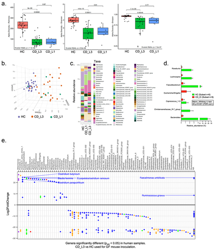
For GF mouse inoculation, microbial communities were generated by pooling samples from the patient groups. Samples were not chosen randomly but rather to approximate the microbiome composition of typical Crohn’s ileocolitis based on the consensus data in published literature, with a focus on seven genera, Roseburia, Lachnospira, Faecalibacterium, Escherichia/Shigella, Coprococcus_1–3, Christensenellaceae_R-7_group, and Bacteroides. shows those genera in samples selected for pooling an inoculation of GF mice. DESeq2 analysis of the selected samples shown in provides a more comprehensive analysis of the bacterial composition of samples from HC and CD_L3 selected for pooling and inoculation.
To test whether the selected samples contained known pathogens that could influence the resulting phenotype in recipient GF mice, pooled inocula from HC and CD_L3 patients as well as stool samples collected from recipient mice at the time of euthanasia (pooled separately from males and females) were analyzed by shotgun metagenomics. Of the 23 viruses or phages that were detected in human inocula, none were detected in colonized mice. Saccharomyces cerevisiae was detected in CD_L3 (relative abundance of 0.43%) but not in HC inocula and was not detected in recipient mice. Of the 309 bacterial species detected in human samples, only 72 were effectively engrafted in GF mice (23.3%) (Supplemental Table S3). Engraftment was higher in the recipients of the HC microbiota (27.9%) than in those of the CD_L3 microbiota (8.6%). Of the 72 engrafted species (present in both donor samples and in mouse recipients), none represented a known human or murine pathogen (Supplemental Tables S3 and S4).
Characterization of microbiota in humanized xGF mice
Although the transferred microbiota did not include known pathogenic species, we identified three species that were entirely absent or below the detection limit in human inocula (HC or CD_L3) but bloomed exclusively in xGF mice colonized with CD_L3. These were Eisenbergiella massiliensis (to 1.64% in female mouse recipients and 3.55% in male recipients), Clostridium_sp_7_2_43FAA (to 0.12% in females and 0.01% in male recipients), and Clostridioides difficile (to 4.74% in females and 3.97% in male recipients) (Supplemental Table S4). Further metagenomic analysis identified two contigs containing a complete C. difficile pathogenicity locus and having high sequence similarity with C. difficile strains Cd10 and Cd11 (NCBI CP037827.1 and CP037822.1, Supplemental Fig. S2). We further confirmed this observation using qPCR (Supplemental Fig. S3). Each mouse that received the CD_L3 pooled microbiome was positive for a toxigenic strain of C. difficile, as indicated by the presence of C. difficile 16S DNA and the tcdA and tcdB genes (Supplemental Fig. S2 and S3).
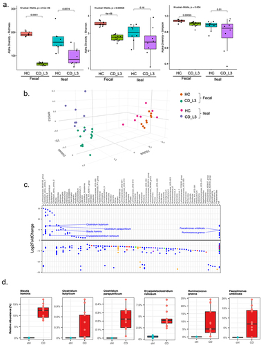
The species diversity of both ileal contents and feces of the humanized mice was characterized using 16S sequencing from samples collected from individual mice at the time of euthanasia. Fecal samples of the recipient mice reflected the differences in community diversity among the human specimens, with richness, Shannon, and Simpson indices significantly lower in CD_L3 recipients (). For ileal contents, only richness reflected a significant difference in alpha diversity (). NMDS analysis of beta diversity showed very distinct clustering of samples from recipients of HC vs CD_L2 microbiota and less dramatic but discernible separation of ileal and fecal samples (). depicts the results of DESeq2 analysis of ASVs in fecal samples from humanized mice (ASVs at p < 0.5 in CD_L3 vs. HC recipients). The relative abundance of selected species identified as having 100% 16S identity with a given ASV is shown in , including Ruminococcus gnavus, a species known for its association with CDCitation41–45 whose abundance was significantly increased in CD_L3 donors and xGF recipients.
Figure 2. Analysis of the established microbiomes in GF mice humanized with HC and CD_L3 fecal samples.
Ileum is not impacted by inoculation of CD-associated fecal microbiota
Fig. S4A depicts unaltered villi and crypt structure in the ileum of xGF mice colonized with HC and CD_L3 microbiota, with no histological evidence of ileitis. Ileal lysozyme, an antimicrobial peptide secreted by Paneth cells in response to pathogensCitation46–48 was not altered between the groups (Fig. S4B). Similarly, no differences were observed in immunostaining for MMP7, an antimicrobial peptide essential for activation of mouse alpha-defensinsCitation49–51 (Fig. S4C), or for MPTX2, a Paneth cell-specific mucosal pentraxinCitation52 (Fig. S4D). A PCA plot comparing the transcriptomic profiles of the ileum of HC and CD_L3 recipients indicated no distinct clustering (Fig. S4E) and confirmed the immunofluorescence (IF) results for Lyz1, Mmp7 and Mptx2 at the mRNA level (Fig. S4F). DESeq2 analysis indicated minimal changes in the gene transcription profile, with the vast majority of altered mRNA being either noninformative or noncoding (Fig. S4G). These observations were confirmed by unbiased histopathology scoring, which did not identify inflammatory changes in the ileum of mice receiving microbiota from CD_L3 patients (Fig. S4H).
CD microbiota in xGF mice induces colonic inflammation
There was no evidence of inflammation in the proximal or distal colon of mice colonized with the HC microbiota (). However, in mice colonized with CD_L3 microbiota, both segments showed evidence of mild to moderate colitis, as indicated by mild to moderate hyperplasia, edema, mild immune cell infiltration, enlarged isolated lymphoid follicles and/or neogenesis of mucosal tertiary lymphoid organs (TLO), typical features of chronic colitis in humans (primarily CD) and mice Citation53–55 (). Higher magnification showed a loss of regular crypt structure as well as damage or loss of the limiting outer epithelial layer surrounding the TLOs (). Inflammation was discontinuous, with no case where the inflammation exceeded the length limits of 50% of the analyzed segment.
Figure 3. Histological features of mild to moderate colitis in xGF mice humanized with HC and CD_L3 fecal microbiota.
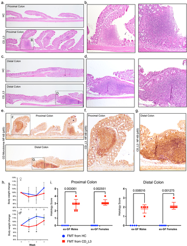
Immunohistochemical (IHC) staining of NF-κB (p65), an established upstream regulator of inflammatory responses, apoptosis, and cytokine signaling, showed high p65 expression within the TLOs of CD_L3 mice, as well as at sites with evident epithelial damage in both segments of the colon (). p65 expression in undamaged or uninflamed regions was comparable in both the HC and CD_L3 mice (not shown). None of the experimental mice had symptoms of diarrhea, rectal prolapse, or rectal bleeding, and we did not observe a significant effect of the CD_L3 FMT on relative body weight change in females, and only a very modest and transient delay on weight gain in males (). Pathology scoring blinded to the experimental design and group assignment confirmed the histological/IHC observations in HC and CD_L3 microbiota recipients, with elevated histological scores in the latter group, regardless of the recipient muse sex ().
Inflammatory signatures in the proximal and distal colon transcriptomes of CD microbiota recipients
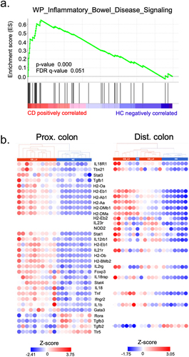
Principal component analysis of the proximal and distal colon transcriptomic profiling, with distinct clustering of samples from HC and CD_L3-associated mice, along with accompanying volcano plots are shown in , respectively. GSEA with gene sets from the KEGG database was used to identify some of the significantly enriched inflammatory pathways. The leading-edge plots shown in (chemokine signaling and leukocyte transmigration) are consistent with the increased immune cell recruitment shown in . We observed enrichment of the GO:BP pathway leukocyte-mediated immunity, with increased expression of TNF, IL-27RA, and CD74 as well as other immune cell-related genes and cytokine receptors (). In the distal colon of CD_L3 recipients, we observed an enrichment in cytokine and chemokine signaling pathways and general upregulation of innate immune signaling-associated transcripts (). Other pathways from the MSigDB Hallmark and KEGG pathway collections that were highly enriched in the proximal colon of CD_L3 recipients, along with a breakdown of genes involved in the IFNγ response, are summarized in . Importantly, the colonic transcriptomic profile of CD_L3 recipients showed an enrichment in the inflammatory bowel disease signaling pathway (Padj = 0.05), with elevated expression more prevalent in the proximal colonic segment, consistent with independent observations pointing to a more proximal disease (). When analyzed in the context of the KEGG IBD pathway (hsa05321), our data point to a picture more consistent with CD than with UC (Fig. S6).
Figure 4. Inflammatory signature of the colonic transcriptome profiling of xGF mice humanized with HC and CD_L3 fecal microbiota.
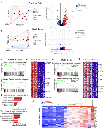
Figure 5. The colonic transcriptomic profile of CD_L3 recipients shows an enrichment in the IBD pathway.
Colitis in CD microbiota recipients is associated with increased mucosal T-cell infiltration and increased epithelial cell expression of CD74 and CD14
IF analysis of CD3 expression was used as an indicator of T-cell infiltration. A significant increase in the number of T cells recruited to both TLO and non-TLO mucosa, including intraepithelial lymphocytes, was observed in the proximal colons of CD_L3 mice compared to HCs (). The increase in T-cell infiltration in the distal colon of CD_L3 mice was less prominent, with no significant changes in the numbers of IELs observed. The plots and data distribution reflect the regional variability of T-cell numbers within the murine CD_L3 tissue and confirm the discontinuous inflammation characteristic of human CD.
Figure 6. Increased T-cell infiltration and increased expression of epithelial CD74 and CD14 in the inflamed colonic mucosa of xGF mice humanized with HC and CD_L3 fecal microbiota.
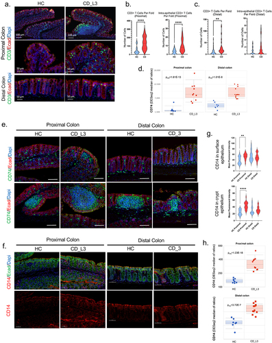
The CD74 gene encodes the invariant chain (Ii), which is critical in the formation and transport of MHC class II peptide complexes for the generation of CD4+ T-cell responses and is upregulated in the epithelium of IBD patientsCitation56–58 where in response to inflammation, it contributes to tissue repair and mucosal healing by acting as the receptor for macrophage migration inhibitory factor (MIF)Citation58–61 depicts elevated Cd74 mRNA expression in the proximal and distal segments of CD_L3 recipients. In the proximal colon of HC recipient mice, CD74-positive cells were limited to the immune cells in the lamina propria and the isolated lymphoid follicles (). The CD recipient proximal and distal colon showed a large increase in the amount of both CD74-positive lamina propria immune cells and increased CD74 expression in the surface epithelium. The observed changes tended to be less dramatic in the distal segment ().
CD14, the LPS-sensing coreceptor of TLR4, is expressed not only by myelomonocyte lineage cells but also by epithelial cells. Although the C-260T polymorphism in the CD14 gene has been associated with IBD in certain patient populations,Citation62 it has also been shown to limit inflammatory responses by improving epithelial barrier function.Citation63 In the proximal colon of mice colonized with HC microbiota, CD14 was detectable at the apical surface of the surface epithelial cells, with slightly less intense staining at the basolateral membranes and low expression in the crypts (). The same segment of the colon in CD_L3-colonized mice exhibited more intense CD14 staining of the surface epithelium, now also extending deeper into the crypts (). In the distal colon, CD14 expression did not differ between the HC and CD_L3 groups. The quantification summary shown in confirmed the interpretation of the IF images. RNA-seq analysis showed a more significant increase in the Cd14 transcript in the proximal colon than in the distal colon of the CD_L3 group (). Interestingly, the change in Cd14 expression in the distal colon of CD_L3 did not correspond with the protein abundance based on the IF data. Changes in mucosal expression of selected genes, including CD74 and CD14 were further validated by qRT-PCR (Fig. S5).
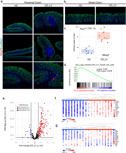
Inflammation in CD microbiota recipients triggers colonic MMP7 expression
The abnormal expression of MMP7, normally a Paneth cell-restricted matrix metallopeptidase, has been associated with colonic inflammation in both CD and UC as well as colorectal cancerCitation64–66 GF mice colonized with HC microbiota did not show detectable MMP7 expression in the proximal or distal colon. However, in the proximal colon of CD_L3-associated mice, secretory cells in the immediate surrounding of the inflamed lymphoid follicles began to express MMP7 (). In the distal colon, the incidence of MMP7-expressing cells was very rare, with no apparent difference between experimental groups (). Increased MMP7 immunoreactivity in the proximal colon of CD_L3 recipient mice corresponded with increased Mmp7 mRNA expression (). Although Paneth cell hyperplasia in the ascending colon and metaplasia in the descending colon have been well documented in IBD patients, they have not been described in rodent models of IBD. Our observations led us to speculate that secretory cells of the mouse proximal colon could be pushed toward differentiation to a Paneth-like cell phenotype as a response to persistent inflammation and the introduction of CD-associated microbes. Previous studies have demonstrated that both goblet and Paneth cells share a common secretory progenitor.Citation67 To further investigate this, we performed gene set enrichment analysis (GSEA) with proximal colon RNA-seq data and identified a cell type signature consistent with the Paneth-like phenotype reported by Gao et al.Citation68 in scRNAseq analysis of the developing human fetal large intestine (MSigDB systematic name M39160; p = .002, FDR q = 0.003) (). Of the associated transcripts, 188 had Padj <0.05, and 127 had a fold change greater than 1.5 (). The heatmap in depicts 13 genes in this subset with a fold change ≥3 (). depicts genes that were also described as Paneth cell signature genes from the PanglaoDB database of murine and human scRNA projects.Citation69
Figure 7. Paneth cell-like signature in the proximal colon of xGF mice humanized with HC and CD_L3 fecal microbiota.
Discussion
Although the role of gut microbiota in the pathogenesis of IBD is beyond debate, the search for individual pathobionts and proinflammatory communities/enterotypes continues. Several studies have demonstrated that fecal transplantation of an IBD-associated dysbiotic community can confer susceptibility to colitis in xGF- or antibiotic-pretreated genetically susceptible or DSS-treated mice.Citation12–15 Our data provide the first evidence that germ-free mice colonized with fecal microbiota from patients with Crohn’s ileocolitis are sufficient to induce mild to moderate chronic colonic inflammation in GF mice, in many respects reminiscent of the CD phenotype. This included a discontinued pattern of colitis, more proximal colonic localization, enlarged isolated lymphoid follicles and/or TLO neogenesis, as well as a transcriptomic pattern consistent with immune dysregulation in CD. To enable the establishment of a stable community while preventing long-term adaptive changes in the human microbiota of mice, we restricted our observations to the three-week post-colonization period. Consequently, we were unable to determine conclusively whether the observed inflammatory changes were transient, occurring during the recovery phase in mice, or if they were indicative of a progressive disease manifesting in its early stages. Given that the mice utilized lacked genetic susceptibility to inflammatory bowel disease (IBD), it appears more probable that the former scenario holds true.
Previous studies that have demonstrated the exacerbation of murine colitis by FMT from IBD patients did not report the efficiency of engraftment of the complex human fecal communities in mice.Citation12–15 Metagenomic compositional analysis in our study suggested only 27.9% engraftment efficiency with FMT from healthy patients and an even lower, 8.6% efficiency with CD microbiota. Low engraftment may be explained by host species differences and lack of genetic IBD susceptibility traits in the murine recipients. Indeed, only 15% of bacterial species in the distal gut microbiota were shown to be shared between humans and mice.Citation70 Fecal sample collection, storage, FMT preparation, and the associated exposure to ambient oxygen could further confound colonization by partial elimination of obligate anaerobes, especially those unable to form spores. The observed much lower engraftment of the CD community compared to the HC community may be explained by the generally lower microbial diversity of these samples, but it is also plausible that the CD microbiome is more unstable or prone to disruption and, as a result, generally more difficult to transfer across species.
Characterization of microbiota from ileal and fecal samples of recipient xGF mice reflected the differences in community diversity among the human specimens. Specifically, fecal contents from CD_L3 recipients had significantly lower richness, Shannon, and Simpson indices of alpha diversity compared to the HC recipients. However, in the ileal contents, only richness reflected a significant difference in alpha diversity between the two groups. Although beta diversity analysis showed altered ileal microbiota composition between CD_L3 and HC recipients, this did not translate to morphological or transcriptional changes consistent with ileitis, which suggests that fecal microbiota is not capable of promoting inflammation in this segment and/or is not able to recapitulate the human ileal community in mice.
Among the bacteria consistently overrepresented in CD_L3 patient samples and recipient mice were Ruminococcus gnavus, Erysipelatoclostridium ramosum, Faecalimonas umbilicate, Blautia hominis, Clostridium butyricum, and C. paraputrificum, with a longer list of 48 ASVs with consistently reduced abundance. Interestingly, the mucin consumer R. gnavus, known for its proinflammatory association with Crohn’s disease and experimental colitisCitation48,Citation71 was dramatically more elevated between mouse recipients of CD_L3 microbiota compared to HC recipients than in the source donor samples. We previously reported that mice lacking Paneth cell lysozyme had significantly elevated R. gnavus colonization.Citation48 These results suggest that the limited IBD-derived microbial community and alteration of the host luminal environment in mice may have established conditions favorable to R. gnavus growth and dominance.
Although no pathogenic bacteria were identified among ASVs concordant between human inoculum and mouse fecal samples, we identified three bacterial species in GF recipients that were below detection levels in human donor samples: Eisenbergiella massiliensis, Clostridium_sp_7_2_43FAA, and Clostridioides difficile. All three have been described in human fecal samplesCitation72–74 but only C. difficile gained notoriety as an infectious cause of pseudomembranous colitisCitation75 and a pathogen that complicates the outcomes in IBD patients and in murine models.Citation76,Citation77 The unexpected expansion of toxigenic tcdA- and tcdB-positive C. difficile in CD_L3 recipient mice is a novel and fascinating observation, as it describes a reduced human microbial community highly capable of promoting its growth. This may be of high relevance to the understanding of the key players among the healthy gut microbial community that limit C. difficile growth and the associated host response, as recently shown in an elegant study by Nagao-Kitamoto et al.Citation78 and contribute to the growing efforts to identify bacteria promoting or restricting pathogen growth in humanized xGF miceCitation79–82 It is difficult, however, to establish whether the pathogenic response in our mice could be solely attributed to C. difficile growth. Although its relative abundance reached ca. 4% in CD_L3 FMT recipients, in human CDI patients and in a mouse CDI model, fecal C. difficile burden can vary dramatically and does not correlate with any measure of illness or outcome.Citation83–85 Moreover, the histological changes observed in the CD_L3 microbiome recipients were not consistent with those reported by others in GF or antibiotic-treated mice, typically with more fulminant disease with diarrhea and the associated high mortality. Because there is no selective treatment for C. difficile, we could not determine the role of C. difficile in our model, and any antibiotic treatment would likely further destabilize the transplanted community and make the results uninterpretable.
CD_L3 FMT was associated with increased epithelial expression of CD14, a CD susceptibility gene and LPS coreceptor, as well as CD74, normally limited to the mucosal immune compartment. CD74, a major histocompatibility complex class II chaperone and a receptor for the cytokine macrophage migration inhibitory factor (MIF), is increased in IBDCitation56,Citation57 and its epithelial cell expression was recently described to contribute to mucosal restitution.Citation58 It is possible that increased CD74 expression in our model limited the inflammatory response and contributed to the relatively mild tissue damage.
MMP7 is one of the markers of small intestinal Paneth cells, where it is responsible for activating α-defensins.Citation86 Although normally absent in a healthy colon, elevated colonic expression of MMP7 has been previously shown in the endothelial cells and infiltrating leukocytes of the inflamed colonic mucosa of IBD patients,Citation66 and it has been associated with epithelial barrier dysfunction.Citation87 However, our IF analysis showed increased but sparse MMP7 immunostaining within the proximal and, to a much lesser extent, distal colonic crypt epithelium, which, we speculate, could represent the precursors leading to Paneth cell metaplasia. Indeed, one of the novel observations in CD_L3-associated xGF mice was a transcriptomic signature of the Paneth cell-like profile in the proximal colon. Paneth cell hyperplasia in the ascending colon and metaplasia in the descending colon are typical features in IBD patients but have not been described in animal models of IBD. It is plausible that the development of the Paneth-like secretory lineage in the inflamed colon is unique to humans due to the associated microbial cues that conventional mice are lacking. Our results point to only a partial Paneth-like signature (e.g., no increase in lysozyme expression), suggesting that additional elements, either host-derived or microbial in origin, were still missing. Future studies will need to address this fascinating concept in more detail.
In summary, our study provides the first evidence that the transfer of a dysbiotic community from CD patients can lead to spontaneous inflammatory changes in the colon of xGF mice. Although there are limitations to the use of humanizing mice with human fecal microbiota, including a limited engraftment of the complex community, it may have also been a fortuitus finding that will allow the field to further narrow down the microbial composition capable of promoting the growth and relative expansion of pathobiont and pathogenic bacteria, which can promote epithelial and immune dysfunction and lead to chronic intestinal inflammation.
Availability of data and materials
All raw data from microbiome analysis and RNAseq transcriptome profiling have been submitted to the NCBI Sequence Read Archive (SRA) database under BioProject PRJNA981221 and BioProject PRJNA980747, respectively.
Supplemental Material
Download Zip (16.8 MB)Acknowledgments
We are grateful to Mrs. Dominika Piątek for her technical assistance with patient fecal sample collection.
Disclosure statement
No potential conflict of interest was reported by the author(s).
Supplementary material
Supplemental data for this article can be accessed online at https://doi.org/10.1080/19490976.2024.2333483
Additional information
Funding
References
- Sartor RB, Wu GD. Roles for intestinal bacteria, viruses, and fungi in pathogenesis of inflammatory bowel diseases and therapeutic approaches. Gastroenterology. 2017;152(2):327–339.e4. doi:10.1053/j.gastro.2016.10.012.
- Graham DB, Xavier RJ. Pathway paradigms revealed from the genetics of inflammatory bowel disease. Nature. 2020;578(7796):527–21. doi:10.1038/s41586-020-2025-2.
- de Lange KM, Moutsianas L, Lee JC, de Lange KM, Lamb CA, Luo Y, Kennedy NA, Jostins L, Rice DL, Gutierrez-Achury J. et al. Genome-wide association study implicates immune activation of multiple integrin genes in inflammatory bowel disease. Nat Genet. 2017;49(2):256–261. doi:10.1038/ng.3760.
- Kuenzig ME, Fung SG, Marderfeld L, Mak JWY, Kaplan GG, Ng SC, Wilson DC, Cameron F, Henderson P, Kotze PG. et al. Twenty-first century trends in the global epidemiology of pediatric-onset inflammatory bowel disease: systematic review. Gastroenterology. 2022;162(4):1147–1159.e4. doi:10.1053/j.gastro.2021.12.282.
- Sartor RB. Therapeutic manipulation of the enteric microflora in inflammatory bowel diseases: antibiotics, probiotics, and prebiotics. Gastroenterology. 2004;126(6):1620–1633. doi:10.1053/j.gastro.2004.03.024.
- D’Haens GR, Geboes K, Peeters M. et al. Early lesions of recurrent Crohn’s disease caused by infusion of intestinal contents in excluded ileum. Gastroenterology. 1998;114:262–267. doi:10.1016/S0016-5085(98)70476-7.
- Dianda L, Hanby AM, Wright NA, Sebesteny A, Hayday AC, Owen MJ. T cell receptor-alpha beta-deficient mice fail to develop colitis in the absence of a microbial environment. Am J Pathol. 1997;150:91–97.
- Sellon RK, Tonkonogy S, Schultz M, Dieleman LA, Grenther W, Balish E, Rennick DM, Sartor RB. Resident enteric bacteria are necessary for development of spontaneous colitis and immune system activation in interleukin-10-deficient mice. Infect Immun. 1998;66(11):5224–5231. doi:10.1128/IAI.66.11.5224-5231.1998.
- Veltkamp C, Tonkonogy SL, De Jong YP, Albright C, Grenther WB, Balish E, Terhorst C, Sartor RB. Continuous stimulation by normal luminal bacteria is essential for the development and perpetuation of colitis in Tgϵ26 mice. Gastroenterology. 2001;120(4):900–913. doi:10.1053/gast.2001.22547.
- Taurog JD, Richardson JA, Croft JT, Simmons WA, Zhou M, Fernández-Sueiro JL, Balish E, Hammer RE. The germfree state prevents development of gut and joint inflammatory disease in HLA-B27 transgenic rats. J Exp Med. 1994;180(6):2359–2364. doi:10.1084/jem.180.6.2359.
- Franzosa EA, Sirota-Madi A, Avila-Pacheco J, Fornelos N, Haiser HJ, Reinker S, Vatanen T, Hall AB, Mallick H, McIver LJ. et al. Gut microbiome structure and metabolic activity in inflammatory bowel disease. Nat Microbiol. 2019;4(2):293–305. doi:10.1038/s41564-018-0306-4.
- Nagao-Kitamoto H, Shreiner AB, Gillilland MG 3rd, Kitamoto S, Ishii C, Hirayama A, Kuffa P, El-Zaatari M, Grasberger H, Seekatz AM. et al. Functional characterization of inflammatory bowel disease–associated gut dysbiosis in gnotobiotic mice. Cell Mol Gastroenterol Hepatol. 2016;2(4):468–481. doi:10.1016/j.jcmgh.2016.02.003.
- Britton GJ, Contijoch EJ, Mogno I, Vennaro OH, Llewellyn SR, Ng R, Li Z, Mortha A, Merad M, Das A. et al. Microbiotas from humans with Inflammatory Bowel disease Alter the Balance of Gut Th17 and RORγt+ regulatory T cells and exacerbate colitis in mice. Immunity. 2019;50(1):212–224.e4. doi:10.1016/j.immuni.2018.12.015.
- Du Z, Hudcovic T, Mrazek J, Kozakova H, Srutkova D, Schwarzer M, Tlaskalova-Hogenova H, Kostovcik M, Kverka M. Development of gut inflammation in mice colonized with mucosa-associated bacteria from patients with ulcerative colitis. Gut Pathog. 2015;7(1):32. doi:10.1186/s13099-015-0080-2.
- Paik J, Meeker S, Hsu CC, Seamons A, Pershutkina O, Snyder JM, Brabb T, Maggio-Price L. Validation studies for germ-free Smad3 -/-mice as a bio-assay to test the causative role of fecal microbiomes in IBD. Gut Microbes. 2020;11(1):21–31. doi:10.1080/19490976.2019.1611151.
- Burich A, Hershberg R, Waggie K, Zeng W, Brabb T, Westrich G, Viney JL, Maggio-Price L. Helicobacter-induced inflammatory bowel disease in IL-10- and T cell-deficient mice. Am J Physiol Gastrointest Liver Physiol. 2001;281(3):G764–78. doi:10.1152/ajpgi.2001.281.3.G764.
- Yu S, Tong K, Zhao Y, Balasubramanian I, Yap GS, Ferraris RP, Bonder EM, Verzi MP, Gao N. Paneth cell multipotency induced by notch activation following injury. Cell Stem Cell. 2018;23(1):46–59.e5. doi:10.1016/j.stem.2018.05.002.
- Caporaso JG, Lauber CL, Walters WA, Berg-Lyons D, Huntley J, Fierer N, Owens SM, Betley J, Fraser L, Bauer M. et al. Ultra-high-throughput microbial community analysis on the Illumina HiSeq and MiSeq platforms. ISME J. 2012;6(8):1621–1624. doi:10.1038/ismej.2012.8.
- Callahan BJ, McMurdie PJ, Rosen MJ, Han AW, Johnson AJA, Holmes SP. DADA2: high-resolution sample inference from Illumina amplicon data. Nat Methods. 2016;13(7):581–583. doi:10.1038/nmeth.3869.
- Wang Q, Garrity GM, Tiedje JM, Cole JR. Naive bayesian Classifier for Rapid Assignment of rRNA Sequences into the new bacterial taxonomy. Appl Environ Microbiol. 2007;73(16):5261–5267. doi:10.1128/AEM.00062-07.
- Quast C, Pruesse E, Yilmaz P, Gerken J, Schweer T, Yarza P, Peplies J, Glöckner FO. The SILVA ribosomal RNA gene database project: improved data processing and web-based tools. Nucleic Acids Res. 2013;41(D1):D590–6. doi:10.1093/nar/gks1219.
- Team RC. R: a language and environment for statistical computing. Vienna, Austria: R Foundation for Statistical Computing; 2021.
- W H. ggplot2: Elegant Graphics for Data Analysis. 2016.
- Love MI, Huber W, Anders S. Moderated estimation of fold change and dispersion for RNA-seq data with DESeq2. Genome Biol. 2014;15(12):550. doi:10.1186/s13059-014-0550-8.
- Bolger AM, Lohse M, Usadel B. Trimmomatic: a flexible trimmer for Illumina sequence data. Bioinformatics. 2014;30(15):2114–2120. doi:10.1093/bioinformatics/btu170.
- Langmead B, Salzberg SL. Fast gapped-read alignment with bowtie 2. Nat Methods. 2012;9(4):357–359. doi:10.1038/nmeth.1923.
- Beghini F, McIver LJ, Blanco-Miguez A, Dubois L, Asnicar F, Maharjan S, Mailyan A, Manghi P, Scholz M, Thomas AM. et al. Integrating taxonomic, functional, and strain-level profiling of diverse microbial communities with bioBakery 3. Elife. 2021;10:10. doi:10.7554/eLife.65088.
- Li D, Liu CM, Luo R, Sadakane K, Lam T-W. MEGAHIT: an ultra-fast single-node solution for large and complex metagenomics assembly via succinct de bruijn graph. Bioinformatics. 2015;31(10):1674–1676. doi:10.1093/bioinformatics/btv033.
- Hyatt D, Chen GL, Locascio PF, Land ML, Larimer FW, Hauser LJ. Prodigal: prokaryotic gene recognition and translation initiation site identification. BMC Bioinf. 2010;11(1):119. doi:10.1186/1471-2105-11-119.
- Aramaki T, Blanc-Mathieu R, Endo H, Ohkubo K, Kanehisa M, Goto S, Ogata H. KofamKOALA: KEGG Ortholog assignment based on profile HMM and adaptive score threshold. Bioinformatics. 2020;36(7):2251–2252. doi:10.1093/bioinformatics/btz859.
- Seemann T. Prokka: rapid prokaryotic genome annotation. Bioinformatics. 2014;30(14):2068–2069. doi:10.1093/bioinformatics/btu153.
- Mistry J, Chuguransky S, Williams L, Qureshi M, Salazar G, Sonnhammer ELL, Tosatto SCE, Paladin L, Raj S, Richardson LJ. et al. Pfam: the protein families database in 2021. Nucleic Acids Res. 2021;49(D1):D412–D419. doi:10.1093/nar/gkaa913.
- Gish W, States DJ. Identification of protein coding regions by database similarity search. Nat Genet. 1993;3(3):266–272. doi:10.1038/ng0393-266.
- Johnson M, Zaretskaya I, Raytselis Y, Merezhuk Y, McGinnis S, Madden TL. NCBI BLAST: a better web interface. Nucleic Acids Res. 2008;36(Web Server):W5–9. doi:10.1093/nar/gkn201.
- Sayers EW, Beck J, Brister JR, Bolton EE, Canese K, Comeau DC, Funk K, Ketter A, Kim S, Kimchi A. et al. Database resources of the National Center for Biotechnology Information. Nucleic Acids Res. 2020;48(D1):D9–D16. doi:10.1093/nar/gkz899.
- Kubota H, Sakai T, Gawad A, Makino H, Akiyama T, Ishikawa E, Oishi K. Development of TaqMan-based quantitative PCR for sensitive and selective detection of toxigenic Clostridium difficile in human stools. PloS One. 2014;9(10):e111684. doi:10.1371/journal.pone.0111684.
- Dobin A, Davis CA, Schlesinger F, Drenkow J, Zaleski C, Jha S, Batut P, Chaisson M, Gingeras TR. STAR: ultrafast universal RNA-seq aligner. Bioinformatics. 2013;29(1):15–21. doi:10.1093/bioinformatics/bts635.
- Mootha VK, Lindgren CM, Eriksson KF, Subramanian A, Sihag S, Lehar J, Puigserver P, Carlsson E, Ridderstråle M, Laurila E. et al. PGC-1α-responsive genes involved in oxidative phosphorylation are coordinately downregulated in human diabetes. Nat Genet. 2003;34(3):267–273. doi:10.1038/ng1180.
- Subramanian A, Tamayo P, Mootha VK, Mukherjee S, Ebert BL, Gillette MA, Paulovich A, Pomeroy SL, Golub TR, Lander ES. et al. Gene set enrichment analysis: a knowledge-based approach for interpreting genome-wide expression profiles. Proc Natl Acad Sci U S A. 2005;102(43):15545–15550. doi:10.1073/pnas.0506580102.
- Silverberg MS, Satsangi J, Ahmad T, Arnott ID, Bernstein CN, Brant SR, Caprilli R, Colombel J-F, Gasche C, Geboes K. et al. Toward an integrated clinical, molecular and serological classification of inflammatory bowel disease: report of a Working Party of the 2005 Montreal World Congress of Gastroenterology. Can J Gastroenterol. 2005;19(Suppl A):5A–36A. doi:10.1155/2005/269076.
- Willing BP, Dicksved J, Halfvarson J, Andersson AF, Lucio M, Zheng Z, Järnerot G, Tysk C, Jansson JK, Engstrand L. et al. A pyrosequencing study in twins shows that gastrointestinal microbial profiles vary with inflammatory bowel disease phenotypes. Gastroenterology. 2010;139(6):1844–1854.e1. doi:10.1053/j.gastro.2010.08.049.
- Joossens M, Huys G, Cnockaert M. et al. Dysbiosis of the faecal microbiota in patients with Crohn’s disease and their unaffected relatives. Gut. 2011;60:631–637. doi:10.1136/gut.2010.223263.
- Hall AB, Yassour M, Sauk J, Garner A, Jiang X, Arthur T, Lagoudas GK, Vatanen T, Fornelos N, Wilson R. et al. A novel ruminococcus gnavus clade enriched in inflammatory bowel disease patients. Genome Med. 2017;9(1):103. doi:10.1186/s13073-017-0490-5.
- Nishino K, Nishida A, Inoue R, Kawada Y, Ohno M, Sakai S, Inatomi O, Bamba S, Sugimoto M, Kawahara M. et al. Analysis of endoscopic brush samples identified mucosa-associated dysbiosis in inflammatory bowel disease. J Gastroenterol. 2018;53(1):95–106. doi:10.1007/s00535-017-1384-4.
- Png CW, Linden SK, Gilshenan KS, Zoetendal EG, McSweeney CS, Sly LI, McGuckin MA, Florin THJ. Mucolytic bacteria with increased prevalence in IBD mucosa augment in vitro utilization of mucin by other bacteria. Am J Gastroenterol. 2010;105(11):2420–2428. doi:10.1038/ajg.2010.281.
- Peeters T, Vantrappen G. The paneth cell: a source of intestinal lysozyme. Gut. 1975;16(7):553–558. doi:10.1136/gut.16.7.553.
- Bel S, Pendse M, Wang Y, Li Y, Ruhn KA, Hassell B, Leal T, Winter SE, Xavier RJ, Hooper LV. et al. Paneth cells secrete lysozyme via secretory autophagy during bacterial infection of the intestine. Science. 2017;357(6355):1047–1052. doi:10.1126/science.aal4677.
- Yu S, Balasubramanian I, Laubitz D, Tong K, Bandyopadhyay S, Lin X, Flores J, Singh R, Liu Y, Macazana C. et al. Paneth cell-derived lysozyme defines the composition of mucolytic microbiota and the inflammatory tone of the intestine. Immunity. 2020;53(2):398–416.e8. doi:10.1016/j.immuni.2020.07.010.
- Ayabe T, Satchell DP, Pesendorfer P, Tanabe H, Wilson CL, Hagen SJ, Ouellette AJ. Activation of paneth cell α-defensins in mouse small intestine. J Biol Chem. 2002;277(7):5219–5228. doi:10.1074/jbc.M109410200.
- Weeks CS, Tanabe H, Cummings JE, Crampton SP, Sheynis T, Jelinek R, Vanderlick TK, Cocco MJ, Ouellette AJ. Matrix metalloproteinase-7 activation of mouse paneth cell pro-α-defensins. J Biol Chem. 2006;281(39):28932–28942. doi:10.1074/jbc.M602041200.
- Mastroianni JR, Costales JK, Zaksheske J, Selsted ME, Salzman NH, Ouellette AJ. Alternative luminal activation mechanisms for paneth cell α-defensins. J Biol Chem. 2012;287(14):11205–11212. doi:10.1074/jbc.M111.333559.
- Haber AL, Biton M, Rogel N, Herbst RH, Shekhar K, Smillie C, Burgin G, Delorey TM, Howitt MR, Katz Y. et al. A single-cell survey of the small intestinal epithelium. Nature. 2017;551(7680):333–339. doi:10.1038/nature24489.
- Van Kruiningen HJ, Colombel JF. The forgotten role of lymphangitis in Crohn’s disease. Gut. 2008;57:1–4. doi:10.1136/gut.2007.123166.
- Sura R, Colombel JF, Van Kruiningen HJ. Lymphatics, tertiary lymphoid organs and the granulomas of Crohn’s disease: an immunohistochemical study. Aliment Pharmacol Ther. 2011;33(8):930–939. doi:10.1111/j.1365-2036.2011.04605.x.
- Lochner M, Ohnmacht C, Presley L, Bruhns P, Si-Tahar M, Sawa S, Eberl G. Microbiota-induced tertiary lymphoid tissues aggravate inflammatory disease in the absence of RORγt and LTi cells. J Exp Med. 2011;208(1):125–134. doi:10.1084/jem.20100052.
- Parikh K, Antanaviciute A, Fawkner-Corbett D, Jagielowicz M, Aulicino A, Lagerholm C, Davis S, Kinchen J, Chen HH, Alham NK. et al. Colonic epithelial cell diversity in health and inflammatory bowel disease. Nature. 2019;567(7746):49–55. doi:10.1038/s41586-019-0992-y.
- Lawrance IC, Fiocchi C, Chakravarti S. Ulcerative colitis and Crohn’s disease: distinctive gene expression profiles and novel susceptibility candidate genes. Human Molecular Genetics. 2001;10(5):445–456. doi:10.1093/hmg/10.5.445.
- Farr L, Ghosh S, Jiang N, Watanabe K, Parlak M, Bucala R, Moonah S. CD74 signaling links inflammation to intestinal epithelial cell regeneration and promotes mucosal healing. Cell Mol Gastroenterol Hepatol. 2020;10(1):101–112. doi:10.1016/j.jcmgh.2020.01.009.
- Borghese F, Clanchy FI. CD74: an emerging opportunity as a therapeutic target in cancer and autoimmune disease. Expert Opin Ther Targets. 2011;15(3):237–251. doi:10.1517/14728222.2011.550879.
- Leng L, Metz CN, Fang Y, Xu J, Donnelly S, Baugh J, Delohery T, Chen Y, Mitchell RA, Bucala R. et al. MIF signal transduction initiated by binding to CD74. J Exp Med. 2003;197(11):1467–1476. doi:10.1084/jem.20030286.
- Maharshak N. et al. CD74 is a survival receptor on colon epithelial cells. World J Gastroenterol. 2010;16(26):3258–3266. doi:10.3748/wjg.v16.i26.3258.
- Wang Z, Hu J, Fan R, Zhou J, Zhong J. Association between CD14 gene C-260T polymorphism and inflammatory bowel disease: a meta-analysis. PloS One. 2012;7(9):e45144. doi:10.1371/journal.pone.0045144.
- Buchheister S, Buettner M, Basic M, Noack A, Breves G, Buchen B, Keubler LM, Becker C, Bleich A. CD14 plays a protective role in experimental inflammatory bowel disease by enhancing intestinal barrier function. Am J Pathol. 2017;187(5):1106–1120. doi:10.1016/j.ajpath.2017.01.012.
- Masaki T, Matsuoka H, Sugiyama M, Abe N, Goto A, Sakamoto A, Atomi Y. Matrilysin (MMP-7) as a significant determinant of malignant potential of early invasive colorectal carcinomas. Br J Cancer. 2001;84(10):1317–1321. doi:10.1054/bjoc.2001.1790.
- Sun DW, Zhang YY, Qi Y, Zhou X-T, Lv G-Y. Prognostic significance of MMP-7 expression in colorectal cancer: a meta-analysis. Cancer Epidemiol. 2015;39(2):135–142. doi:10.1016/j.canep.2015.01.009.
- Rath T, Roderfeld M, Halwe JM, Tschuschner A, Roeb E, Graf J. Cellular sources of MMP-7, MMP-13 and MMP-28 in ulcerative colitis. Scand J Gastroenterol. 2010;45(10):1186–1196. doi:10.3109/00365521.2010.499961.
- Almohazey D, Lo YH, Vossler CV, Simmons AJ, Hsieh JJ, Bucar EB, Schumacher MA, Hamilton KE, Lau KS, Shroyer NF. et al. The ErbB3 receptor tyrosine kinase negatively regulates paneth cells by PI3K-dependent suppression of Atoh1. Cell Death Differ. 2017;24(5):855–865. doi:10.1038/cdd.2017.27.
- Gao S, Yan L, Wang R, Li J, Yong J, Zhou X, Wei Y, Wu X, Wang X, Fan X. et al. Tracing the temporal-spatial transcriptome landscapes of the human fetal digestive tract using single-cell RNA-sequencing. Nat Cell Biol. 2018;20(6):721–734. doi:10.1038/s41556-018-0105-4.
- Franzen O, Gan LM, Bjorkegren JLM. PanglaoDB: a web server for exploration of mouse and human single-cell RNA sequencing data. Database (Oxford). 2019;2019. doi:10.1093/database/baz046.
- Ley RE, Backhed F, Turnbaugh P, Lozupone CA, Knight RD, Gordon JI. Obesity alters gut microbial ecology. Proc Natl Acad Sci U S A. 2005;102(31):11070–11075. doi:10.1073/pnas.0504978102.
- Henke MT, Kenny DJ, Cassilly CD, Vlamakis H, Xavier RJ, Clardy J. Ruminococcus gnavus, a member of the human gut microbiome associated with Crohn’s disease, produces an inflammatory polysaccharide. Proc Natl Acad Sci U S A. 2019;116(26):12672–12677. doi:10.1073/pnas.1904099116.
- Togo AH, Khelaifia S, Bittar F, Maraninchi M, Raoult D, Million M. ‘Eisenbergiella massiliensis’, a new species isolated from human stool collected after bariatric surgery. New Microbes New Infect. 2016;13:15–16. doi:10.1016/j.nmni.2016.05.015.
- Qin N, Zheng B, Yao J, Guo L, Zuo J, Wu L, Zhou J, Liu L, Guo J, Ni S. et al. Influence of H7N9 virus infection and associated treatment on human gut microbiota. Sci Rep. 2015;5(1):14771. doi:10.1038/srep14771.
- Hall IOTE. Intestinal flora in newborn infants with a description of a new pathogenic anaerobe, bacillus difficilis. Am J Dis Child. 1935;49(2):390. doi:10.1001/archpedi.1935.01970020105010.
- Smits WK, Lyras D, Lacy DB, Wilcox MH, Kuijper EJ. Clostridium difficile infection. Nat Rev Dis Primers. 2016;2(1):16020. doi:10.1038/nrdp.2016.20.
- Boeriu A, Roman A, Fofiu C, Dobru D. The Current knowledge on clostridioides difficile infection in patients with inflammatory bowel diseases. Pathogens. 2022;11(7):11. doi:10.3390/pathogens11070819.
- Zhou F, Hamza T, Fleur AS, Zhang Y, Yu H, Chen K, Heath JE, Chen Y, Huang H, Feng H. et al. Mice with inflammatory bowel disease are susceptible to clostridium difficile infection with severe disease outcomes. Inflamm Bowel Dis. 2018;24(3):573–582. doi:10.1093/ibd/izx059.
- Nagao-Kitamoto H, Leslie JL, Kitamoto S, Jin C, Thomsson KA, Gillilland MG, Kuffa P, Goto Y, Jenq RR, Ishii C. et al. Interleukin-22-mediated host glycosylation prevents clostridioides difficile infection by modulating the metabolic activity of the gut microbiota. Nat Med. 2020;26(4):608–617. doi:10.1038/s41591-020-0764-0.
- Collins J, Auchtung JM, Schaefer L, Eaton KA, Britton RA. Humanized microbiota mice as a model of recurrent Clostridium difficile disease. Microbiome. 2015;3(1):35. doi:10.1186/s40168-015-0097-2.
- Auchtung JM, Preisner EC, Collins J, Lerma AI, Britton RA. Identification of simplified microbial communities that inhibit clostridioides difficile infection through Dilution/Extinction. mSphere. 2020;5(4). doi:10.1128/mSphere.00387-20.
- Liu L, Kirst ME, Zhao L, Li E, Wang GP. Microbiome resilience despite a profound loss of minority microbiota following clindamycin challenge in humanized gnotobiotic mice. Microbiol Spectr. 2022;10(1):e0196021. doi:10.1128/spectrum.01960-21.
- Lesniak NA, Schubert AM, Flynn KJ, Leslie JL, Sinani H, Bergin IL, Young VB, Schloss PD. The gut bacterial community potentiates clostridioides difficile infection severity. mBio. 2022;13(4):e0118322. doi:10.1128/mbio.01183-22.
- El Feghaly RE, Stauber JL, Deych E, Gonzalez C, Tarr PI, Haslam DB. Markers of intestinal inflammation, not bacterial burden, correlate with clinical outcomes in clostridium difficile infection. Clin Infect Dis. 2013;56(12):1713–1721. doi:10.1093/cid/cit147.
- AbdelKhalek A, Narayanan SK. Comparison between symptomatic and asymptomatic mice after clostridioides difficile infection reveals novel inflammatory pathways and contributing microbiota. Microorganisms. 2022;10(12):10. doi:10.3390/microorganisms10122380.
- Daquigan N, Seekatz AM, Greathouse KL, Young VB, White JR. High-resolution profiling of the gut microbiome reveals the extent of Clostridium difficile burden. npj Biofilms Microbiomes. 2017;3(1):35. doi:10.1038/s41522-017-0043-0.
- Wilson CL, Ouellette AJ, Satchell DP, Ayabe T, Lopez-Boado YS, Stratman JL, Hultgren SJ, Matrisian LM, Parks WC. Regulation of Intestinal α-Defensin Activation by the metalloproteinase matrilysin in innate Host defense. Science. 1999;286(5437):113–117. doi:10.1126/science.286.5437.113.
- Xiao Y, Lian H, Zhong XS, Krishnachaitanya SS, Cong Y, Dashwood RH, Savidge TC, Powell DW, Liu X, Li Q. et al. Matrix metalloproteinase 7 contributes to intestinal barrier dysfunction by degrading tight junction protein claudin-7. Front Immunol. 2022;13:1020902. doi:10.3389/fimmu.2022.1020902.