
ABSTRACT
Premature infants lack a normal intestinal microbial community and also at risk of perinatal hypoxic-ischemic (HI) brain injury, which is considered to be one of the major factors for motor, sensory, and cognitive deficits. We hypothesized that neonatal gut microbiota composition modulated the immune reaction and severity of neonatal H-I brain injury. Neonatal C57BL/6J mouse pups were exposed to H-I protocol consisting of permanent left carotid artery ligation, followed by 8% hypoxia for 60 min. Microbial manipulation groups included 1) antibiotic treatment, E18 (maternal) to P5; 2) antibiotic treatment E18 to P5 + E. coli gavage; 3) antibiotic treatment E18 to P5 + B. infantis gavage; and 4) saline to pups with dams getting fresh water. The extent of brain injury and recovery was measured on MRI. Edematous injury volume was significantly higher in E. coli group than that in B. infantis group and in fresh water group. Gene expression in brains of pro-inflammatory cytokines (IL1β, IL6, IL2, TNF-α and toll-like receptors 2–6) were elevated to a greater extent in the E. coli group at P10, no injury, and at P13, 72 hours after H-I relative to sham control and B. infantis groups. Significant effects of microbiome and brain injury and interaction of these factors were found in abundance of major phyla. The neuroinflammatory response and brain injury after neonatal hypoxia-ischemia are affected by intestinal microbiota, providing opportunities for therapeutic intervention through targeting the early colonization and development of the gut microbiota.
1. Introduction
During the perinatal period, microbes from the mother and the surrounding environment colonize the gastrointestinal tract of infants, until a dense, complex bacterial community is established. Premature infants are exposed to a hostile extra-uterine environment and to unnatural microbiota in the hospital that results in abnormal intestinal colonization.Citation1 In addition, premature infants often lack normal commensal bacterial communities, due to frequent abdominal deliveries and lack of maternal breast feeding and skin-to-skin interaction. Colonization of beneficial bacteria such as Lactobacilli and Bifidobacteria is often delayed in preterm infants and is only found in relatively low numbers during the first few weeks of life, whereas potentially pathogenic bacteria such as E. coli, Clostridia, and Staphylococci are found in higher numbers.Citation2,Citation3 Considering that one of the most pronounced effects of gut microbiota is on maturation of the infant immune system,Citation4 variations in immune responses in premature infants may reflect the influence of abnormal gut microbiota.Citation5
Preterm infants, particularly those born at less than 32 weeks post-conception age and/or with a birth-weight less than 1500 g are at risk for adverse neurological outcomes, including cognitive and behavioral deficits.Citation6,Citation7 The most important risk factors leading to neurologic deficits in the newborn period include hypoxia and maternal inflammation.Citation8,Citation9 Inflammation in neonatal brain and unbalanced immune responses play a pivotal role in the extent of damage and recovery after perinatal hypoxic-ischemic (H-I) brain injury.Citation9–11 It has been suggested that an unbalanced, accentuated pro-inflammatory response in neonates, associated with an impaired anti-inflammatory capability,Citation12 plays a major role in brain injury.Citation11,Citation13
Perinatal hypoxic–ischemic brain damage is a major cause of acute mortality and chronic neurologic morbidity in infants and children and often is associated with inflammation. Inflammation occurs before, during, and after the brain injury, and modulates the vulnerability to and development of brain injury.Citation9 The effects of inflammation and ischemia are likely modulated by early gut microbial composition through immune, endocrine, and neural pathways.Citation14 A prime example of the interaction between gut dysbiosis and brain injury in premature infants is necrotizing enterocolitis (NEC).Citation15 The key features of the disease include formula gavage, hypoxia episodes, and TLR4 signaling.Citation16 Survivors of NEC suffer brain volume loss, demyelination and develop profound neurological impairments.Citation17 Specific probiotics, including Bifidobacterium, were effective in preventing NEC in very-low-birth-weight infants.Citation18
Considering the link between gut microbiome, systemic inflammation, and hypoxia, we hypothesized that neonatal gut microbiota modulates the extent of brain injury and recovery after hypoxic-ischemic insult via inflammatory pathways. To test this hypothesis, we manipulated gut microbial composition in an established murine model of perinatal H-I. We created several scenarios of neonatal gut dysbiosis with bacterial depletion by maternal antibiotic administration, followed by gavage transfaunation with either pathogenic or commensal bacteria and examined the relationship between the inflammatory response and the severity of brain injury and recovery.
2. Results
We examined the effect of microbial manipulation on neonatal mouse gut by maternal antibiotic treatment, followed by a gavage of either Escherichia coli (“E. coli” group), Bifidobacterium longum infantis (“B. infantis”), or sterile saline (“Antibiotics”) and fresh water controls, on H-I brain injury and neuroinflammatory response.
2.1. Spatial distribution and temporal evolution of brain edema after H-I injury
Unilateral H-I resulted in brain edema that could be unambiguously delineated on T2-weighted MR images as hyperintense areas at 24 and 72 h after H-I insult ().
Figure 1. Temporal evolution of brain injury after neonatal mouse H-I injury on serial T2-weighted MR images. Hyperintensity regions (arrowheads), indicative of edema, were apparent on serial imaging at 24 (A) and 72 h (B) after H-I insult. Hyperintensity regions were manually outlined in cortex (red), hippocampus (green), and striatum (blue). B.
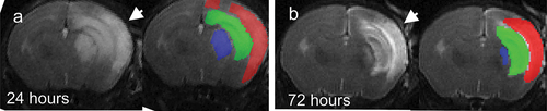
The severity of H-I brain injury on MRI was stratified by hyperintense volume as 1) no injury (0 mm2), 2) moderate (<30 mm2), and 3) severe (>30 mm2). Between 4 and 22% of animals in our study did not exhibit apparent changes on T2-weighted images after H-I, representing minimal to no injury. The occurrence of cases with no injury on MRI was not different between the microbiota treatment group (χ2 = 2.593, df = 3, p = 0.459). A pairwise comparison of outcome groups revealed a smaller proportion of animals in the severe group in the B. infantis group relative to the E. coli group at 24 and 72 h (p = .0036 and p = .0016, Fisher’s exact test). No other significant differences in severity outcome on MRI were found.
In summary, we observed that the distribution of the severity of brain injury was typical for this neonatal mouse H-I modelCitation19 and found a decrease in the proportion of animals with severe brain injury in the B. infantis group.
2.2. H-I edema volume was the largest in E. coli and the smallest in B. infantis groups
The analysis of variance showed that the effect of microbiota treatment on total edema volume was significant at 24 h, F(3,55) = 5.03, p = .0037, and at 72 h after H-I, F(3,55) = 6.07, p = .0012. The total volume of edema was significantly larger in E. coli group relative to B. infantis group on post-hoc analysis at 24 (p = .0015) and at 72 h (p = .0005) and relative to the Fresh water group at 72 h (p = .048). There was no significance in total edema volume between the other groups. When compared between brain regions, the decrease of edema in B. infantis group relative to E. coli group was significant only in cerebral cortex: F(3,55) = 4.11, p = .011 at 24 h and F(3,55) = 3.88, p = .014 at 72 h ().
Figure 2. Edema volumes at 24 and 72 h after H-I. A,B – regional edema volume in cortex, hippocampus and striatum. C,D. Change of edema volume from 24 to 72 h after H-I. No difference was found between microbiota treatment groups in absolute or relative amount of edema volume change. *- significant difference between group means (ANOVA with Tukey-Kramer post-hoc tests).
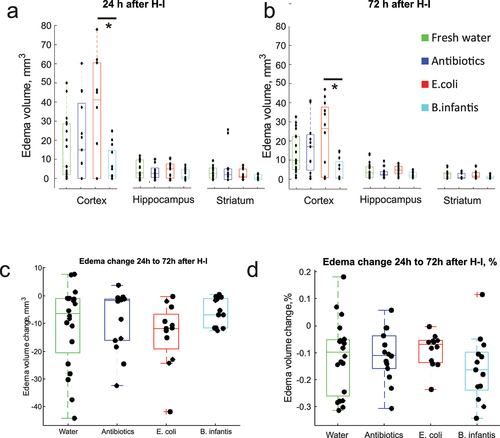
In summary, we observed a larger edema volume in E. coli group, predominantly due to an injury to the cerebral cortex.
2.3. Brain recovery after H-I injury between 24 and 72 h was not different between microbiota groups
Serial MRI measurements revealed that the edematous brain volume decreased between 24 and 72 h in most cases (). The initial H-I cellular injury, manifested as edema at 24 h, may resolve without tissue loss. However, hyperintense areas on T2-weighted images at 72 h represent the tissue with irreversible injury that will predominantly undergo necrosis, as confirmed previously by histology.Citation20 There was no difference between the microbiota treatment groups in edema volume change in absolute units (one-way ANOVA F(3,55) = 1.03, p = . 38), or when the change is expressed as a fraction of initial edema volume (one-way ANOVA F(3,55) = 0.58, p = .65).
2.4. Gene expression of pro-inflammatory cytokines increase in E. coli group brains at P10, no injury, and at P13, after H-I
We examined the gene expression of pro- and anti-inflammatory cytokines in the whole brain tissue with real-time PCR on separate cohorts on animals at two time points: P10, no injury and at P13, 72 h after H-I injury.
At P10, expression of pro-inflammatory cytokines TNFα was not different between microbiota groups (F(3,42) = 2.37, p = .083, ). At P13, 72 h after H-I, expression of TNFα was higher in Fresh water and E. coli groups (F(4,63) = 4.63, p < .001) relative to the sham control group. Noticeably, TNFα expression did not change with H-I in B. infantis and Antibiotics groups.
Figure 3. Pro-inflammatory cytokines gene expression before and after H-I across microbiota treatment groups. *-p < 0.05, ** - p < 0.01 on post-hoc tests.
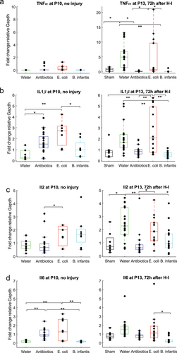
The effect of microbiota treatment on IL1β gene expression was significant at P10, F(3,42) = 7.75, p = 3.13e-04. IL1β expression was elevated in Antibiotics and in E. coli groups (). At P13, 72 h after H-I, the effect of microbiota treatment on IL1β gene expression was also significant (F(4,63) = 9.04, p < .001). IL1β expression was elevated in E. coli group relative to sham and Fresh water group, while it was lower in B. Infantis group relative to Fresh water group.
At P10, the expression of pro-inflammatory cytokine IL2 was higher in E. coli groups relative to Antibiotics group (F(3,42) = 4.09, p < .01, ). At P13, 72 h after H-I, expression of IL2 was higher in fresh water and E. coli groups (F(4,63) = 9.11, p < .001). At P10, expression of pro-inflammatory cytokine IL6 was higher in Antibiotics and E. coli groups relative to Fresh water and B. infantis groups (F(3,42) = 11.33, p < .001, ). At P13, 72 h after H-I, expression of IL6 was higher in E. coli group relative to B. infantis group.
A two-way ANOVA revealed that there was a statistically significant interaction between the effects of gut microbial treatment and H-I factors for all examined pro-inflammatory cytokines expressions: TNFα (F(3, 107) = 6.05, p < .001), IL1β (F(3, 107) = 6.25, p < .001), IL 2 (F(3, 107) = 6.25, p < .000), IL6 (F(3, 107) = 4.48, p < .001). Simple main effects of the microbial treatment modulated the expression of proinflammatory cytokines TNFα (F(1, 107) = 15.39, p < .001) and IL6 (F (1, 107) = 3.13, p = .031) after H-I with a large, 2- to 3-fold, increase in Fresh water and E. coli groups ().
In summary, microbiota manipulation altered the expression of pro-inflammatory cytokines at P10, with a significant increase in the E.coli group. After H-I, at P13, the expression of the pro-inflammatory cytokine was increased relative to the age-matched sham controls, in Fresh water and E.coli group, but not in Antibiotics and B.infantis group.
2.5. Anti-inflammatory cytokines gene expression increase in E.Coli group before and after H-I
At P10, ANOVA revealed that the effect of microbial treatment on expression of anti-inflammatory cytokine TGFβ was significant (F (3,42) = 50.8, p < .001, ), with increased expression of TGFβ and Antibiotics groups in post-hoc analysis. At P13, 72 h after H-I, expression of TGFβ was decreased in the Antibiotics and B. infantis groups relative to Fresh water (F(4,63) = 10.26, p < .001).
Figure 4. Anti-inflammatory cytokines gene expression before and after H-I in different microbiota treatment groups. *-p < 0.05, ** - p < 0.01 on post-hoc tests.
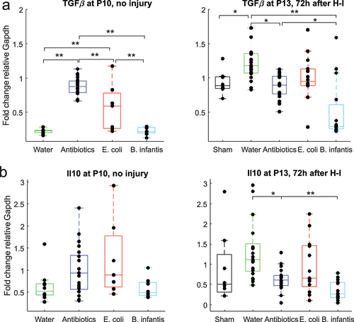
At P10 ANOVA revealed that the effect of microbial treatment on expression of anti-inflammatory cytokine IL10 was significant (F(3,42) = 2.85, p = .048, ), but there was no difference between the group on post-hoc analysis. At P13, 72 h after H-I, expression of IL10 was decreased in Antibiotics and B. infantis groups relative to Fresh water (F(4,63) = 5.75, p < .001).
A two-way ANOVA revealed that there was a statistically significant interaction between the effects of gut microbial treatment and H-I for all examined anti-inflammatory cytokines expressions: TGFβ (F(3, 107) = 19.7, p < .001), IL10 (F(3, 107) = 6.11, p < .001), indicating that the increased anti-inflammatory cytokines with H-I was only in Fresh water and E. coli groups. Simple main effects of H-I factor were significant for expression of TGFβ (F(1, 107) = 62.36, p < .001), indicating increase of TGFβ expression after H-I, but was not significant for IL10 (F(1, 107) = 0.24, p = 0.62).
In summary, we observed an increased expression of anti-inflammatory cytokine at P10 and P13 relative to the Fresh water group except B.infantis group.
2.6. Microbial treatment contributed to pro-inflammatory cytokine expression independently of initial injury after H-I
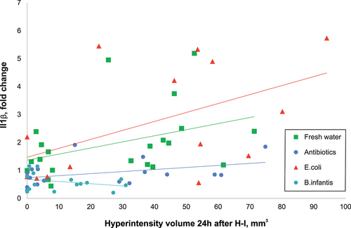
Cytokine expressions increased with the volume of injury after H-I (). To examine whether microbiota composition contributes to the neuroinflammatory reaction independently of injury severity, gene expression of cytokines was modeled using ANCOVA with injury volume at 24 h as a covariate. Coefficients of the ANOVA for each studied cytokine are presented in Supplementary Table S1. The E. coli group had the largest positive, and B. infantis had the largest negative significant intercepts in the linear models, which explains the levels of pro-inflammatory IL1β independently of the extent of H-I brain injury.
Figure 5. Pro-inflammatory cytokine IL1β increased proportionally with the extent of H-I injury. The linear regression slopes of the increase were significantly lower in antibiotic group relative to fresh water and E. coli groups, and even lower in B. infantis group.
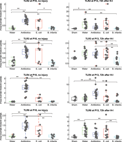
Two-way ANOVA analysis revealed that both H-I and microbiota treatment and their interaction were significant factors for all TLR expressions, as summarized in Supplementary Table S2. Expression of all studied TLRs in brains before H-I was increased in Antibiotics and E. coli groups relative to the Fresh water and B. infantis groups (). After H-I, the expression of all TLRs increased several folds relative to pre-H-I values and shams in Fresh water, Antibiotics and E. coli groups without difference between the groups. Remarkably, the expression of TLRs in B. infantis group remained on the same level as in shams, approximately at pre-H-I levels.
Figure 6. Gene expression of TLRs before and after H-I in different microbiota treatment groups. *-p < 0.05, ** - p < 0.01 based on post-hoc tests.
In summary, cytokine expressions increased with the volume of injury after H-I, with the largest slope in the E.coli group.
2.7. Activation of astroglia and microglia activation after H-I is decreased in B. infantis groups
To test whether the differential expression of brain cytokines after H-I in microbiota groups corresponded to the level of reactive gliosis, which is known to mediate neuroinflammatory response,Citation21 brain sections were stained for GFAP and Iba-1 markers to identify activated astro- and microglia cells (). The number of GFAP and Iba-1 positive reactive glia cells was very low in sham controls (), there was a dramatic increase in the number of reactive glia cells in the hemisphere ipsilateral to the carotid ligation (). Microbial treatment was a significant factor in the number of reactive astrocytes (F(3, 12) = 10.15, p = .0013), and activated microglia cells (F(3, 12) = 8.89, p = .0022), . Post-hoc Tukey’s multiple comparisons test revealed significantly smaller number reactive astroglia in the B.infantis group relative to the Fresh water (p = .005), Antibiotic (p = .0077), and E.coli (p = .0016) groups. Similarly, the number of activated microglia cells were lower in B.infantis group relative to the Fresh water (p = .0087) and E.coli (p = .0016) groups. The number of reactive glia cells was significantly lower in the cortex contralateral to the carotid ligation side, but slightly higher than in the sham, with no difference between microbiota groups.
Figure 7. Activation of astro glia and microglia activation after H-I.
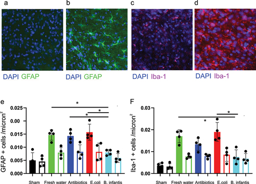
In summary, reactive astro- and microgliosis was observed in the ipsilateral hemisphere in all microbial treatment groups and was significantly smaller in the B.infantis group.
2.8. Small intestines injury after H-I
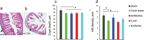
To examine whether microbial manipulation and neonatal gut dysbiosis may result in a selective gut susceptibility to hypoxia during H-I insult, terminal ileum morphology was examined on H&E staining. Small intestine injury after H-I was evident by structural changes (). Crypt depth decreased at 72 h after H-I in all microbiota groups relative to sham controls (one-way ANOVA F(4,24) = 3.30, p = .031, ). This decrease was not different between microbiota groups. Villi density was lower in the Fresh water and E. coli groups after H-I relative to the sham controls ((4,24) = 9.04, p < .001). No difference after H-I was found in villi length between the microbiota groups (F(4,24) = 0.79, p = .51).
Figure 8. Small intestine injury after hypoxia – ischemia. Hematoxylin and eosin staining of terminal ileum of controls (A) and mouse pups in the fresh water group 72 h after H-I (B). Decreased thickness of lamina propria is indicated by arrows. Crypt depth (C) and villi density (C) were decreased after H-I in all microbiota groups. *- p < 0.05 post-hoc group comparisons with control group.
In summary, injury in small intestines was observed in all microbiota groups after H-I.
2.9. Serum pro-inflammatory cytokines increase after global hypoxia without brain injury in all microbiota groups
To test whether global H-I injury to organs other than brain contributed to the increase in serum cytokine levels independently of brain injury, we subjected all microbial manipulation groups to 8% hypoxia protocol at P10 but did not perform carotid artery ligation surgery. With this regimen, we expected no brain injury, confirmed by the absence of cell injury in non-ligated ipsilateral hemisphere in a similar neonatal hypoxia-ischemia mice model.Citation22 Animals were euthanized and serum cytokines concentrations were measured in mice subjected to global hypoxia in age-matched normoxia controls.
Serum concentrations of IL1β, IL2, IL4, and interferon-γ, with and without hypoxia, were lower than the ELISA assay sensitivity (<3 pg/mL). A one-way ANOVA revealed a significant effect of microbial treatment on expression of pro-inflammatory cytokine IL6 in the group without hypoxia (F(3,17) = 3.22, p = .042, ), with the increase of IL6 in E. coli relative to Fresh water group (post-hoc p = .031). No difference between microbiota treatment groups was found in IL6 after hypoxia (ANOVA F(3,16) = 0.81). No difference between microbiota treatment groups was found in serum concentration of TNFα before hypoxia (F(3,17) = 2.31, p = 0.11) or after hypoxia (F(3,16) = 1.02, p = 0.40) as well as in serum concentration of IL10 before (F(3,17) = 0.95, p = 0.43) and after hypoxia (F(3,16) = 2.49, p = .09).
Figure 9. Cytokines serum concentrations before and after global hypoxia without carotid ligation. *-p < 0.05, ** - p < 0.01 on post-hoc tests.
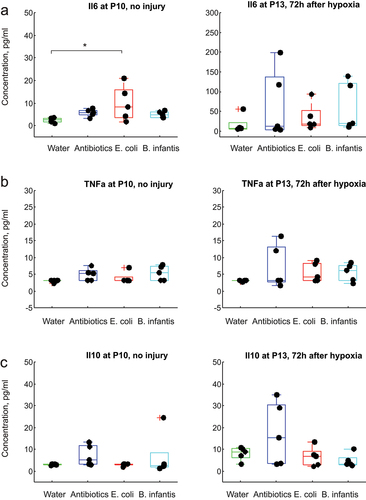
A two-way ANOVA revealed a significant effect of hypoxia on serum concentration of IL6 (F(1,33) = 9.56, p = .004), resulting in elevated IL6 in all microbiota groups except fresh water, . No other significant effects of microbiome treatment or hypoxia or interaction between them were found in pro-inflammatory TNFα and anti-inflammatory IL10 (.
In summary, the level of circulating cytokines was increased after global H-I without brain injury that may be an independent factor determining neonatal brain injury severity.
2.10. Changes in neonatal gut microbiome composition with transfaunation and after H-I
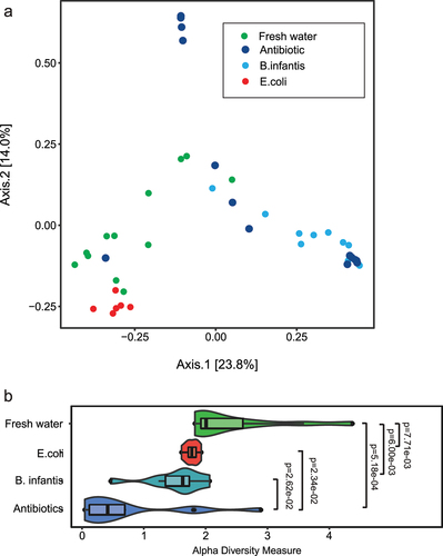
Indexes of mouse pup microbiome biodiversity were examined using 16S rRNA gene sequencing of colon microbiota at P10, no injury and at P13, 72 h after H-I. There was a distinct separation of E. coli and water groups using principal component analysis with Bray-Curtis dissimilarity measure (, ANOSIM, R = 0.762; p = .001), while the B. infantis and antibiotics groups had a large overlap. The Shannon index of alpha-diversity was significantly lower in Antibiotics group relative to Fresh water group, indicating the successful depletion of microbiota in neonatal digestive tract after maternal antibiotic treatment until P10. Alpha-diversity increased with microbial transfaunation with E. coli and B. infantis by P10 after the maternal antibiotics treatment but did not reach the level of the Fresh water group.
Figure 10. Indexes of mouse pup microbiome biodiversity after neonatal gavage transfaunation using 16S rRNA gene sequencing before H-I at P10. A. Beta diversity PCoA plots with bray-curtis dissimilarity measure, showing separation of gut bacterial communities with different microbial treatment. B. Shannon index of alpha-diversity was significantly lower in the Antibiotics group relative to fresh water group, but no difference between E. coli and B. infantis groups.
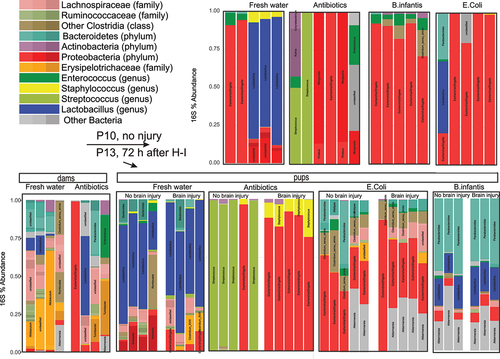
Microbial composition was apparently different between microbiota treatment groups at P10, no injury, as shown on the distribution of operational taxonomic units (OTU) in large intestine samples (). Fresh water controls had a large abundance of Lactobacillus genus relative to the other microbiota treatment groups. By P13, all mice, except Antibiotics group, showed an increase of diversity in 16s abundance, similar to the previously described trend in neonatal gut microbiome development.Citation14 We also separated groups with and without brain injury to examine possible interactions. After the insult, pups with brain injury in Antibiotics group had a larger proportion of Escherichia/Shigella family. Pups in B. infantis group had a larger proportion of Bacteroidetes and Lactobacillus ().
Figure 11. Taxonomic distribution in OTUs in colon samples obtained from mouse pups before (P10) and after IH (P13). The samples after H-I were sorted onto by the extent of brain injury.
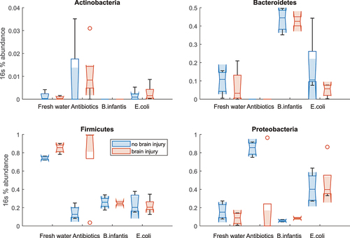
We further analyze OTU abundance changes between microbial treatment groups in pups with and without brain injury on MRI in four major phyla (). Two-way ANOVA revealed no effects of microbiome and brain injury on Actinobacteria () and a significant main effect of microbiome F(3,25) = 35.7, p < .001 in abundance of Bacteroidetes phylum (). The relative abundance of Bacteroidetes was significantly higher in B. infantis group regardless of brain injury factor. Two-way ANOVA revealed significant main effects of microbiome F(3,25) = 16.41, p < .001 and brain injury factors F(1,25) = 7.52, p = .01, and their interactions F(3,25) = 6.52, p = .002 in abundance of Firmicutes (). Significant main effects of microbiome F(3,25) = 8.97, p < .001 and brain injury factors F(1,25) = 4.59, p = .04, and their interactions F(3,25) = 5.48, p = .004 were also found in abundance of Proteobacteria (). The significant effect of brain injury factor was driven primarily by with higher Firmicutes and low Proteobacteria in antibiotic group.
Figure 12. 16s abundance for the four most abundant phyla in colon samples in pups with and without brain injury at P13 after H-I.
In summary, we observed a complex interaction between H-I insult, brain injury, and microbial composition in neonatal guts.
3. Discussion
The major finding of the study was that neonatal gut microbiota modulated the extent of brain injury and neuroinflammatory response after H-I in a neonatal mouse model. Transfaunation of mouse pups early after birth with pathogenic bacteria E. coli significantly increased the H-I-induced brain injury relative to saline control pups from dams receiving fresh water. The extent of brain injury in pups, transfaunated with commensal bacteria B. infantis, remained similar to the saline control. Expression of proinflammatory cytokines in the brain was higher in E. coli group before and after H-I and was associated with the larger H-I brain injury, while B. infantis significantly decreased extent of H-I injury and lowered pro-inflammatory cytokines before and after H-I. This increase of pro-inflammatory cytokines occurred in conjunction with elevated expression of TLR in brains of E. coli groups. Elevated pro-inflammatory cytokines closely corresponded to the level of reactive astro- and microgliosis and was significantly smaller in the B.infantis group. We did not find evidence that gut microbial composition differentially affect pro- and anti-inflammatory cytokine increase after global hypoxia without brain injury. Microbial manipulation and brain injury were also significant factors affecting microbiota gut composition, indexes of biodiversity and relative abundance of major bacterial phyla in neonatal intestines at 72 h after H-I.
In the current study, we determined that the exposure to opportunistic pathogenic (E. coli) or commensal (B. infantis) bacteria modulated inflammatory responses before and after hypoxic-ischemic brain injury. Bacteria strains were chosen to provide pro- (E. coli) and anti-inflammatory (B. infantis) effects on brain based on published data. Both taxa are common both to human and conventional mouse gut flora and typically present in hospital environment. E. coli is associated with necrotizing enterocolitis (NEC) in premature infants.Citation15 Inoculation of germ free parent mice with enteropathogenic E. coli increased pro-inflammatory cytokines IL1β and IL6 in plasma of offspring.Citation16 Transfaunation with B. infantis increased the expression anti-inflammatory cytokines IL-10 and TGF-β1 in mouse colon tissue,Citation17 and decreased IL1β in a rat colitis model.Citation17 Bifidobacterium is the dominant bacterial genus in normal neonatal intestinal flora (commensal bacteria)Citation18,Citation23 and has received attention for preventing NEC.Citation24 Bifidobacterium strains increased anti-inflammatory cytokines IL-4, IL-10 in inoculated germ-free miceCitation25 and improved cognition in BALB/c mice.Citation26
In neonates, systemic modulation of inflammation has been shown to affect the extent of brain hypoxic-ischemic injury.Citation27,Citation28 Bacterial exposure and systemic inflammation in neonates have been modeled with lipopolysaccharide (LPS) injections, which dramatically increased H-I brain injury.Citation28 Sensitization or protective preconditioning to H-I depended on the timing of LPS injection: brain injury was decreased if LPS had been administered 24 h before H-I and increased in a shorter or longer period before H-I.Citation29 In our study, we observed an increase of pro-inflammatory cytokines in blood and in brains in E. coli group, associated with an increased injury volume on MRI, suggesting primary sensitization of neonatal brains to H-I due to change in neonatal gut microbial composition. E. coli group had also increased TLRs expression in brains before H-I, suggestive of elevated bacterial component presence in the pups’ systemic circulation.
There was a large, several fold increase of TLRs 2, 3, 4, and 6 and proinflammatory cytokines TNF-α, IL1β, IL2 gene expression in brains after H-I, in accordance with previous reports.Citation30,Citation31 Expression of the proinflammatory cytokine was proportional to the extent of injury, but higher for the E. coli group compared to the Fresh water group given the same amount of the injury. Markedly, transfaunation with B. infantis resulted in the least amount of brain injury and expression of TLRs and pro-inflammatory cytokines before and after H-I across all groups, which suggests a neuroprotective effect of this microbiota manipulation due to decreased sensitization and inflammatory response to H-I. Taken together, we have demonstrated that proinflammatory E. coli is associated with increased brain injury and neuroinflammation before and after H-I injury, whereas commensal B. infantis had protective effects on brain injury and decreased neuroinflammation after H-I.
We examined whether the differential expression of pro- and anti-inflammatory cytokines was related to changes in the intestinal wall integrity due to dysbiosis caused by microbiota manipulations or due to global hypoxia injury to organs other than brain, including intestines. Both scenarios are relevant to clinical conditions of dysbiosis and exposure to intermittent hypoxic episodes in preemies or birth asphyxia in term infants and may result in damage to intestines.Citation32 We did find histopathological evidence of structural injury in ileum after H-I, including crypt depth and villi density decrease in all microbiota groups relative to Fresh water sham controls, especially in E. coli group. At the same time, the level of circulating pro-inflammatory cytokine IL6 was elevated in the experiment of global hypoxia without brain injury, indicating that hypoxic injury to the other organs may contribute independently to H-I brain injury. Possible mechanisms could involve inflammation and activation of HIF and other molecules after global hypoxia that can have an impact on cytokine expression, possibly in gut tissue, but in other sites as well.Citation33
Microbiota manipulations in this study altered the normal pattern of microbial colonization in the intestine of neonatal mice, that is typically characterized by a large presence of Lactobacillus.Citation34 Maternal antibiotics administration dramatically reduced bacterial load and biodiversity of intestinal microbiome, as reported before.Citation14 The presence of a complex and diverse intestinal flora was found to be functionally important for regulating intestinal mucosal and systemic immune responsesCitation35 and provide resistance to viral and bacterial infections, including E. coli.Citation5,Citation14 While transfaunation with E. coli and B. infantis in our study increased alpha diversity measures compared to Fresh water controls, the composition of the microbiota was different between these groups, which may explain the difference in the extent of H-I injury and magnitude of neuroinflammatory response. We also found significant effects of microbiota composition and H-I brain injury on 16s abundance of major phyla, suggesting that H-I injury in the brain affects microbial composition in the bowl, as observed in adult mice,Citation36 but this observation requires additional data.
Our study included maternal antibiotic administration until P5 to model dysbiosis in offspring. While antibiotics were essentially eliminated from the pups’ systemic circulation before H-I insult and were unlikely to directly affect the neuroinflammatory response to H-I as demonstrated in post-H-I insult antibiotic administration,Citation27,Citation37 they had a long-lasting effect by depleting microbiota and dramatically reducing microbiome biodiversity. The situation of developmental dysbiosis is clinically relevant since infants are often treated with systemic antibiotics.Citation38 While mouse pups after maternal antibiotics, followed by saline gavage in our study, demonstrated a decrease of pro-inflammatory cytokines IL1b, IL2, TNF-α, and also significant increase of TGFβ in brains after H-I, there was no difference in initial injury size or the dynamics of injury size in this group relative to fresh water controls. Therefore, early antibiotic treatment resulting in microbiota depletion and low gut microbial biodiversity did not reduce susceptibility of infant brain to H-I injury, but may affect the long-term recovery.
The study has several limitations and over simplifications. The experimental groups defined by microbial manipulation were intended to model conditions of “pathogenic versus commensal” bacteria load, which is likely an over simplification. E. coli indeed is a normal component of neonatal gut microbiota and may be beneficial, along with other bacteria, including Bifidobacterium, to the maturation of normal antibody response to infection.Citation39 However, E. coli is known as an opportunistic pathogen, and its presence in large amounts has been associated with necrotizing enterocolitis in premature infants.Citation15 Transfaunation of E. coli in large amounts in the immature mouse intestines in our study was intended to create dysbiosis with increased systemic inflammation.Citation16
Second, the assessment of neuroinflammation before and after H-I insult at was conducted at different time points, P10 and P13, on separate cohorts of animals. However, there was no significant difference found between the Fresh water group at P10 and the Fresh water group with sham surgery at P13, indicating minimal confounding effect of age on expression of the studied cytokines and justifying the use of 2-way ANOVA to examine the effect of H-I and microbiota factors using two age groups.
Third, TGF-β was referred to as an anti-inflammatory cytokine, as commonly refereed in the perinatal neuroinflammation field,Citation11 although it has both a pro- and an anti-inflammatory action, participating in the resolution of chronic neuroinflammation.Citation9
Fourth, antibiotics from the mother’s milk may be absorbed in the pups’ intestine and affect the response to H-I insult. However, we expected that the systemic concentration of antibiotics in pups to decrease to minimal values during 6 days from cessation of maternal antibiotic administration at P5 before H-I insult at P10, and do not provide anti-inflammatory and neuroprotective effect as described in rodent models with antibiotics administration around the time of H-I.Citation27,Citation37
Fifth, we did not use a carotid ligation normoxia group. Carotid ligation alone (without hypoxia) in the Vannucci model in mice and rats does not cause permanent cell death.Citation8 Instead, the confounding inflammatory effects of the surgery were controlled in the sham group where incision was made but the vessels were not manipulated.
In adults, the microbiome is now considered as an established modulator of secondary neuroinflammation and regeneration after experimental strokeCitation40 and in human patients.Citation41 In adult mice, microbiota modulation with antibiotics was protective against stroke via a mechanism that was dependent on IL-17.Citation42 Multiple mechanisms linking microbiota as well as the associated metabolites have been proposed to play a role in pathogenesis of ischemic stroke, including oxidative stress, apoptosis, and neuroinflammation.Citation43 The current study examined the role of the microbiome in the regulation of neuroinflammation, but the other factor affecting infant brain susceptibility to hypoxia-ischemia due to gut-brain interaction remains to be discovered. Infant microbiota is increasingly recognized as a critical contributor to brain cognitive development.Citation44–47 Simple strategies, like oral administration of specific probiotics (such as B. infantis) may be neuroprotective to decrease the volume and risk of brain damage after perinatal hypoxic ischemic brain injury.
4. Materials and methods
Timeline of the experiments is shown in and experimental groups and sample numbers are in Supplementary Table S3.
Figure 13. Time line of the experiments and experimental groups. The study groups are formed by a combination of two experimental factors: 1) microbial manipulation (fresh water, saline, E.Coli, B. infantis) and 2) hypoxia (normoxia, hypoxia, carotid ligation + hypoxia), at 2 time points. Each group had the same outcome measures, listed in the bottom row.
4.1. Manipulation of intestinal microbiota in neonatal mice
C57BL/6J mice were originally obtained from Jackson Laboratory (Bar Harbor, Maine) and bred in house. Time pregnant mice were maintained in autoclaved plastic cages with autoclaved food and water. Starting day E15 of pregnancy (5 days before term delivery) mice received sterile drinking water mixed with five antibiotics: (ampicillin, gentamicin, metronidazole, vancomycin, neomycin, all 1 mg/ml, from Sigma-Aldrich). This regiment has been shown to deplete bacterial load in neonates by at least a magnitude of 16sRNA copies/mg of intestinal content.Citation14 Depletion of neonatal gut microbiota mimics abnormal gut flora development that may occur after cesarean section or antibiotic application in neonates and also equalizes the colonization opportunities with microbial manipulations. Antibiotic water was changed weekly. The dams continued to receive antibiotic containing drinking water after the pups were born until P5. After P5, sterile drinking water was given to the dams until the experiment was finished.
At P5, littermates were randomly assigned to microbial treatment groups and received a gavage suspension of either Escherichia coli (“E. coli” group, n = 28), or Bifidobacterium longum infantis (“B. infantis” group, n = 23), or sterile saline (“Antibiotics” group, n = 44). Naïve pups in the control group were fed by dams receiving fresh water (“Fresh water” group, n = 37) throughout the experiment. The bacteria were obtained from the American Type Culture Collection (E. coli ATCC 10,798 and B. infantis ATCC 15,697). Bacteria were plated on agar plates and selected colonies free of contamination were propagated in tryptic soy broth type E. The bacteria concentration was measured using spectrophotometer (Siemens Micro Scan Turbidity Meter). All vials with bacteria were adjusted to reach the 0.5 Mcfarland units that approximates 1.5 × 10⁸ cell count density per 1 mL, by adding more bacteria cells with a cotton swab. The vials were spun down; pellets with cells were collected and stored in −80°C until use. Before each feeding, cells were re-suspended and each pup received 100 µL of gavage suspension containing 1.5 × 10⁸ CFU of bacteria or saline at P5, P7, P9, and P11 using a silicone gavage tube.
Fecal samples of the pups were collected from the pups’ colons at P10 and P13 (before and after H-I insult, correspondingly) and stored at −80°C for sequencing analysis. The intestinal contents were processes at the University of Chicago Duchossois Family Institute facility for genomic DNA extraction and subsequent 16S rRNA gene sequencing on the Illumina MiSeq platform. Data processing was conducted using R statistical software version 3.6.2 and the R package DADA2 version 1.14.1 pipeline.Citation48 Specifically, reads were first trimmed at 180 bp for both forward and reverse read to remove low-quality nucleotides. Chimeras were detected and removed using the default consensus method in the DADA2 pipeline. Then, ASVs with lengths between 300 and 360 base pairs were kept and deemed as high-quality ASVs. Taxonomy of the resultant ASVs wase assigned to the genus level using the RDP database with a minimum bootstrap confidence score of 50. Multiple sequence alignment of all ASVs was performed to generate a neighbor-join phylogenetic tree using the R package “msa” (v1.18.0) and “ape” (v5.3). Indexes of microbiome alpha (Shannon) diversity or overall β-diversity were calculated.
4.2. Neonatal brain hypoxia-ischemia
At age P10, neonatal mice of both sexes from each litter were subjected to H-I insults using the Vannucci model adapted for neonatal mice.Citation22,Citation49 Briefly, the left carotid artery was permanently ligated with a double knot silk suture 7–0 under isoflurane anesthesia. Lidocaine was added to the wound for local analgesia. The wound was sutured, and pups returned to dam for recovery and nursing for 3 h, followed by 60 min of hypoxia with 8% O2 at 37°C. Temperature during H-I was maintained by a servo-controlled heating blanket with temperature probe inside hypoxic chamber (Homeothermic Blanket System, Harvard apparatus, MA). Sham controls from the Fresh water group underwent surgery with carotid artery exposed but not ligated and did not receive hypoxia. Mortality rate due to the surgical manipulation and hypoxia was between 11.8–18.2% and not different between microbiota treatment groups. Therefore, experimental groups were “Fresh water +H-I”, n = 31, “”Antibiotics + H-I”, n = 44, E.coli+H-I”, n = 28, “B.infantis + H-I”, n = 23, “Fresh water +sham H-I”, n = 6.
4.3. Neonatal global hypoxia
To evaluate the systemic inflammatory response to hypoxia independently of brain injury, a separate set of sham operated animals with carotid artery exposed but not ligated (n = 4 in each microbiota group, i.e. “Fresh water”, “Antibiotics”, E.coli”, “B.infantis”) underwent 60 min of hypoxia with 8% O2 at 37°C. There was no mortality in this experiment. Blood samples were obtained 4 h after hypoxia exposure using carotid artery incision under isoflurane anesthesia and spanned down. Serum was frozen for cytokine measurements by multiplex ELISA.
4.4. Injury volume on MR imaging
At 24 and 72 h after H-I, serial MRI data were acquired for all pups. During the imaging, mice were anesthetized with 1% isoflurane in air and kept warm on a heated water blanket. MR images were acquired on a 9.4 Tesla scanner (Bruker Biospin, Billerica, MA) using a Bruker mouse receive-only surface coil and a 72 mm quadrature transmitter coil. T2-weighted images were acquired using a fast spin echo RARE sequence with TE/TR 40/3000 ms, 4 signal averages, echo train length 8, in-plane resolution 0.12 × 0.12 mm. Twenty coronal slices with thicknesses 0.7 mm were placed to cover the whole brain. To increase throughput, pups were scanned in pairs. Hyperintense areas indicating injured brain regions (edema) were manually outlined on each slice using ITK-SNAP 3.2 (itksnap.org). Volumes of edematous areas in the cortex, hippocampus, striatum, as well as total edematous volumes were calculated.
4.5. RNA isolation and real-time PCR
Only the animals with hyperintense regions, detected either on T2-weighted MRI at 24 h or 72 h (Supplementary Table S3), were included in the further analysis after H-I: Fresh water group, n = 16, Antibiotics group, n = 14, E. coli group, n = 13, B. infantis group, n = 8. Brain and ileum tissues were collected and individually frozen on dry ice and stored at −80°C before use. The ipsilateral (left) brain hemisphere tissue was homogenized in Qiazol Lysis Reagent (cat. # 79306 Qiagen, Germantown, MD) on ice for extraction of total RNA. NanoDrop (ThermoFisher Scientific, USA) instrument was used to measure RNA quantity and quality. 500 ng of isolated total RNA was used to synthesize cDNA using an RT2 First Strand Kit from QIAGEN. cDNA was amplified by polymerase chain reaction (PCR) with Quantitect Sybr Green PCR Kit (Qiagen, Germantown, MD, cat. # 204143) on an Applied Biosystems GeneAmp 5700 real-time quantitative PCR instrument. The total reaction volume of 25 μL consisting of 1.0 ul RT product (cDNA). Primers for TLR2–6, IL1b, IL2, IL6, IL8, IL10, TNF-α, TGFβb, GAPDH for Sybr Green Assay were predesigned and ordered from Integrated DNA Technologies, Inc., USA. For each primer, the final concentration was 0.4 μm and annealing temperature was adjusted. Gene expression was normalized to the housekeeping gene GAPDH and expressed as a fold change of experimental controls using delta-delta Ct method.
4.6. Inflammatory cytokine measurements by multiplex ELISA
Blood serum samples were used for cytokine analysis. Protein levels of cytokines TNF-α, IL-1β, IL-2, IL-6, IL-10, interferon-γ were assessed using a MILLIPLEX® Mouse Cytokine/Chemokine magnetic bead panel (MCYTOMAG-70, Millipore) according to the manufacturer’s instructions. Standard curves were generated using the specifics standards supplied by the manufacturer. Samples were analyzed on a MAGPIX® system (Millipore) using the MILLIPLEX® Analyst 5.1 software (Millipore).
4.7. Histological assessment of glia activation in brains after H-I
Pups were euthanized at P13, 72 h after H-I by transcardial perfusion with saline solution (0.9% NaCl) followed by 4% paraformaldehyde (PFA). Brains were removed and post-fixed in 4% PFA overnight, soaked in 30% sucrose solution for cryoprotection, frozen on dry ice and cryosectioned 40 μm in the coronal plane. For identification of astrocytes, a primary anti-glial fibrillary acidic protein antibody (anti-GFAP, #G9269, 1:200, Sigma-Aldrich) and for identification of microglia the Iba-1 antibody (anti-iba-1, ab5076, 1:200, Abcam) was used. The secondary antibodies were Alexa 488 anti-rabbit (1:1000, Molecular Probes, Invitrogen) and Alexa Fluor 594 (1:1000, TermoFisher Scientific), respectively. The slides were sealed with mounting medium containing DAPI (Vector Laboratories, Inc. H-1200) and coverslipped. Three consecutive brain sections, 100 μm apart, were imaged using a Nikon Eclipse 80i microscope and 40× magnification at the level of dorsal hippocampus – anterior thalamus. Four to six rectangular 60 × 60 μm regions of interest were randomly placed on the cerebral cortex bilaterally. The number of reactive astrocytes and activated microglia, identified using morphological criteriaCitation22 on immunostaining and with nuclei co-localization on DAPI stain (), were counted and averaged between sections and region of interest, separately for ipsilateral and contralateral to H-I injury site.
4.8. Histological assessment of intestinal injury
After euthanizing pups on P13, 1 cm samples of terminal ileal tissue were harvested, rinsed with saline, and fixed in 4% PFA. Coded frozen cross-sections (20 μm thick) were stained with hematoxylin and eosin by standard protocols. Lamina propria thickness in crypts, villi length and density per ileum cross-section circumference was measured by micrometry under 10×magnification in a blinded manner; at least 10 measurements from non-overlapping well oriented areas were taken from each sample. Five samples were taken from the Fresh water sham group and all microbiota groups after H-I.
4.9. Statistical analysis
Effects of microbiome treatment on brain injury volume on MRI, cytokines and TLRs gene expression was tested using a one-way ANOVA, followed by post-hoc Tukey-Kramer group comparisons as implemented in the Matlab R2018a (Natick, MA) statistical toolbox. A two-way ANOVA was used to test the main effects of microbiota group and H-I factors and their interactions on cytokines expression. An ANCOVA was used to test significance of microbiota group on cytokines gene expression (y) with H-I injury volume at 24 h as a covariate (x), using a model y = (α + αi) + βx + ε, where αi was the intercept for each microbiota group. Distribution of injury severity outcomes between microbiota treatment groups was calculated using a Chi-squared and Fisher’s exact test in Matlab.
Authors’ contributions
A. Drobyshevsky, M. Caplan – conceptualization, analysis, writing, S. Synowiec, I. Goussakov, J. Lu, R. Fabres – data acquisition. All authors reviewed the manuscript.
Availability of data and materials
All sequencing data are available on NCBI (bioproject ID: PRJNA1001342).
Ethics approval
The study received approval by the Institutional Animal Care and Use Committee of NorthShore University HealthSystem.
Supplemental Tables clean.docx
Download MS Word (17.5 KB)Acknowledgments
D. Gascoigne– review & editing.
Disclosure statement
No potential conflict of interest was reported by the author(s).
Supplementary material
Supplemental data for this article can be accessed online at https://doi.org/10.1080/19490976.2024.2333808
Additional information
Funding
References
- Schwiertz A, Gruhl B, Löbnitz M, Michel P, Radke M, Blaut M. Development of the intestinal bacterial composition in hospitalized preterm infants in comparison with breast-fed, full-term infants. Pediatr Res. 2003;54(3):393–21. doi:10.1203/01.PDR.0000078274.74607.7A.
- Stark PL, Lee A. The bacterial colonization of the large bowel of pre-term low birth weight neonates. J Hyg (Lond). 1982;89(1):59–67. doi:10.1017/S0022172400070546.
- Rubaltelli FF, Biadaioli R, Pecile P, Nicoletti P. Intestinal flora in breast- and bottle-fed infants. J Perinat Med. 1998;26(3):186–191. doi:10.1515/jpme.1998.26.3.186.
- Guarner F, Malagelada JR. Gut flora in health and disease. Lancet. 2003;361(9356):512–519. doi:10.1016/S0140-6736(03)12489-0.
- Gonzalez-Perez G, Hicks AL, Tekieli TM, Radens CM, Williams BL, Lamousé-Smith ESN. Maternal antibiotic treatment impacts development of the neonatal intestinal microbiome and antiviral immunity. J Immunol. 2016;196(9):3768–3779. doi:10.4049/jimmunol.1502322.
- Larroque B, Ancel P-Y, Marret S, Marchand L, André M, Arnaud C, Pierrat V, Rozé J-C, Messer J, Thiriez G. et al. Neurodevelopmental disabilities and special care of 5-year-old children born before 33 weeks of gestation (the EPIPAGE study): a longitudinal cohort study. Lancet. 2008;371(9615):813–820. doi:10.1016/S0140-6736(08)60380-3.
- Woodward LJ, Edgin JO, Thompson D, Inder TE. Object working memory deficits predicted by early brain injury and development in the preterm infant. Brain. 2005 Sep 8;128(Pt 11):2578–2587. doi:10.1093/brain/awh618.
- Volpe JJ. Neurology of the newborn. Vol. xiv. 5th ed. Philadelphia: Saunders/Elsevier; 2008. p. 1094.
- Hagberg H, Mallard C, Ferriero DM, Vannucci SJ, Levison SW, Vexler ZS, Gressens P. The role of inflammation in perinatal brain injury. Nat Rev Neurol. 2015;11(4):192–208. doi:10.1038/nrneurol.2015.13.
- Fleiss B, Tann CJ, Degos V, Sigaut S, Van Steenwinckel J, Schang A-L, Kichev A, Robertson NJ, Mallard C, Hagberg H. et al. Inflammation-induced sensitization of the brain in term infants. Develop Med Child Neuro. 2015;57(Suppl 3):17–28. doi:10.1111/dmcn.12723.
- Jin C, Londono I, Mallard C, Lodygensky GA. New means to assess neonatal inflammatory brain injury. J Neuroinflammation. 2015;12(1):180. doi:10.1186/s12974-015-0397-2.
- Schultz C, Temming P, Bucsky P, Göpel W, Strunk T, Härtel C. Immature anti-inflammatory response in neonates. Clin Exp Immunol. 2004;135(1):130–136. doi:10.1111/j.1365-2249.2004.02313.x.
- Chalak LF, Sánchez PJ, Adams-Huet B, Laptook AR, Heyne RJ, Rosenfeld CR. Biomarkers for severity of neonatal hypoxic-ischemic encephalopathy and outcomes in newborns receiving hypothermia therapy. J Pediatr. 2014;164(3):468–474.e1. doi:10.1016/j.jpeds.2013.10.067.
- Deshmukh HS, Liu Y, Menkiti OR, Mei J, Dai N, O’Leary CE, Oliver PM, Kolls JK, Weiser JN, Worthen GS. et al. The microbiota regulates neutrophil homeostasis and host resistance to Escherichia coli K1 sepsis in neonatal mice. Nat Med. 2014;20(5):524–530. doi:10.1038/nm.3542.
- Coggins SA, Wynn JL, Weitkamp JH. Infectious causes of necrotizing enterocolitis. Clin Perinatol. 2015;42(1):133–154, ix. doi:10.1016/j.clp.2014.10.012.
- Sudo N, Chida Y, Aiba Y, Sonoda J, Oyama N, Yu X-N, Kubo C, Koga Y. Postnatal microbial colonization programs the hypothalamic–pituitary–adrenal system for stress response in mice. Journal Of Physiology. 2004;558(1):263–275. doi:10.1113/jphysiol.2004.063388.
- Zhou L, Liu D, Xie Y, Yao X, Li Y. Bifidobacterium infantis Induces protective colonic PD-L1 and Foxp3 regulatory T cells in an acute murine experimental model of inflammatory bowel disease. Gut Liver. 2018;13(4):430–439. doi:10.5009/gnl18316.
- Stewart CJ, Ajami NJ, O’Brien JL, Hutchinson DS, Smith DP, Wong MC, Ross MC, Lloyd RE, Doddapaneni H, Metcalf GA. et al. Temporal development of the gut microbiome in early childhood from the TEDDY study. Nature. 2018;562(7728):583–588. doi:10.1038/s41586-018-0617-x.
- Wu D, Martin LJ, Northington FJ, Zhang J. Oscillating-gradient diffusion magnetic resonance imaging detects acute subcellular structural changes in the mouse forebrain after neonatal hypoxia-ischemia. J Cereb Blood Flow Metab. 2019;39(7):1336–1348. doi:10.1177/0271678X18759859.
- Ashwal S, Tone B, Tian HR, Chong S, Obenaus A. Comparison of two neonatal ischemic injury models using magnetic resonance imaging. Pediatr Res. 2007;61(1):9–14. doi:10.1203/01.pdr.0000251612.16069.4b.
- McAdams RM, Juul SE. The role of cytokines and inflammatory cells in perinatal brain injury. Neurol Res Int. 2012;2012:1–15. doi:10.1155/2012/561494.
- Albertsson A-M, Bi D, Duan L, Zhang X, Leavenworth JW, Qiao L, Zhu C, Cardell S, Cantor H, Hagberg H. et al. The immune response after hypoxia-ischemia in a mouse model of preterm brain injury. J Neuroinflammation. 2014;11(1):153. doi:10.1186/s12974-014-0153-z.
- Turroni F, Peano C, Pass DA, Foroni E, Severgnini M, Claesson MJ, Kerr C, Hourihane J, Murray D, Fuligni F. et al. Diversity of bifidobacteria within the infant gut microbiota. PloS One. 2012;7(5):e36957. doi:10.1371/journal.pone.0036957.
- Repa A, Thanhaeuser M, Endress D, Weber M, Kreissl A, Binder C, Berger A, Haiden N. Probiotics (Lactobacillus acidophilus and bifidobacterium infantis) prevent NEC in VLBW infants fed breast milk but not formula [corrected]. Pediatr Res. 2015;77(2):381–388. doi:10.1038/pr.2014.192.
- Menard O, Butel M-J, Gaboriau-Routhiau V, Waligora-Dupriet A-J. Gnotobiotic mouse immune response induced by Bifidobacterium sp. strains isolated from infants. Appl Environ Microbiol. 2008;74(3):660–666. doi:10.1128/AEM.01261-07.
- Savignac HM, Tramullas M, Kiely B, Dinan TG, Cryan JF. Bifidobacteria modulate cognitive processes in an anxious mouse strain. Behav Brain Res. 2015;287:59–72. doi:10.1016/j.bbr.2015.02.044.
- Lai JCY, Svedin P, Ek CJ, Mottahedin A, Wang X, Levy O, Currie A, Strunk T, Mallard C. Vancomycin is protective in a neonatal mouse Model of Staphylococcus epidermidis-potentiated hypoxic-ischemic brain injury. Antimicrob Agents Chemother. 2020;64(3):64(3. doi:10.1128/AAC.02003-19.
- Wang X, Stridh L, Li W, Dean J, Elmgren A, Gan L, Eriksson K, Hagberg H, Mallard C. Lipopolysaccharide sensitizes neonatal hypoxic-ischemic brain injury in a MyD88-dependent manner. J Immunol. 2009;183(11):7471–7477. doi:10.4049/jimmunol.0900762.
- Eklind S, Mallard C, Arvidsson P, Hagberg H. Lipopolysaccharide induces both a primary and a secondary phase of sensitization in the developing rat brain. Pediatr Res. 2005;58(1):112–116. doi:10.1203/01.PDR.0000163513.03619.8D.
- Mallard C, Wang X, Hagberg H. The role of Toll-like receptors in perinatal brain injury. Clin Perinatol. 2009;36(4):763–72, v–vi. doi:10.1016/j.clp.2009.07.009.
- Shrivastava K, Llovera G, Recasens M, Chertoff M, Giménez-Llort L, Gonzalez B, Acarin L. Temporal expression of cytokines and signal transducer and activator of transcription factor 3 activation after neonatal hypoxia/ischemia in mice. Dev Neurosci. 2013;35(2–3):212–225. doi:10.1159/000348432.
- Ballance WA, Dahms BB, Shenker N, Kliegman RM. Pathology of neonatal necrotizing enterocolitis: a ten-year experience. J Pediatr. 1990;117(1):S6–13. doi:10.1016/S0022-3476(05)81124-2.
- Bartels K, Grenz A, Eltzschig HK. Hypoxia and inflammation are two sides of the same coin. Proc Natl Acad Sci USA. 2013;110(46):18351–18352. doi:10.1073/pnas.1318345110.
- Pantoja-Feliciano IG, Clemente JC, Costello EK, Perez ME, Blaser MJ, Knight R, Dominguez-Bello MG. Biphasic assembly of the murine intestinal microbiota during early development. ISME J. 2013;7(6):1112–1115. doi:10.1038/ismej.2013.15.
- Lamouse-Smith ES, Tzeng A, Starnbach MN, Bereswill S. The intestinal flora is required to support antibody responses to systemic immunization in infant and germ free mice. PloS One. 2011;6(11):e27662. doi:10.1371/journal.pone.0027662.
- Moreno-Indias I, Torres M, Sanchez-Alcoholado L, Cardona F, Almendros I, Gozal D, Montserrat JM, Queipo-Ortuño MI, Farré R. Normoxic recovery mimicking treatment of sleep apnea does not reverse intermittent hypoxia-induced bacterial dysbiosis and low-grade endotoxemia in mice. Sleep. 2016;39(10):1891–1897. doi:10.5665/sleep.6176.
- Barks JDE, Liu Y, Wang L, Pai MP, Silverstein FS. Repurposing azithromycin for neonatal neuroprotection. Pediatr Res. 2019;86(4):444–451. doi:10.1038/s41390-019-0408-6.
- Clark RH, Bloom BT, Spitzer AR, Gerstmann DR. Reported medication use in the neonatal intensive care unit: data from a large national data set. Pediatrics. 2006;117(6):1979–1987. doi:10.1542/peds.2005-1707.
- de Koff EM, van Baarle D, van Houten MA, Reyman M, Berbers GAM, van den Ham F, Chu MLJN, Sanders EAM, Bogaert D, Fuentes S. et al. Mode of delivery modulates the intestinal microbiota and impacts the response to vaccination. Nat Commun. 2022;13(1):6638. doi:10.1038/s41467-022-34155-2.
- Singh V, Sadler R, Heindl S, Llovera G, Roth S, Benakis C, Liesz A. The gut microbiome primes a cerebroprotective immune response after stroke. J Cereb Blood Flow Metab. 2018;38(8):1293–1298. doi:10.1177/0271678X18780130.
- Singh V, Roth S, Llovera G, Sadler R, Garzetti D, Stecher B, Dichgans M, Liesz A. Microbiota dysbiosis controls the neuroinflammatory response after stroke. J Neurosci. 2016;36(28):7428–7440. doi:10.1523/JNEUROSCI.1114-16.2016.
- Benakis C, Brea D, Caballero S, Faraco G, Moore J, Murphy M, Sita G, Racchumi G, Ling L, Pamer EG. et al. Commensal microbiota affects ischemic stroke outcome by regulating intestinal γδT cells. Nat Med. 2016;22(5):516–523. doi:10.1038/nm.4068.
- Long J, Wang J, Li Y, Chen S. Gut microbiota in ischemic stroke: where we stand and challenges ahead. Front Nutr. 2022;9:1008514. doi:10.3389/fnut.2022.1008514.
- Lu J, Lu L, Yu Y, Oliphant K, Drobyshevsky A, Claud EC. Early preterm infant microbiome impacts adult learning. Sci Rep. 2022;12(1):3310. doi:10.1038/s41598-022-07245-w.
- Aswendt M, Green C, Sadler R, Llovera G, Dzikowski L, Heindl S, Gomez de Agüero M, Diedenhofen M, Vogel S, Wieters F. et al. The gut microbiota modulates brain network connectivity under physiological conditions and after acute brain ischemia. iScience. 2021;24(10):103095–103095. doi:10.1016/j.isci.2021.103095.
- Lu J, Claud EC. Connection between gut microbiome and brain development in preterm infants. Dev Psychobiol. 2019;61(5):739–751. doi:10.1002/dev.21806.
- Chu C, Murdock MH, Jing D, Won TH, Chung H, Kressel AM, Tsaava T, Addorisio ME, Putzel GG, Zhou L. et al. The microbiota regulate neuronal function and fear extinction learning. Nature. 2019;574(7779):543–548. doi:10.1038/s41586-019-1644-y.
- Callahan BJ, McMurdie PJ, Rosen MJ, Han AW, Johnson AJA, Holmes SP. DADA2: High-resolution sample inference from Illumina amplicon data. Nat Methods. 2016;13(7):581–583. doi:10.1038/nmeth.3869.
- Rice JE, Vannucci RC, Brierley JB. The influence of immaturity on hypoxic-ischemic brain damage in the rat. Ann Neurol. 1981;9(2):131–141. doi:10.1002/ana.410090206.