
ABSTRACT
Helicobacter pylori (H. pylori) causes a diversity of gastric diseases. The host immune response evoked by H. pylori infection is complicated and can influence the development and progression of diseases. We have reported that the Group 2 innate lymphocytes (ILC2) were promoted and took part in building type-2 immunity in H. pylori infection-related gastric diseases. Therefore, in the present study, we aim to clarify how H. pylori infection induces the activation of ILC2. It was found that macrophages were necessary for activating ILC2 in H. pylori infection. Mechanistically, H. pylori infection up-regulated the expression of indoleamine 2,3-dioxygenase (IDO) in macrophages to induce M2 polarization, and the latter secreted the alarmin cytokine Thymic Stromal Lymphopoietin (TSLP) to arouse ILC2.
1. Introduction
Helicobacter pylori (H. pylori) is a parasitic gram-negative bacillus in the gastric mucosa. Its infection is associated with atrophic gastritis, peptic ulcer, gastric cancer, and gastric mucosa-associated lymphoid tissue lymphoma.Citation1 H. pylori can modulate the Th1/Th2 and Th17/Treg immune balance, which represent the balance between pro-inflammatory and anti-inflammatory effects, and regulate mucosal immune homeostasis.Citation2–4 As for the Th1/Th2 balance, a strong type-1 immunity can be observed in the early phase of H. pylori infection, and persistent H. pylori infection can promote the secretion of Th2 cytokines, activate the type-2 immunity in the gastric mucosa.Citation5 Recently, we reported that in the progression of the H. pylori infection associated “inflammatory-cancer” chain (atrophic gastritis, atypical hyperplasia, and gastric cancer), the expression of the Th2 cell-specific transcription factor GATA-3, accompanied by Th2 cytokines IL-4, IL-5, IL-13, were gradually increased in gastric mucosa and peripheral blood of patients infected with H. pylori.Citation6,Citation7
Th2 cytokines are derived from Th2 cells and the Group 2 innate lymphocytes (ILC2). The ILC2 subset is one of the three major subsets of innate lymphoid cells. ILC2 primarily reside in the mucosal barrier, mesenteric lymph nodes, spleen, and peripheral blood, mediating the mutual promotion between innate immunity and adaptive immunity.Citation8–10 Like Th2 cells, ILC2 express GATA-3 and secretes type 2 cytokines in response to IL-25, IL-33, and Thymic Stromal Lymphopoietin (TSLP).Citation9,Citation11 In the early stage of infection, ILC2 have been activated while adaptive immunity has not yet been established. ILC2 produces Th2 cytokines, which rapidly promote the establishment of adaptive type-2 immunity. Then, Th2 cells secrete abundant cytokines and act on ILC2 to form a positive interaction loop, which exerts a cascade effect.Citation12 Our previous study found for the first time that the ratio of ILC2 cells and Th2 cytokines increased significantly after H. pylori infection. To determine whether ILC2 played a role in this process, Th2 cells were cleared from Peripheral Blood Mononuclear Cells (PBMC), and H. pylori infection still promoted the secretion of Th2 cytokines.Citation6 Accordingly, ILC2 grew and activated in H. pylori infection, but the underlying mechanisms remain unraveled.
Macrophages are also one of the essential mediator cells in the early immune response to invading pathogens. They participate in acute and chronic inflammation when encountering H. pylori.Citation13 M2 macrophages and ILC2 are involved in the mutual promotion, coordination, and development of type-2 immunity.Citation14–17 A predominant Th2 and M2-like phenotype was found in patients with gastric cancer.Citation18,Citation19 It was tempting to speculate that M2 polarization of macrophages may have something to do with the activation of ILC2 induced by H. pylori infection.
This study provides evidence that M2 macrophages participate in ILC2 activation induced by H. pylori infection. Moreover, the inner interaction between ILC2 and M2 macrophages was explored, and TSLP derived from macrophages was identified as the essential signal in the process. These findings provide a deeper understanding of the innate immune response in H. pylori infection.
2. Materials and methods
2.1. Bacterial culture, cell culture, and coculture system
The H. pylori strains and culture were performed as previously described.Citation6 Among them, the H. pylori strains of the East Asian type (E), Western type SS1 (W), and ATCC43504 are CagA+ strains. Animal experiments were performed with H. pylori strains of E, W, and CagA− type (C). H. pylori ATCC43504 was used for all of the in vitro experiments, and the multiplicity of infection (MOI) is 50 if not specified otherwise.
The human gastric epithelial cell line GES-1 and the human acute monocytic leukemia cell line THP-1 were purchased from the Chinese Academy of Sciences Cell Bank. GES-1, THP-1, or freshly isolated human PBMC were grown in RPMI-1640 (Gibco, #11875119) containing 10% fetal bovine serum (Every Green, #13011–8611), 100 U/ml penicillin, and 100 mg/ml streptomycin in a 37°C incubator with 5% CO2. Penicillin and streptomycin were withdrawn when cells were cocultured with H. pylori.
A transwell chamber (pore size = 0.4 μm) was used for the coculture system. 1 × 10Citation4 GES-1 cells were planted overnight on the upper chamber of cell culture inserts, then live H. pylori ATCC43504 (MOI = 50) or H. pylori lysates (ultrasonic crushing of H. pylori ATCC43504) were added into the upper chamber, and 1 × 10⁶ fresh isolated PBMC were separately seeded into the lower culture plates. After coculture for 24 hours, PBMC, GES-1, and supernatants from the lower chamber were collected for further analysis.
2.2. PBMC isolation and ILC2 sorting.
The blood samples for PBMC isolation were derived from peripheral blood of healthy volunteers aged 18–75. They had negative results of both H. pylori serology and13C-urease breath test, with normal routine laboratory results, free of gastrointestinal symptoms, and without past medical history. All specimens were collected with informed consent, and the research program was approved by the Ethics Committee of the Second Xiangya Hospital, Central South University. Heparinized peripheral blood diluted with 1: 1 phosphate-buffered saline (PBS) was layered over an equal volume of Ficoll and centrifuged as per the manufacturer’s instructions. Fluorescent cell sorting was used to separate ILC2, which were defined as Lin−GATA-3+, Lin−CD127+CRTH2+.Citation20,Citation21 Recombinant human IL-2 (R&D, #BT-002-050), IL-25 (R&D, #1258-IL-025), and IL-33 (R&D, #3625-IL-010) were used to activate ILC2 as a positive control.
2.3. Construction and identification of M0, M1, M2 macrophages models
THP-1 was induced by PMA (100 ng/ml, 72 h, Sigma, #16561-29-8) to construct an M0 macrophage model. After successful induction, the M0 surface molecular markers (CD68 and CD11b) were analyzed by flow cytometry (data not shown).
IL-4 (Biolegend, #574006) and IL-13 (Biolegend, #571106) were added to the cell culture medium (10 ng/ml, 48 h). The M1 macrophages (CD11b+CD86+) and M2 macrophages (CD11b+CD206+) were detected by flow cytometry. The preliminary results showed that the method we used could induce the formation of more than 90% of M2 macrophages (data not shown).
2.4. Animal model
Twenty 4–6 weeks old male C57BL/6 mice were purchased from the Department of Laboratory Animal, Third Xiangya Hospital of Central South University (Changsha, China). Live H. pylori were suspended in PBS to 109 CFU/ml. After fasting for 24 h, mice were inoculated with H. pylori at a volume of 300 μl by intragastric gavages (once every two days and lasting for a week), respectively. Mice were divided into 5 groups according to different H. pylori strains: mice group with no treatment (N), mice group treated with PBS (NC), mice group treated with H. pylori strain of East Asian type (E), mice group treated with H. pylori strain of Western type SS1 (W), mice group treated with CagA− H. pylori strain (C). All mice were dieted with the same food and sterile water. The mice were sacrificed 6 weeks, 12 weeks, or 24 weeks after intragastric gavages. Rapid urease test (RUT) and pathological biopsy were employed for detecting H. pylori infection. Splenocytes were obtained for the preparation of lymphocyte suspensions. Medical ethics protocol was approved by the Ethics Committee of the Second Xiangya Hospital, Central South University.
2.5. Extraction of mice bone marrow cells
After being sacrificed, the mice’s tibias were isolated. The epiphyseal was cut off, and the bone marrow fluid was rinsed with normal saline. After filtering with a 70 μm cell screen, the single-cell suspension was prepared.
2.6. Extraction of mice peritoneal macrophages
After being sacrificed, the mice were intraperitoneally injected with 5 ml RPMI-1640, and the abdomen was gently rubbed for 2–3 min. The syringe was pumped back to extract a suspension rich in macrophages. The extracted cells were resuspended in complete medium and cultured in a 5% CO2 incubator at 37°C for 2 hours. The supernatants were discarded and rinsed 3 times with PBS, and the adherent cells were monolayers of macrophages. After adding an appropriate amount of trypsin for digestion, the medium was added to the complete medium and pipetted into a single cell suspension by pipette, then washed 3 times with PBS and collected.
2.7. Extraction of mice spleen lymphocytes
After removal, the spleen was gently ground on a 70 μm cell sieve to prepare a single-cell suspension. Then, the cell suspension was carefully and slowly added to the liquid surface of the separation solution. After centrifugating at 2500 r/min for 25 min, the second layer of cloud-like lymphocyte layer was aspirated and resuspended in PBS. After centrifugating at 1500 r/min for 5 min, the spleen lymphocytes were collected.
2.8. Neutralization of TSLP
Anti-TSLP mAb (Biolegend, #512206) was added to the culture system (20 μg/ml, 24 h) in vitro experiment.
2.9. Flow cytometry analysis
After the cleavage of red blood cells by Red Blood Cell Lysis Buffer (BD Pharmingen, San Jose, CA, USA), lymphocyte suspensions were obtained and washed in PBS. Then the cells were fixed, permeabilized, and stained at 4°C with antibodies before analysis. Antibodies used are listed in Table S1.
2.10. Quantitative real-time PCR (qRT-PCR).
qRT-PCR was performed as previously described.Citation6 The expression of mRNAs was normalized to GAPDH, while the expression of miRNAs was normalized to U6. Gene expression were calculated by the 2−ΔΔCt method.Citation22 Primers were listed in Table S2.
2.11. Western blot
Proteins were harvested using RIPA lysis buffer (Beyotime, #P0013B), then separated with 10% SDS-PAGE, transferred onto PVDF membrane, and blocked with 5% skim milk powder at room temperature for 2 h. The PVDF membranes were incubated with primary antibodies (listed in Table S1) at 4°C overnight and then incubated with HRP-labeled secondary antibodies. ECL System (Azure Biosystems, Dublin, CA, USA) was used to develop the films.
2.12. Cell transfection
siRNAs were purchased from Ribobio Biotech (Guangzhou, China). The siRNAs and negative controls were transfected into cells using Lipofectamine 2000 (Invitrogen, #11668030) for 72 h. The siRNAs sequences: si-h-IDO1 GAAAGAGTTGAGAAGTTAA; si-h-IDO2 GGAGCTACCATCTGCAAAT; si-h-IDO3 GAACGGGACACTTTGCTAA.
The inhibitor and mimics of miR-27a-3p were purchased from GenePharma (Suzhou, China). M2 macrophages were constructed as described above and then transfected using TransIT-X2 (Mirusbio, #MIR 6003) for 72 h. Inhibitor sequence: S: GCGGAACUUAGCCACUGUGAA; Mimics sequence: S: UUCACAGUGGCUAAGUUCCGC, AS: GGAACUUAGCCACUGUGAAUU.
2.13. Enzyme-linked immunosorbent assay (Elisa) for cytokines
The levels of IL-4 (Elabscience, #E-EL-H0101c), IL-5 (Elabscience, #E-EL-H0191c), IL-13 (Elabscience, #E-EL-H0104c), and TSLP (Elabscience, #E-EL-H1598c; Jonln, #JL20618-96T) in the cell supernatants were measured by the ELISA kit following the manufacturer’s instructions. Concentration was calculated according to the standard curve.
2.14. Statistical analysis
Each experiment was repeated at least 3 times. Data were analyzed with GraphPad Prism 8.0. Data normality was confirmed by the Shapiro – Wilk normality test and data were presented as the Means ± standard deviations. Student’s t-test was used to analyze the difference between two groups. For multiple comparisons, one-way ANOVA was performed, followed by Dunnett correction. Correlations were analyzed using Spearman’s correlation. p < .05 was considered as statistical significance.
3. Results
3.1. ILC2 activation caused by H. pylori infection requires macrophages
The proliferation and activation of ILC2 are mainly affected by the transcription factor GATA-3 in bone marrow, stimulation of mucosal barrier by pathogens, and interaction with other immune cells.Citation23,Citation24 Therefore, we first detected the ILC2 and GATA-3+ cells ratio in the bone marrow of mice infected with H. pylori for 12 weeks and 24 weeks, respectively. Data showed no significant differences between the H. pylori infection groups and the negative control group (Figure S1). Therefore, the activation of ILC2 in H. pylori infection is not exerted by affecting GATA-3 in the bone marrow.
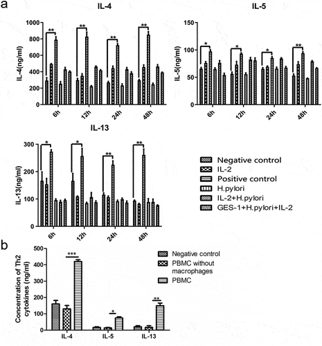
Stimulation of the mucosal barrier by pathogens is another critical factor facilitating the proliferation and activation of ILC2.Citation24 When infection occurs, the mucosal barrier releases “alarmin” proteins such as IL-25, IL-33, and TSLP, which can activate ILC2 and help to trigger the type-2 immunity process.Citation25 In a previous study, we proved that H. pylori infection promoted ILC2 in the gastric mucosa or PBMC. Here, we further sorted out ILC2 from the PMBC and cocultured them with H. pylori or together with GES-1. Elisa results showed that the level of Th2 cytokines in the supernatants did not increase when stimulating ILC2 with H. pylori or H. pylori-infected GES-1 cells, regardless of different time points or different MOI (, Data only showed the MOI of 50). These observations revealed that H. pylori did not activate ILC2 directly and some other immune cells may serve as intermediaries. As classic participants in innate immunity, macrophages also respond immediately at the early stage of disease. Interestingly, in the coculture system (H. pylori, GES-1, and PBMC), the Th2 cytokines of the supernatants increased. When macrophages were removed, no significant changes were observed (). These findings suggested that ILC2 activation caused by H. pylori infection requires macrophages.
Figure 1. Macrophages were required in the activation of ILC2 induced by H. pylori infection. (a) ILC2 sorted out from PBMC were cocultured with H. pylori or together with GES-1. The IL-2 +IL-25 +IL-33 stimulating group was used as positive control. Elisa showed the level of Th2 cytokines (IL-4, IL-5, and IL-13) in the supernatants; (b) PBMC removing or remaining macrophages were cocultured with GES-1 and H. pylori ATCC43504 for 24 hours. The Th2 cytokines in the supernatants were detected by Elisa. p values were determined by one-way ANOVA, Dunnett-adjusted; error bars, SD (n = 3 per group); *p < .05, **p < .01, ***p < .001.
3.2. H. pylori infection promoted macrophages M2 polarization to activate ILC2
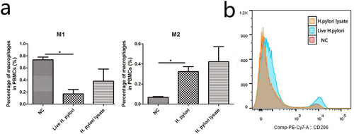
To investigate how macrophages help to activate ILC2, we extracted mice peritoneal macrophages when mice were infected with H. pylori for 12 weeks and 24 weeks. Compared with the negative control group, the M2/M1 ratio of macrophages was increased in H. pylori infection groups, especially in the 24-week groups (). Although, the total number of peritoneal macrophages among groups showed no significant differences (Figure S2). Meanwhile, GES-1 stimulating with live H. pylori or H. pylori lysate was cocultured with human PBMC. Data showed that the percentage of M1 macrophages was reduced while the percentage of M2 macrophages was augmented in the live H. pylori group but not the H. pylori lysate group (). Consistently, the expression of CD206, a M2 macrophage marker, was up-regulated in PBMC when stimulated with live H. pylori (). These results demonstrated that H. pylori infection promoted macrophages M2 polarization.
Figure 2. H. pylori infection promotes M2 polarization in peritoneal macrophages. H. pylori-infected mice were sacrificed at the 12th week or 24th week. (a) Flow cytometry was used to detect the peritoneal macrophages, and the extraction efficiency of mice peritoneal macrophages was more than 80%. (b, d) the expression of CD206 in mice peritoneal macrophages. (c, e) the ratio of M2/M1 macrophages. N: mice group with no treatment, NC: mice group treated with PBS, E: mice group treated with H. pylori strain of East Asian type, W: mice group treated with H. pylori strain of Western type SS1, C: mice group treated with CagA- H. pylori. p values were determined by one-way ANOVA, dunnett-adjusted; error bars, SD (n = 6 per group); *p < .05.
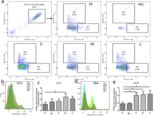
Figure 3. H. pylori infection promotes M2 polarization of macrophages in PBMC. In the H. pylori or the lysate of H. pylori coculture system: (a)The percentages of M1 and M2 macrophages were analyzed with flow cytometry. (b) The expression of CD206 in PBMC. NC: negative control. p values were determined by one-way ANOVA, Dunnett-adjusted; error bars, SD (n = 3 per group); *p < .05.
Since M2 macrophages have been proven to promote and cooperate with ILC2 in type-2 immunity,Citation16,Citation17 we analyzed the percentages of ILC2 and M2 macrophages in the coculture system (H. pylori, GES-1, and PBMC), and a positive correlation was found between the two (R2 = 0.914, p=.011, Figure S3a). As for H. pylori-infected mice, the percentage of ILC2 in spleen lymphocytes and the percentage of M2 macrophages in peritoneal cavity were positively corrected too (R2 = 0.6835, p < .001, Figure S3b). These results indicated that H. pylori infection activated ILC2 by promoting macrophages M2 polarization.
3.3. Indoleamine 2,3-dioxygenase (IDO) and TSLP were involved in the “H. pylori infection- M2 polarization- ILC2 activation” pathological process
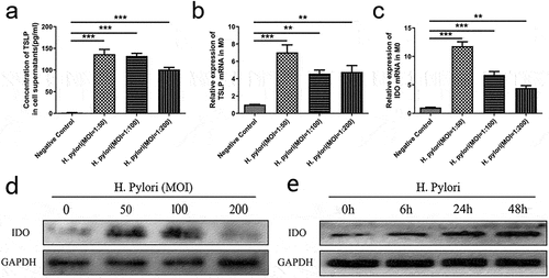
Next, we explored the mechanism by which H. pylori infection induced M2 polarization. We conducted gene expression profiling of M0 macrophages, which were stimulated by H. pylori for 24 hours. A total of 22 genes were significantly up-regulated and 16 genes were significantly down-regulated (Figure S4 & Table S3). Among them, IDO and TSLP aroused our interest. IDO has been known to induce macrophages M2 polarization, and TSLP is one of the well-known classical “alarmin” to activate ILC2. Further experiments showed that after H. pylori infection, the concentration of TSLP in cell supernatants and the mRNA expression of TSLP in M0 macrophages were enriched (). The expression of IDO was also up-regulated both in mRNA and protein levels (). Undoubtedly, IDO and TSLP were involved in the “H. pylori infection- M2 polarization- ILC2 activation” pathological process.
Figure 4. H. pylori infection promotes the expression of IDO and TSLP in macrophages. M0 macrophages were infected with H. pylori ATCC43504. (a) The concentration of TSLP in the supernatants. (b, c) the mRNA expression of TSLP and IDO. (d) The expression of IDO protein at different MOI. (e) The expression of IDO protein at different time points (MOI = 50). MOI: multiplicity of infection. p values were determined by one-way ANOVA, Dunnett-adjusted; error bars, SD (n = 3 per group); **p < .01, ***p < .001.
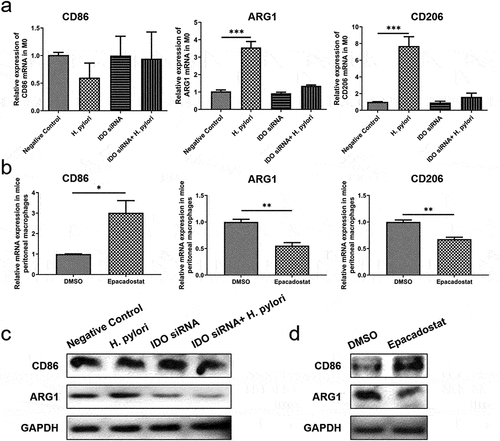
Next, we knocked down IDO in M0 macrophages with specific siRNA and then cocultured them with H. pylori for 24 hours. Flow cytometric analysis showed that H. pylori infection increased the proportion of M2 macrophages and the concentration of TSLP, while the transfer of siIDO counteracted this effect (). The mRNA and protein expression of M1 molecular marker CD86 was not affected by H. pylori or siIDO. In contrast, the promotion effect of H. pylori on M2 markers ARG1 and CD206 was weakened by siIDO (). Then, the peritoneal macrophages from H. pylori SS1 infected mice (12 weeks after infection) were treated with Epacadostat (IDO inhibitor, 50 nM for 48 h). We found that IDO inhibiting significantly up-regulated M1 marker and down-regulated M2 markers (). These results declared that IDO was required for the induction and maintenance of macrophage M2 polarization in H. pylori infection.
Figure 5. The expression of IDO is vital for macrophage M2 polarization in H. pylori infection. M0 macrophages were transfected with IDO siRnas and cocultured with H. pylori ATCC43504 for 24 hours. (a-c) flow cytometry was used to analyze the proportion of M2 macrophages (a, b) and the ratio of M2/M1 macrophages (c). (d) The concentration of TSLP in the supernatants. NC: negative control. p values were determined by one-way ANOVA, Dunnett-adjusted; error bars, SD (n = 3 per group); **p < .01, ***p < .001.
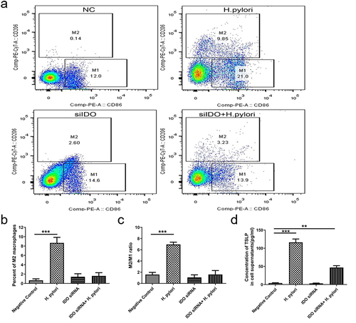
Figure 6. The influence of IDO on the expression of M1/M2 markers. (a, b) the mRNA expression of M1/M2 markers in M0 macrophages (a) and mice peritoneal macrophages (b). (c, d) the protein expression of M1/M2 markers in M0 macrophages (c) and mice peritoneal macrophages (d). p values were determined by one-way ANOVA followed by Dunnett correction (a) or Student’s t-test (c); error bars, SD (n = 3 per group); *p < .05, **p < .01, ***p < .001.
3.4. TSLP played a critical role in the activation of ILC2 in H. pylori infection
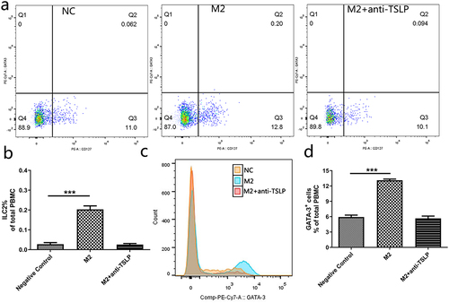
As described above, TSLP might be the central member to mediate ILC2 activation in H. pylori infection. In order to verify this, M2 macrophages were treated with anti-TSLP and then cocultured with PBMC. We found M2 macrophages boosted the proportion of ILC2 and GATA-3+ cells in PBMC. However, anti-TSLP treatment abolished this effect completely (), which proved that TSLP was the key player in M2 macrophages-mediated ILC2 activation.
Figure 7. TSLP secreted by M2 macrophages increases the proportion of ILC2 in PBMC. (a) Three groups were cocultured with H. pylori: negative control group (NC), M2 macrophage group (M2), and anti-TSLP group (M2+anti-TSLP). (b) The proportion of ILC2 in PBMC. (c) The count of GATA-3+ cells in PBMC. (d) The proportion of GATA-3+ cells in PBMC. NC: negative control. p values were determined by one-way ANOVA, Dunnett-adjusted; error bars, SD (n = 3 per group); ***p < .001.
Hence, we intended to dig out how H. pylori infection up-regulated the expression of TSLP in macrophages. Since microRNAs are potent genetic regulators, miR-27a-3p and miR-1956 came into view. They were the top two down-regulated miRNAs in the gene expression profiling of H. pylori-infected M0 macrophages (Figure S4 & Table S3). Target prediction using Targetscan (http://www.targetscan.org/) indicated that TSLP could be targeted by miR-27a-3p (Figure S5a). Then, we infected M0 macrophages with H. pylori in different MOI and found that miR-27a-3p was negatively correlated with the expression of TSLP (Figure S5b). We consequently transfected M2 macrophages with miR-27a-3p inhibitors or mimics. Anticipatedly, knocking down miR-27a-3p raised the mRNA expression and the protein secretion of TSLP, while transfection of mimics curbed it (Figure S5c-e). Based on this, we proposed that H. pylori infection inhibited the expression of miR-27a-3p in macrophages, thus boosting the production and secretion of its target protein TSLP and then promoting the activation of ILC2.
4. Discussion
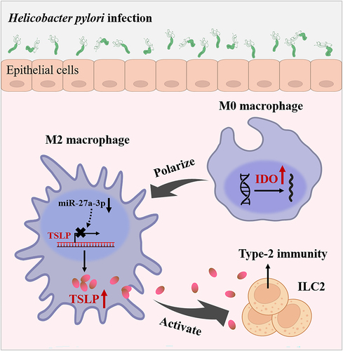
Our previous study demonstrated that the activation of ILC2 drove skewed type-2 immunity in H. pylori infection-associated gastric disease.Citation6 In this research, we described a novel mechanism involving macrophage M2 polarization as an essential procedure promoting ILC2 activating in H. pylori infection. In addition to that, IDO and TSLP were identified as the fat parts of this process ().
Figure 8. A schematic chart of H. pylori infection-induced ILC2 activation mechanism.
The role of ILC2 in H. pylori infection has come into focus over the past few years. Stomach ILC2 proliferation and IL-5 secretion provoked IgA-mediated H. pylori elimination and gastric inflammation restraint in the early phase of H. pylori infection.Citation26 We have also observed the proliferation of ILC2 as well as the activation of the type-2 immunity in gastric tissue. ILC2 and its mediated type-2 immunity play a vital role in mucosal healing and homeostasis. Additionally, some human populations with equipollent H. pylori infection rates but a high gut parasite burden have a lower incidence of gastric cancer.Citation27 Thus, we share views with some researchers that mechanisms promoting type-2 immunity can limit H. pylori-induced gastric pathology.Citation28
It is reported that the number of macrophages in the gastric mucosa of H. pylori infection is significantly increased.Citation29 Moreover, some studies also showed that macrophages extracted from the peritoneal cavity of H. pylori-infected mice or H. pylori-stimulated macrophages in vitro may play a role in regulating the transformation of Th1/Th2 immune balance.Citation30,Citation31 In our system, we removed the macrophages from PBMC and cocultured the remaining cells with H. pylori in vitro. As anticipated, ILC2 could not be activated after the removal of macrophages, which suggests the indispensable role of macrophages.
Macrophages can be polarized to M1 or M2 in different microenvironments. Studies have found that the molecular marker CD163 of M2 macrophages was significantly increased in H. pylori + gastric cancer tissues compared with that in H. pylori − tissues.Citation32,Citation33 Our findings indicated that H. pylori infection could promote the polarization of macrophages to M2. We also confirmed that the proportion of ILC2 was positively correlated with the proportion of M2 macrophages after H. pylori infection. This is in line with a recent report that M2 macrophages promoted ILC2 expansion and mucous metaplasia in early-life rhinovirus infections.Citation17 These observations, together with our findings presented here, indicate that M2 polarization of macrophages is involved in the activation of ILC2 induced by H. pylori infection.
One thing to note here is that in our model, H. pylori infection conducted to M2 phenotype in a CagA-independent manner (). Our finding is in keeping with a previous study that the mRNA expression of the M2 marker CD163 was higher in H. pylori-infected relative to noninfected patients, while no significant difference was found in H. pylori CagA+ relative to H. pylori CagA− patients.Citation29
Multiple signals are involved in the M2 polarization of macrophages.Citation34 Herein, we demonstrated for the first time that IDO, the rate-limiting enzyme of tryptophan catabolism, contributed to the H. pylori infection-induced M2 polarization. Various pathogens such as viruses,Citation35,Citation36 bacteria,Citation37,Citation38 fungi,Citation39,Citation40 and parasitesCitation41 could activate IDO in macrophages and modulate inflammation. IDO-expressing macrophages were deemed to skew differentiation toward the M2 phenotype.Citation37 Our results are consistent with previous studies that elevated IDO attributed to the predominance of M2-like phenotype in lepromatous leprosyCitation42 and Schistosoma japonicumCitation41 infection. However, some studies reported that Fusobacterium nucleatumCitation43 and Aspergillus fumigatusCitation39 infection induced IDO expression and M1 polarization. Such differences may result from the species and virulence of pathogens; the conditions, MOI, and course of infection. Anyhow, IDO is critical for regulatory macrophage function and mediating inflammatory response in pathogen infection.
Furthermore, we found that TSLP secreted by macrophages is requisite to promote ILC2 as anti-TSLP neutralized this effect. Like IL-25 and IL-33, TSLP is a classic “alarmin” to activate ILC2.Citation44 We also found out that miR-27a-3p is a potential regulator of TSLP, since transfection of inhibitors or mimics influenced the secretion of TSLP in M2 macrophages.
In conclusion, our study highlights that M2 macrophages contribute to the activation of ILC2 in H. pylori infection. We demonstrate that H. pylori infection can induce high expression of IDO and M2 polarization of macrophages, which subsequently secret TSLP and finally activate ILC2 ().
Author contribution
Ruyi Peng performed the experiments and drafted the manuscript. Canxia Xu designed the study. Linfang Zhang and Xiaoming Liu assisted with data analysis. Dongzi Peng and Xingcen Chen assisted with the animal experiments and data analysis. Deliang Liu revised the manuscript. Rong Li designed the study and checked the manuscript. All authors read and approved the final version of the manuscript.
Supplemental Material
Download Zip (4.2 MB)Acknowledgments
We thank Professor Xie Yong (Department of Gastroenterology, First Affiliated Hospital of Nanchang University, Jiangxi, China) for donating the H. pylori strains SS1 (Western type) and ATCC43504 to us.
Disclosure statement
No potential conflict of interest was reported by the author(s).
Data availability statement
The authors confirm that the data supporting the findings of this study are available within the article and its supplementary materials.
Supplementary material
Supplemental data for this article can be accessed online at https://doi.org/10.1080/19490976.2024.2347025
Additional information
Funding
References
- Uno Y. Prevention of gastric cancer byHelicobacter pylori eradication: a review from Japan. Cancer Med. 2019;8(8):3992–14. doi:10.1002/cam4.2277. PMID: 31119891.
- Jafarzadeh A, Larussa T, Nemati M, Jalapour S. T cell subsets play an important role in the determination of the clinical outcome of Helicobacter pylori infection. Microb Pathog. 2018;116:227–236. doi:10.1016/j.micpath.2018.01.040. PMID: 29407232.
- Zhang H, Dai Y, Liu Y, Wu T, Li J, Wang X, Wang W. Helicobacter pylori colonization protects against chronic experimental colitis by regulating Th17/Treg balance. Inflamm Bowel Dis. 2018;24(7):1481–1492. doi:10.1093/ibd/izy107. PMID: 29788098.
- Lehours P, Ferrero RL. Review: Helicobacter: inflammation, immunology, and vaccines. Helicobacter. 2019;24(Suppl S1):e12644. doi:10.1111/hel.12644. PMID: 31486236.
- Codolo G, Fassan M, Munari F, Volpe A, Bassi P, Rugge M, Pagano F, D’Elios MM, de Bernard M. HP-NAP inhibits the growth of bladder cancer in mice by activating a cytotoxic Th1 response. Cancer Immunol Immunother. 2012;61(1):31–40. doi:10.1007/s00262-011-1087-2. PMID: 21833592.
- Li R, Jiang XX, Zhang LF, Liu XM, Hu TZ, Xia XJ, Li M, Xu CX. Group 2 innate lymphoid cells are involved in Skewed type 2 immunity of gastric diseases induced by Helicobacter pylori infection. Mediators Inflamm. 2017;2017:4927964. doi:10.1155/2017/4927964. PMID: 29138530.
- Liu X, Cao K, Xu C, Hu T, Zhou L, Cao D, Xiao J, Luo L, Guo Y, Qi Y. GATA-3 augmentation down-regulates Connexin43 in Helicobacter pylori associated gastric carcinogenesis. Cancer Biol Ther. 2015;16(6):987–996. doi:10.1080/15384047.2015.1030552. PMID: 25901741.
- Barrow AD, Colonna M. Innate lymphoid cell sensing of tissue vitality. Curr Opin Immunol. 2019;56:82–93. doi:10.1016/j.coi.2018.11.004. PMID: 30529190.
- Klose CS, Artis D. Innate lymphoid cells as regulators of immunity, inflammation and tissue homeostasis. Nat Immunol. 2016;17(7):765–774. doi:10.1038/ni.3489. PMID: 27328006.
- Huang J, Fu L, Huang J, Zhao J, Zhang X, Wang W, Liu Y, Sun B, Qiu J, Hu X, et al. Group 3 innate lymphoid cells protect the Host from the uropathogenic Escherichia coli infection in the bladder. Adv Sci (Weinh). 2022;9(6):e2103303. doi:10.1002/advs.202103303. PMID: 35018740.
- Naito M, Kumanogoh A. Group 2 innate lymphoid cells and their surrounding environment. Inflamm Regen. 2023;43(1):21. doi:10.1186/s41232-023-00272-8. PMID: 36941691.
- Annunziato F, Romagnani C, Romagnani S. The 3 major types of innate and adaptive cell-mediated effector immunity. J Allergy Clin Immunol. 2015;135(3):626–635. doi:10.1016/j.jaci.2014.11.001. PMID: 25528359.
- Na YR, Kim SW, Seok SH. A new era of macrophage-based cell therapy. Experimental & Molecular Medicine. 2023;55(9):1945–1954. doi:10.1038/s12276-023-01068-z. PMID: 37653035.
- Sakemura RL, Hefazi M, Cox MJ, Siegler EL, Sinha S, Hansen MJ, Stewart CM, Feigin JM, Manriquez RC, Schick KJ, et al. AXL inhibition improves the antitumor activity of chimeric antigen receptor t cells. Cancer Immunol Res. 2023;11(9):1222–1236. doi:10.1158/2326-6066.CIR-22-0254. PMID: 37378662.
- Dang B, Gao Q, Zhang L, Zhang J, Cai H, Zhu Y, Zhong Q, Liu J, Niu Y, Mao K, et al. The glycolysis/HIF-1α axis defines the inflammatory role of IL-4-primed macrophages. Cell Rep. 2023;42(5):112471. doi:10.1016/j.celrep.2023.112471. PMID: 37149865.
- Glaubitz J, Wilden A, Golchert J, Homuth G, Volker U, Broker BM, Thiele T, Lerch MM, Mayerle J, Aghdassi AA, et al. In mouse chronic pancreatitis CD25(+)FOXP3(+) regulatory T cells control pancreatic fibrosis by suppression of the type 2 immune response. Nat Commun. 2022;13(1):4502. doi:10.1038/s41467-022-32195-2. PMID: 35922425.
- Han M, Breckenridge HA, Kuo S, Singh S, Goldsmith AG, Li Y, Kreger JE, Bentley JK, Hershenson MB. M2 macrophages promote IL-33 expression, ILC2 expansion and mucous metaplasia in response to early life rhinovirus infections. Front Immunol. 2022;13:952509. doi:10.3389/fimmu.2022.952509. PMID: 36032072.
- Li W, Zhang X, Wu F, Zhou Y, Bao Z, Li H, Zheng P, Zhao S. Gastric cancer-derived mesenchymal stromal cells trigger M2 macrophage polarization that promotes metastasis and EMT in gastric cancer. Cell Death Disease. 2019;10(12):918. doi:10.1038/s41419-019-2131-y. PMID: 31801938.
- Yang P, Qiu G, Wang S, Su Z, Chen J, Wang S, Kong F, Lu L, Ezaki T, Xu H. The mutations of Th1 cell-specific T-box transcription factor may be associated with a predominant Th2 phenotype in gastric cancers. Int J Immunogenet. 2010;37(2):111–115. doi:10.1111/j.1744-313X.2010.00899.x. PMID: 20193034.
- Bartemes KR, Kephart GM, Fox SJ, Kita H. Enhanced innate type 2 immune response in peripheral blood from patients with asthma. J Allergy Clin Immunol. 2014;134(3):671–678.e4. doi:10.1016/j.jaci.2014.06.024. PMID: 25171868.
- Licona-Limon P, Kim LK, Palm NW, Flavell RA. TH2, allergy and group 2 innate lymphoid cells. Nat Immunol. 2013;14(6):536–542. doi:10.1038/ni.2617. PMID: 23685824.
- Xi Y, Shen Y, Wu D, Zhang J, Lin C, Wang L, Yu C, Yu B, Shen W. CircBCAR3 accelerates esophageal cancer tumorigenesis and metastasis via sponging miR-27a-3p. Mol Cancer. 2022;21(1):145. doi:10.1186/s12943-022-01615-8. PMID: 35840974.
- Vivier E, van de Pavert SA, Cooper MD, Belz GT. The evolution of innate lymphoid cells. Nat Immunol. 2016;17(7):790–794. doi:10.1038/ni.3459. PMID: 27328009.
- Walker JA, McKenzie AN. Development and function of group 2 innate lymphoid cells. Curr Opin Immunol. 2013;25(2):148–155. doi:10.1016/j.coi.2013.02.010. PMID: 23562755.
- Hammad H, Lambrecht BN. Barrier epithelial cells and the control of type 2 immunity. Immunity. 2015;43(1):29–40. doi:10.1016/j.immuni.2015.07.007. PMID: 26200011.
- Satoh-Takayama N, Kato T, Motomura Y, Kageyama T, Taguchi-Atarashi N, Kinoshita-Daitoku R, Kuroda E, Di Santo JP, Mimuro H, Moro K, et al. Bacteria-induced group 2 innate lymphoid cells in the stomach provide immune protection through induction of IgA. Immunity. 2020;52(4):635–649.e4. doi:10.1016/j.immuni.2020.03.002. PMID: 32240600.
- Holcombe C. Helicobacter pylori: the African enigma. Gut. 1992;33(4):429–431. doi:10.1136/gut.33.4.429. PMID: 1582581.
- Buzzelli JN, Chalinor HV, Pavlic DI, Sutton P, Menheniott TR, Giraud AS, Judd LM. IL33 is a stomach alarmin that initiates a skewed th2 response to injury and infection. Cell Mol Gastroenterol Hepatol. 2015;1(2):203–221.e3. doi:10.1016/j.jcmgh.2014.12.003. PMID: 28210674.
- Michalkiewicz J, Helmin-Basa A, Grzywa R, Czerwionka-Szaflarska M, Szaflarska-Poplawska A, Mierzwa G, Marszalek A, Bodnar M, Nowak M, Dzierzanowska-Fangrat K. Innate immunity components and cytokines in gastric mucosa in children with Helicobacter pylori infection. Mediators Inflamm. 2015;2015:176726. doi:10.1155/2015/176726. PMID: 25948881.
- Gobert AP, Verriere T, Asim M, Barry DP, Piazuelo MB, de Sablet T, Delgado AG, Bravo LE, Correa P, Peek RJ, et al. Heme oxygenase-1 dysregulates macrophage polarization and the immune response to Helicobacter pylori. J Immunol. 2014;193(6):3013–3022. doi:10.4049/jimmunol.1401075. PMID: 25108023.
- Krakowiak MS, Noto JM, Piazuelo MB, Hardbower DM, Romero-Gallo J, Delgado A, Chaturvedi R, Correa P, Wilson KT, Peek RJ. Matrix metalloproteinase 7 restrains Helicobacter pylori-induced gastric inflammation and premalignant lesions in the stomach by altering macrophage polarization. Oncogene. 2015;34(14):1865–1871. doi:10.1038/onc.2014.135. PMID: 24837365.
- Fu XL, Duan W, Su CY, Mao FY, Lv YP, Teng YS, Yu PW, Zhuang Y, Zhao YL. Interleukin 6 induces M2 macrophage differentiation by STAT3 activation that correlates with gastric cancer progression. Cancer Immunol Immunother. 2017;66(12):1597–1608. doi:10.1007/s00262-017-2052-5. PMID: 28828629.
- Yamaguchi T, Fushida S, Yamamoto Y, Tsukada T, Kinoshita J, Oyama K, Miyashita T, Tajima H, Ninomiya I, Munesue S, et al. Tumor-associated macrophages of the M2 phenotype contribute to progression in gastric cancer with peritoneal dissemination. Gastric Cancer. 2016;19(4):1052–1065. doi:10.1007/s10120-015-0579-8. PMID: 26621525.
- Cutolo M, Campitiello R, Gotelli E, Soldano S. The role of M1/M2 macrophage polarization in rheumatoid arthritis synovitis. Front Immunol. 2022;13:867260. doi:10.3389/fimmu.2022.867260. PMID: 35663975.
- Suchard MS, Savulescu DM. Nicotinamide pathways as the root cause of sepsis – an evolutionary perspective on macrophage energetic shifts. FEBS J. 2022;289(4):955–964. doi:10.1111/febs.15807. PMID: 33686748.
- Liu WL, Lin YH, Xiao H, Xing S, Chen H, Chi PD, Zhang G. Epstein-Barr virus infection Induces Indoleamine 2,3-Dioxygenase expression in human monocyte-derived macrophages through p38/Mitogen-activated protein kinase and NF-κB pathways: impairment in T cell functions. J Virol. 2014;88(12):6660–6671. doi:10.1128/JVI.03678-13. PMID: 24696473.
- Rao MJ, Parasa VR, Lerm M, Svensson M, Brighenti S. Polarization of human monocyte-derived cells with vitamin d promotes control of mycobacterium tuberculosis infection. Front Immunol. 2019;10:3157. doi:10.3389/fimmu.2019.03157. PMID: 32038652.
- Popov A, Abdullah Z, Wickenhauser C, Saric T, Driesen J, Hanisch FG, Domann E, Raven EL, Dehus O, Hermann C, et al. Indoleamine 2,3-dioxygenase–expressing dendritic cells form suppurative granulomas following listeria monocytogenes infection. J Clin Invest. 2006;116(12):3160–3170. doi:10.1172/JCI28996. PMID: 17111046.
- Jiang N, Zhang L, Zhao G, Lin J, Wang Q, Xu Q, Li C, Hu L, Peng X, Yu F, et al. Indoleamine 2,3-dioxygenase regulates macrophage recruitment, polarization and phagocytosis in aspergillus fumigatus keratitis. Invest Ophthalmol Vis Sci. 2020;61(8):28. doi:10.1167/iovs.61.8.28. PMID: 32692841.
- Yu F, Jiang W, Zhang L, Jiang N. IDO regulates macrophage functions by inhibiting the CCL2/CCR2 signaling pathway in fungal keratitis. Cornea. 2023;42(8):1005–1015. doi:10.1097/ICO.0000000000003309. PMID: 37155343.
- Ran L, Yu Q, Zhang S, Xiong F, Cheng J, Yang P, Xu JF, Nie H, Zhong Q, Yang X, et al. Cx3cr1 deficiency in mice attenuates hepatic granuloma formation during acute schistosomiasis by enhancing the M2-type polarization of macrophages. Dis Model Mech. 2015;8:691–700. doi:10.1242/dmm.018242. PMID: 26035381.
- de Mattos BM, SPR D, Andrade PR, Ferreira H, de Andrade SB, Da PDOJ, Assis TQ, de Toledo-Pinto TG, de Lima BO, CNJ D, et al. Indoleamine 2,3-dioxygenase and iron are required for mycobacterium leprae survival. Microbes Infect. 2017;19(11):505–514. doi:10.1016/j.micinf.2017.06.006. PMID: 28684130.
- Xue Y, Xiao H, Guo S, Xu B, Liao Y, Wu Y, Zhang G. Indoleamine 2,3-dioxygenase expression regulates the survival and proliferation of fusobacterium nucleatum in THP-1-derived macrophages. Cell Death Disease. 2018;9(3):355. doi:10.1038/s41419-018-0389-0. PMID: 29500439.
- Han M, Rajput C, Hong JY, Lei J, Hinde JL, Wu Q, Bentley JK, Hershenson MB. The innate cytokines IL-25, IL-33, and TSLP cooperate in the induction of type 2 innate lymphoid cell expansion and mucous metaplasia in rhinovirus-infected immature mice. J Immunol. 2017;199(4):1308–1318. doi:10.4049/jimmunol.1700216. PMID: 28701507.