
ABSTRACT
Adherent-invasive Escherichia coli (AIEC) strain LF82, isolated from patients with Crohn’s disease, invades gut epithelial cells, and replicates in macrophages contributing to chronic inflammation. In this study, we found that RstAB contributing to the colonization of LF82 in a mouse model of chronic colitis by promoting bacterial replication in macrophages. By comparing the transcriptomes of rstAB mutant- and wild-type when infected macrophages, 83 significant differentially expressed genes in LF82 were identified. And we identified two possible RstA target genes (csgD and asr) among the differentially expressed genes. The electrophoretic mobility shift assay and quantitative real-time PCR confirmed that RstA binds to the promoters of csgD and asr and activates their expression. csgD deletion attenuated LF82 intracellular biofilm formation, and asr deletion reduced acid tolerance compared with the wild-type. Acidic pH was shown by quantitative real-time PCR to be the signal sensed by RstAB to activate the expression of csgD and asr. We uncovered a signal transduction pathway whereby LF82, in response to the acidic environment within macrophages, activates transcription of the csgD to promote biofilm formation, and activates transcription of the asr to promote acid tolerance, promoting its replication within macrophages and colonization of the intestine. This finding deepens our understanding of the LF82 replication regulation mechanism in macrophages and offers new perspectives for further studies on AIEC virulence mechanisms.
Introduction
Crohn’s disease (CD) is a chronic inflammatory intestine disease. The prevalence of CD is 322 and 318 per million in Europe and North America, respectively, and the number of new cases is increasing dynamically in developing countries.Citation1–3 There is no cure for CD; despite medical treatment, nearly half of the patients who require therapy develop refractory disease. CD is associated with characteristic changes in the intestinal microbiome, including expansion of adherent-invasive Escherichia coli (AIEC). Penetration of the epithelial barrier, survival and replication in macrophages are important steps in AIEC expansion in patients with CD. In macrophages, AIEC encounters acidic, oxidative, genotoxic, and proteotoxic stresses.Citation4,Citation5 Hence, it is important for AIEC to combat the harsh environment. According to current reports, the mechanism that promotes AIEC intracellular replication mainly consists of three aspects: damaging the host cell autophagy function,Citation6–13 forming a replication niche by assembling biofilm-like communities to prevent clearing by autolysosome,Citation14–16 and tolerating the acid attack of macrophages.Citation7,Citation8
The two-component system (TCS) is a key strategy for pathogens to adapt to their living environments and contributes to bacterial survival during infection. The TCS consists of two proteins, one being the histidine kinase (HK) responsible for detecting environmental changes and the other being the response regulator (RR) that activates or represses downstream genes in response to the signal received from HK. RstAB comprises RstA (HK) and RstB (RR). The functions of RstAB in pathogenic bacteria have been extensively studied. RstAB promotes motility in Salmonella typhimurium,Citation17 Photobacterium damselae subsp. damselaeCitation18 and Vibrio alginolyticus,Citation19 but represses motility in Clostridioides difficile.Citation20 RstAB is essential for the adhesion of V. alginolyticus to mucusCitation19 and Edwardsiella ictaluri Citation21 to catfish skin and the invasion of Salmonella typhimuriumCitation17 to HeLa cells. RstAB promotes antibiotic resistance in Yersinia pseudotuberculosis Citation22 and P. damselae subsp. damselae.Citation18 RstAB is also important for the colonization in animal models in avian pathogenic E. coli (APEC) strain E058,Citation23 S. enterica,Citation24 P. damselae subsp. damselae,Citation18 and E. ictaluri .Citation21 However, it remains unclear whether RstAB contributes to AIEC virulence.
Replication in macrophages is essential for AIEC LF82 expansion in patients with CD.Citation25,Citation26 LF82 establishes a replication niche by assembling biofilm-like communities called intracellular biofilm communities (IBCs), which protect it from phagolysosomal attacks for long-term survival and replicate in macrophages.Citation8 Curli fibers are the main extracellular matrix composed of IBCs.Citation27 The structural components and assembly factors of the curli fibers are encoded by two operons: csgBA and csgDEFG. CsgD controls the transcription of both operons as a master regulator of curli fiber production.Citation28–30 csgD is activated by three factors (OmpR, RstA, and IHF) and repressed by two factors (CpxR and H-NS) in E. coli.Citation29,Citation30 RstA regulates csgD expression by directly binding to the RstA-box on the promotor of csgD, and activating csgD only under acidic conditions in E.coli .Citation29 Adaptation to an acidified macrophage environment is crucial for intracellular survival and replication of LF82.Citation31,Citation32 The acid shock protein Asr, encoded by the asr gene, strongly supports the growth of E. coli under acidic conditions.Citation33 A previous study has shown that RstA is involved in asr activation, as asr expression is reduced when rstA is deleted.Citation34 RstA also contributes to acid tolerance during the gut colonization in enterohemorrhagic E.coli (EHEC) O157:H7 by regulating asr expression.Citation35 However, whether RstAB regulates csgD and asr in LF82 and whether it contributes to bacterial replication in macrophages is unclear.
In this study, we found that RstAB contributes to the virulence of LF82 by comparing the colonization of rstAB mutant and wild-type (WT) in a mouse model of chronic colitis. We found that RstAB contributes to LF82 virulence by promoting intracellular bacterial replication in macrophages. Hence, we investigated the mechanisms by which RstAB affects LF82 replication by comparing the transcriptomes of rstAB mutant and WT when infected macrophages. Differentially expressed genes (DEGs) in LF82 were identified and combined with the RstA-box motif on the promoters of DEGs to study the regulatory mechanism of RstAB. We found that RstAB directly activated intracellular biofilm community formation gene csgD and acid shock gene asr to promote LF82 replication in macrophages. Finally, we demonstrated that in response to acidic conditions within macrophages, RstAB activates csgD and asr, thereby contributing to LF82 pathogenesis. Our work contributes to the understanding of the regulatory function of RstAB in AIEC, particularly in relation to virulence, potentially influencing the future management and treatment of CD.
Results
RstAB promotes LF82 virulence in chronic colitis mice
To investigate whether RstAB contributes to LF82 virulence, a mouse model of dextran sulfate sodium (DSS) salt (w/v = 2%)-induced chronic colitis was established (). We recorded model data for chronic colitis; normally fed mice gained weight over time and stabilized after 35 days. DSS-treated mice lost 7.11% weight after three cycles of DSS administration, and the body weight recovered to a level similar to that of normally fed mice when DSS was replaced with water (Figure S1a). Intestinal edema in mice treated with DSS resulted in an increase in the weight of the cecum-connected colon to approximately 0.1 g (Figure S1b) and decreased the colon length to approximately 2 cm compared with that in normally fed mice (Figure S1c,d). The above characteristics exhibited the same trend as previously reported,Citation36 indicating that the construction of a mouse model of chronic colitis was successful.
Figure 1. RstAB promotes LF82 virulence in chronic colitis mice.
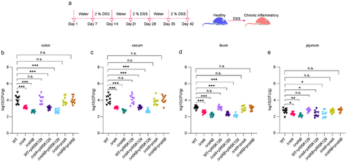
Next, we constructed rstA and rstAB deletion mutants (ΔrstA and ΔrstAB), the complementary strains (ΔrstA+prstA and ΔrstAB+prstAB), and the control strains (WT+pWSK129, ΔrstA+pWSK129, and ΔrstAB+pWSK129). C57BL/6 mice were infected with ΔrstA, ΔrstAB, ΔrstA+prstA, ΔrstAB+prstAB, WT+pWSK129, ΔrstA+pWSK129, ΔrstAB+pWSK129, and WT, the bacterial titers in the mouse colon, cecum, ileum, and jejunum were calculated at 24 h post infection (p.i.). The bacterial titers of the colon, cecum, ileum, and jejunum were significantly lower of ΔrstA or ΔrstAB than that of WT in chronic colitis mice (). ΔrstA+prstA and ΔrstAB+prstAB restored the reduced bacterial titers of ΔrstA and ΔrstAB to a level that resembles WT in chronic colitis mice (). The bacterial titers of WT+pWSK129 colonizing the colon, cecum, ileum, and jejunum were not significantly different from those of WT mice (). ΔrstA+pWSK129 and ΔrstAB+pWSK129 failed to restore the colonization ability of ΔrstA and ΔrstAB in the colonized colon, cecum, ileum, and jejunum (), indicating that pWSK129 alone does not affect the colonization ability of LF82. This suggests that RstAB is essential for the colonization of LF82 in the intestine of mice with chronic colitis. ΔrstA and ΔrstAB grow as well as WT in LB and RPMI 1640 medium (Figure S2a,b), eliminating the possibility of the decreased LF82 colonization of ΔrstA or ΔrstAB in vivo to be due to a growth defect. These results suggest that RstAB is required by LF82 to effectively colonize in the intestine of mice with chronic colitis, thereby contributing to LF82 virulence.
RstAB promotes the replication of LF82 in macrophages
Macrophages in the mucous lamina propria of patients with CD are the main sites of LF82 replication, which is the basis for LF82 to invade deeper tissues across the intestinal barrier.Citation25 Therefore, it is important to investigate the mechanisms underlying LF82 replication in macrophages. The expression of rstA and rstB was upregulated in LF82-infected Thp-1 or Raw 264.7 cells,Citation37 suggesting that the expression of rstA and rstB is induced when LF82 survives in macrophages. Hence, we speculated that the decreased colonization of ΔrstA and ΔrstAB in chronic colitis mice is caused by reduced LF82 replication in macrophages. We investigated the expression pattern of rstA and rstB in LF82 when infected Raw 264.7 cells for 1, 6, and 24 h. The results showed that rstA was upregulated by 6.40-fold, 13.38-fold, and 17.09-fold, and rstB was upregulated by 6.40-fold, 8.56-fold, and 13.10-fold at 1, 6, and 24 h p.i. (), which is consistent with a previous study.Citation8 These results suggest that the upregulation of rstA and rstB may help LF82 adapt to the intracellular environment of macrophages.
Figure 2. RstAB promotes the replication of LF82 in macrophages.

To investigate whether RstAB affects the replication of LF82 in macrophages, we infected Raw 264.7 cells with WT, ΔrstA, ΔrstAB, WT+pWSK129, ΔrstA+pWSK129, ΔrstAB+pWSK129, ΔrstA+prstA, and ΔrstAB+prstAB to assay the ability of intracellular replication at 6 and 24 h p.i. The replication of ΔrstA and ΔrstAB in Raw 264.7 cells were reduced by 1.4-fold and 2.3-fold at 6 h p.i. ( and Table S1). The replication of ΔrstA and ΔrstAB was reduced by 2.5-fold and 3.0-fold compared with that of WT at 24 h p.i. ( and Table S1). A more significant reduction of replication was obtained in ΔrstA and ΔrstAB-infected murine bone marrow-derived macrophages (MBMM) from chronic colitis mice. The replication of ΔrstA and ΔrstAB in MBMM was reduced by 2.6-fold and 3.7-fold at 6 h p.i. and reduced by 4.2-fold and 4.0-fold compared with that of WT at 24 h p.i., respectively ( and Table S2). ΔrstA+prstA and ΔrstAB+prstAB restored the differences to similar levels of WT (). The ability of replication of WT+pWSK129 in Raw 264.7 cells and MBMM was not significantly different compared with that of WT (). ΔrstA+pWSK129 and ΔrstAB+pWSK129 failed to restore the replication ability of ΔrstA and ΔrstAB in Raw 264.7 cells and MBMM (), indicating that pWSK129 alone does not affect the replication of LF82 in macrophages. The confocal microscopy results showed the same trend as the intracellular replication results (). These results indicate that RstAB promotes the intracellular replication of LF82 in macrophages.
These results suggest that by promoting LF82 replication in macrophages, rstAB contributes to LF82 colonization in mice with chronic colitis, thereby enhancing LF82 virulence.
RstAB promotes LF82’s replication in macrophages by regulating the expression of csgD and asr
To understand the downstream genes regulated by RstAB of LF82 in macrophages, the transcriptome of WT and ΔrstAB when infected Raw 264.7 cells for 1 h was performed by high-throughput Illumina RNA-sequencing (RNA-seq). The transcriptomic results showed that 83 genes were significantly differently expressed in the ΔrstAB group compared with that in the WT group, and 33 and 50 genes were categorized as up and downregulated in ΔrstAB (Table S3), respectively (fold change > 2 and p value < .05), indicating that RstAB acts as both an activator and a repressor in LF82 when infected macrophages. DEGs in the ΔrstAB compated with WT were classified using the NCBI Clusters of Orthologous Groups (COG) functional categories annotation system. The COG categories that were significantly enriched in the ΔrstAB group of upregulated genes were primarily associated with cell motility, signal transduction mechanisms, extracellular structures, replication, recombination, and repair. COG categories significantly enriched in the list of downregulated genes in the ΔrstAB group included post-translational modification, protein turnover, chaperones, translation, ribosomal structure and biogenesis, transcription, energy production, and conversion (). These results indicate that the impact of RstAB on LF82 gene expression is bidirectional, providing a basis for studying the mechanism of action of RstAB on the replication of LF82 in macrophages at the transcriptional level. We randomly selected seven DEGs to validate the result of the transcriptome by quantitative real-time PCR (qRT-PCR). The qRT-PCR results correlated well with the transcriptomic data, indicating that the transcriptome results were robust and valid ().
Figure 3. RstAB promotes LF82’s replication in macrophages by regulating the expression of csgD and asr.
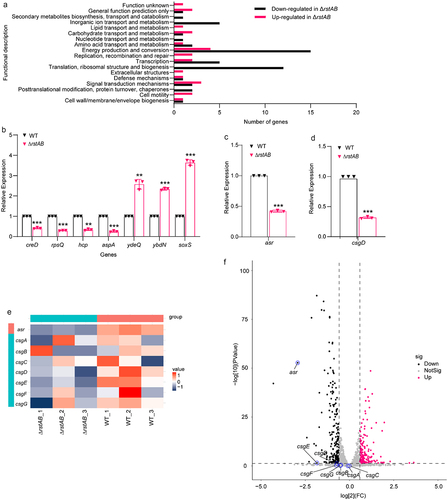
csgD and asr were found in the DEGs known to be regulated by RstAB in E. coli K12, with RstA-box sequences in their promoters,Citation21 suggesting that RstA may bind to the promoters of csgD and asr in LF82. The expression of csgD and asr in WT and ΔrstAB when infected Raw 264.7 cells at 1 h p.i. were verified using qRT-PCR. The results showed that csgD and asr were downregulated in ΔrstAB when infected cells compared with WT (), which is consistent with the transcriptomic data (). We also verified the expression of csg gene cluster by qRT-PCR. Results showed that the expression of csgA, csgB, csgC, csgE, csgF and csgG was down-regulated in ΔrstAB when infected Raw 264.7 cells compared with that in WT (Figure S3). These results indicated that RstAB may positively regulate the expression of csgD and asr in LF82 upon infected macrophages.
RstA regulates biofilm formation of LF82 by directly activates the expression of csgD
csgD is a key transcriptional response regulator that controls curli fiber production and contributes to biofilm formation.Citation38 As the RstA-box is present in the csgD gene promoter, an electrophoretic mobility shift assay (EMSA) was used to verify whether RstA directly binds to the csgD promoter. The results showed that with increasing concentrations of RstA protein, slowly migrating bands were observed for the csgD promoter region (gray value of bound DNA bands from left to right are 219.3, 222.9), but not rpoS (negative control) under the same conditions (). Deletion or scrambling of the RstA-box in the csgD promoter region completely abolished the binding of RstA to the csgD promoter (). These data suggest that RstA binds to the csgD promoter through the RstA-box to activate the expression of csgD.
Figure 4. RstA regulates biofilm formation of LF82 by directly activates the expression of csgD.
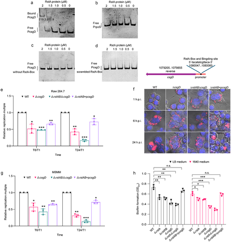
AIEC LF82 forms IBCs within phagolysosomes, which helps LF82 resist killing by phagolysosomes to survive, replicate, and provide a nutrient niche.Citation8 Next, we investigated whether RstAB-activated csgD contributes to the intracellular replication of LF82 in macrophages. We constructed ΔcsgD, rstAB and csgD double deletion (ΔrstABΔcsgD), and ΔrstAB+pcsgD mutants. Replication of ΔcsgD in Raw 264.7 cells was 2.0-fold and 2.3-fold decreased, and ΔrstABΔcsgD was 2.1-fold and 6.0-fold decreased compared with WT at 6 and 24 h p.i., and ΔrstAB+pcsgD was 1.5-fold and 1.4-fold decreased compared with WT at 6 and 24 h p.i., respectively ( and Table S4). In MBMM, the replication of ΔcsgD was 1.8-fold and 3.2-fold decreased, and ΔrstABΔcsgD was 2.3-fold and 7.5-fold compared with that of WT at 6 and 24 h p.i., and ΔrstAB+pcsgD were 1.6-fold and 1.4-fold decreased compared with that of WT at 6 and 24 h p.i., respectively ( and Table S5). The confocal microscopy results showed a consistent trend (). The reduced replication ability of ΔcsgD to similar extent as that of ΔrstABΔcsgD compared with WT at 6 h p.i. (). This difference was more pronounced in the ΔrstABΔcsgD than ΔcsgD at 24 h p.i. (). Additionally, the ΔrstAB+pcsgD could not fully restore LF82‘s replication ability in macrophages (), suggesting that csgD is not the sole downstream virulence gene of RstAB. These results suggest that csgD affects intracellular replication and is regulated by RstAB; however, RstAB affects intracellular replication not only by regulating csgD.
CsgD is an important factor in the construction of an LF82-specific biofilm matrix and facilitates IBC formation within macrophages. To evaluate the effect of RstAB in biofilm formation by regulating csgD in LF82, a crystal violet staining assay was performed to quantify biofilm formation by WT, ΔrstA, ΔrstAB, ΔcsgD, ΔrstABΔcsgD, and ΔrstAB+pcsgD. Biofilm formation was significantly decreased in ΔrstA, ΔrstAB, ΔcsgD, and ΔrstABΔcsgD compared with that of WT, and ΔrstAB+pcsgD restored the biofilm formation ability of LF82 in LB and RPMI 1640 medium (). Moreover, the formation of biofilm of ΔrstABΔcsgD was similar to that of ΔcsgD (), indicating that RstAB influenced biofilm formation only through regulating csgD. These results suggest that RstA promotes LF82 biofilm formation by activating csgD.
These data indicate that RstA contributes to LF82 biofilm formation by directly activating csgD expression and promoting the replication of LF82 in macrophages.
RstAB regulates the acid stress response of LF82 by directly activates the expression of asr
Asr is an acid shock protein encoded by the asr gene. The EMSA results showed that with increasing concentrations of RstA protein, slowly migrating bands were observed for asr promoter (gray value of bound DNA bands from left to right are 238.1, 219.8), whereas no retarded bands were observed for the negative control (), indicating that RstA binds to asr promoter in vitro. Deletion or scrambling of the RstA-box in the asr promoter region completely abolished the binding of RstA to the asr promoter (). These data suggest that RstA binds to the asr promoter through the RstA-box to activate the expression of asr.
Figure 5. RstAB regulates the acid stress response of LF82 by directly activates the expression of asr.
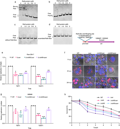
The acidic environment in macrophages is required for the replication of LF82.Citation31 Next, we constructed Δasr, rstAB and asr double deletion strain (ΔrstABΔasr), and ΔrstAB+pasr mutants to investigate whether RstAB-activated asr contributes to intracellular replication of LF82 in macrophages. Replication of Δasr in Raw 264.7 cells was 1.9-fold and 2.3-fold decreased, and ΔrstABΔasr was 2.1-fold and 3.8-fold compared with that of WT at 6 and 24 h p.i., and ΔrstAB+pasr was 1.7-fold and 1.5-fold decreased compared with that of WT at 6 and 24 h p.i., respectively ( and Table S6). In MBMM, the replication of Δasr was 1.5-fold and 2.2-fold decreased, and ΔrstABΔasr was 2.0-fold and 3.4-fold decreased compared with that of WT at 6 and 24 h p.i., and ΔrstAB+pasr was 1.3-fold and 1.3-fold decreased compared with that of WT at 6 and 24 h p.i., respectively ( and Table S7). Confocal microscopy revealed a consistent trend (). These results indicate that RstAB-activated asr contributes to LF82 intracellular replication. The replication ability of Δasr is weaker than that of WT (). This difference is more pronounced in the ΔrstABΔasr strain (). Additionally, the ΔrstAB+pasr could not fully restore LF82‘s replication ability in macrophages (), suggesting that asr is not the sole downstream virulence gene of RstAB. These results indicate that asr affects intracellular replication and is regulated by RstAB; however, RstAB affects intracellular replication not only by regulating asr.
To further investigate whether RstAB-activated asr helps LF82 in tolerating the acidic environment within macrophages, we conducted an acid tolerance assay. The tolerance of ΔrstA, ΔrstAB, ΔrstABΔasr and Δasr to low pH decreased drastically within 2 h, and ΔrstABΔasr was weaker than Δasr, ΔrstAB+pasr could not fully restore the reduced bacteria survival rate compared to WT (). These results indicated that RstA regulates asr to promote the survival of LF82 under acidic conditions, whereas RstAB promotes LF82 acidic tolerance not only by regulating asr.
RstAB responds to acidic signal in macrophages and promotes intracellular replication of LF82
In our previous study, we found that the expression of rstA and rstB was significantly upregulated in the transcriptome under acid treatment conditions, mimicking the acidic environment of macrophages, indicating that acid may activate the expression of rstA and rstB.Citation39 qRT-PCR was performed to verify the transcriptome results. rstA and rstB were significantly upregulated by 3.4-fold and 2.1-fold in the acid-treated group compared with the control group, respectively (). Next, we investigated whether the acidic environment in macrophages regulates the expression of rstA and rstB by comparing the expression of rstA and rstB in LF82-infected normal and acid-neutralized macrophages. NH4Cl and chloroquine (CQ) were used to neutralize the acid within the macrophages.Citation31,Citation32 Acid neutralization with 30 mM NH4Cl or 10 μM CQ reduced the upregulated expression of rstA and rstB compared with the control group (), indicating RstAB response to acidic conditions in LF82-infected macrophages.
Figure 6. RstAB responds to acidic signal in macrophages and promotes intracellular replication of LF82.
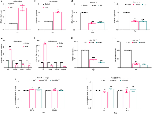
To determine whether RstAB regulates csgD and asr expression in response to acidic conditions, we assessed the mRNA levels of csgD and asr in LF82 cells cultured in acidic and neutral RPMI 1640 medium. qRT-PCR analysis showed that acid treatment increased the expression of csgD and asr in the WT (). However, rstA, rstB, and rstAB mutants abolished the upregulation of csgD and asr induced by acid (). These results indicated that acid is the signal that RstB senses to activate the transcription of csgD and asr. Moreover, the expression of csgD and asr in ΔrstA and ΔrstAB was also downregulated within Raw 264.7 cells compared with that of WT (), indicating that RstAB activates csgD and asr expression in LF82 within Raw 264.7 cells. These results suggest that, in response to acidic conditions in macrophages by RstB, LF82 RstAB actively regulates the expression of csgD and asr.
The acid-responsive TCS EvgAS activates PhoPQ via the small protein B1500, and PhoP activates transcription of rstA in a manner potentiated by EvgAS in E. coli .Citation40,Citation41 To investigate whether RstB directly or indirectly responds to acidic conditions, we constructed evgAS and phoPQ mutants (ΔevgAS and ΔphoPQ) and evgASphoPQ double mutant (ΔevgASΔphoPQ). Result showed that either ΔevgAS, ΔphoPQ mutant, or ΔevgASΔphoPQ reduces the up-regulation of asr and csgD under acidic condition (Figure S4A, B), indicating that acidic condition activates EvgAS or PhoPQ to regulate the asr and csgD. Combines with the above results, it suggests that in addition to RstB directly sensing the acidic conditions and regulating the expression of asr and csgD, EvgAS or PhoPQ also senses the acidic conditions and indirectly regulates the expression of asr and csgD.
We further verified whether acidic conditions in macrophages affect RstAB-dependent LF82 replication. Our previous study demonstrated that the neutralization of the acidic environment in macrophages with NH4Cl or CQ resulted in a reduction in LF82 replication by 3.3-fold and 4.2-fold, respectively.Citation39 Whereas the replication ability of ΔrstA or ΔrstAB was unaffected in NH4Cl or CQ-treated Raw 264.7 cells compared with WT (). These results indicate that RstAB promotes the replication of LF82 in response to the acidic environment of macrophages.
Discussion
RstAB is a critical TCS that plays a vital role in various pathogenic functions in bacteria. RstAB is a positive regulator of key virulence factors including damselysin, phobalysin P, and phobalysin C cytotoxins, in the marine pathogen P. damselae subsp. damselae.Citation18,Citation42 However, the signal by which RstAB responds to P. damselae subsp. damselae to affect virulence has not been clarified. RstA contributes to APEC E058 virulence by affecting chicken macrophage survival. RstA promotes APEC E058 to cause systemic infection in a chicken infection model.Citation23 Deletion of rstAB increases the sensitivity of APEC to acidic conditions, as RstA positively regulates the acid resistance operons hdeABD and gadABE .Citation43 RstA contributes to the bacterial colonization of EHEC O157:H7 in vivo, by indirectly regulating LEE gene expression to promote bacterial adhesion in vitro and directly regulating the acid tolerance genes asr and hdeA .Citation35 In this study, we found that RstAB promotes the replication of AIEC LF82 in macrophages, thereby contributing to its colonization in the intestine of mice with chronic colitis. Combined with studies of RstAB in various bacterial virulences, this indicates that RstAB is an essential virulence-regulatory TCS that responds to environmental cues.
Biofilms are bacterial aggregates embedded in a matrix of exopolysaccharides, DNA, and proteins that can shield bacteria from the immune system and antibiotics,Citation44,Citation45 leading to persistent and recurrent clinical infections.Citation46 RstA negatively regulates biofilm formation in EHEC O157: H7 by controlling c-di-GMP biosynthesis.Citation35 RstA also inhibits biofilm formation by S. enterica Typhimurium.Citation24 In Acinetobacter baumannii, the RstA homology protein BfmR contributes to biofilm formation by activating the expression of the Csu pili chaperone – usher assembly system.Citation47 Our results demonstrate that RstA contributes to biofilm formation in AIEC LF82 by directly activating csgD expression. These studies suggest that RstA either positively or negatively regulates biofilm formation in different pathogenic bacteria.Citation35,Citation48,Citation49
Biofilm formation is a novel pathogenic feature of AIEC in the mucosa of patients with CD.Citation8 AIEC infection causes intestinal dysbiosis by altering the microbiome, forming thick biofilms on the epithelium,Citation50 and assembling IBCs within host cells to protect from phagolysosomal attack.Citation8 Intracellular LF82 produces an extra-bacterial matrix that acts as a biofilm to control IBC formation.Citation8 IBC also forms in the uropathogenic E. coli (UPEC)-infected bladder epithelial cell cytosol.Citation51 The formation of IBCs provides a replication site for bacteria and contributes to recurrent urinary tract infections.Citation52 AIEC and UPEC belong to the phylogenetic group B2,Citation53 but the sites of IBC formation are different. UPEC forms IBCs in the cytosol of infected bladder epithelial cells, and AIEC forms IBCs inside phagolysosome.Citation54 These results indicate that IBC formation is a crucial step in the intracellular pathogen life cycle. CsgD is the main regulator of curli fiber formation. Curli fibers are the main extracellular matrix that compose biofilms.Citation55 The matrix of UPEC IBC contained curli fibers; hence, we speculated that curli fibers may also contribute to AIEC IBC formation. We found that RstAB contributes to LF82 biofilm formation by directly activating csgD expression and promoting the replication of LF82 in macrophages. These findings suggest that curli fibers are crucial for bacterial biofilm formation inside host cells.
AIEC survives in mature phagolysosomes and proliferates in low pH and protease environment.Citation31,Citation32,Citation56 S. enterica Typhimurium is an intracellular pathogen that survives within Salmonella-containing vacuoles (SCVs), and the acidic conditions in SCVs is the signal for the expression of the pathogenicity island 2 type III secretion system and its secreted effectors.Citation57–59 RstA contributes to Salmonella virulence by activating the expression of STM1485 (the homolog of asr).Citation33,Citation60 STM1485 is required to prevent the degradation of Salmonella in SCV and facilitate type III secretion system assembly, promoting intracellular bacterial replication in human epithelial cells and murine macrophages, thus affecting Salmonella virulence.Citation61 In this study, we found that the deletion of rstAB led to the downregulation of asr gene expression in macrophages and that the deletion of asr gene weakened the acid tolerance and replication ability of LF82 in macrophages. These studies indicate that tolerance to the acidic environment within host cell vacuoles is a common strategy for the survival and replication of intracellular pathogenic bacteria.
We demonstrated that RstAB promoted the replication of AIEC LF82 in macrophages by enhancing LF82 biofilm formation and acid tolerance, which are necessary for its virulence of LF82. The replication results of single mutants (ΔcsgD and Δasr) and triple mutants (ΔrstABΔcsgD and ΔrstABΔasr) in RAW 264.7 cells and MBMM were at the same level at 6 h p.i., whereas the replication of triple mutants (ΔrstABΔcsgD and ΔrstABΔasr) was attenuated compared with single mutants (ΔcsgD and Δasr) at 24 h p.i. These results indicate that RstA regulates downstream replication-associated genes and contributes to LF82 replication. To investigate whether there are other genes regulated by RstA, we complemented the ΔrstAB with the ectopic expression of csgD or asr. The replication results showed that neither csgD nor asr fully compensated for the decreased replication capacity of ΔrstAB. RstAB regulates genes other than csgD and asr in E. coli K-12,Citation34 suggesting that RstA regulates different downstream genes in various regulation patterns. Whether RstAB regulates additional genes in AIEC LF82 requires further investigation.
Materials and methods
Strains and plasmids
The bacterial and plasmids used in this study are listed in Table S7. E. coli LF82 O83:H1 was used as the WT strain. Gene deletion mutants were generated using the λ Red recombinase system supported by the pSim17 plasmid (blasticidin-resistant) encoding three proteins (Exo, Beta, and Gam) that are required for homologous recombination.Citation62,Citation63 A WT strain harboring the pSim17 plasmid was established using electrotransformation. Then, DNA fragments composed sequentially (5′→3′) of an upstream 39–45 bp sequence of the target gene, the chloramphenicol resistance gene sequence, and the downstream 39–45 bp reverse complementary sequence of the target gene were amplified using PCR. The chloramphenicol-resistant pKD3 plasmid was used as the template for PCR, and primers were designed to cover 39–45 bp homologous arm sequences. Next, DNA fragments were introduced into competent bacterial cells via electrotransformation. Finally, the cells were cultured at 37°C for 2 h, and the suspensions of recovered bacteria were spread onto agar plates containing 25 μg/mL of chloramphenicol to obtain single mutant colonies. To validate the correctness of the mutation, the target mutation loci of single chloramphenicol-resistant colonies were PCR-amplified and preliminarily identified using agarose gel electrophoresis. Further identification was performed by Sanger sequencing. The ΔrstAΔrstB (ΔrstAB) double mutant was established based on a chloramphenicol-resistant ΔrstA mutant by introducing a kanamycin-resistance gene (from pKD4 plasmid) to replace the rstB gene.
We used pWSK129 for rstA and rstAB complementation, and pTrc99A for csgD and asr complementation. To express and purify the RstA protein with a 6×His-tag, the rstA fusion gene was cloned into the pET-28a (+) between the NdeI and XhoI sites, and the constructed plasmid were transformed into E. coli BL21 (DE3). Plasmid pUC57 carrying red fluorescent protein (mCherry) was used for confocal microscopy. All the constructed plasmids and strains were validated using the methods described for the identification of mutant strains. The plasmids, strains, and primers used in this study are listed in Table S7 and Table S8.
Bacteria were cultured overnight at 37°C in LB medium. When necessary, appropriate antibiotics were added: ampicillin, 50 μg/mL; kanamycin, 50 μg/mL; chloramphenicol, 25 μg/mL; gentamicin, 20 μg/mL, or 100 μg/mL.
RNA-seq
The LF82 was inoculated in LB medium at a ratio of 1:1000 for overnight culture (approximately 16 h), and the overnight bacteria were inoculated in LB medium at a ratio of 1:100 for 6 h and centrifuged at 5,500 × g for 5 min. The supernatant was discarded to collect the bacteria and washed three times with RPMI 1640 medium containing 10% fetal bovine serum (FBS), incubated at 37°C for 30 min. Raw 264.7 cells were infected with LF82 at a multiplicity of infection (MOI) of 100:1 for 20 min, and then extracellular bacteria were killed with 100 μg/mL gentamicin for 40 min. Samples were collected to extract total RNA.
Total RNA was isolated using the TRIzol reagent (Invitrogen) according to the manufacturer’s instructions. The concentration and purity of the extracted RNA were detected using a Nanodrop 2000, the integrity of the RNA was detected using agarose gel electrophoresis, and the RNA integrity number was detected using an Agilent 2100. To ensure that the single database construction can meet the requirements of total RNA amount of ≥0.5 μg, concentration ≥ 45 ng/μL, and OD260/280 between 1.8 and 2.2. For library preparation, we used an amount ≥5 µg, concentration ≥ 800 ng/μL, and OD 260/280 between 1.8 and 2.2 of total RNA per sample. rRNA was depleted from the total RNA using a Ribo-off rRNA depletion kit (Vazyme, Nanjing, China), and libraries were constructed and analyzed by Majorbio, Inc. (Shanghai, China). The eukaryotic cell data in each sample were removed, and the results of prokaryotic sequencing were mapped to the LF82 reference genome to obtain gene annotation and expression results. DEGs in ΔrstAB mutant compared with WT invading Raw 264.7 cells were identified using the DESeq R package. The resulting p values were adjusted using the Benjamini – Hochberg test to control for the false discovery rate. Genes with an adjusted p < .05 were considered differentially expressed. All the significant DEGs are listed in Table S2.
qRT-PCR
RNA samples were isolated using TRIzol (Invitrogen), reverse transcribed using a PrimeScript RT reagent kit (Takara, Shiga, Japan), and processed for qRT-PCR. qRT-PCR was performed using the Applied Biosystems 7500 Real-Time PCR System and SYBR Green PCR Master Mix (Applied Biosystems). The fold change in the expression of the target gene relative to that of the housekeeping gene (16s) was determined using the 2−ΔΔCt method.Citation64 At least three biological replicates were used for each qRT-PCR. All oligonucleotides used for qRT-PCR are listed in Table S3.
Cell culture and macrophage replication
Raw 264.7 cells, a murine macrophage cell line, were cultured with RPMI 1640 medium (containing 10% FBS and 1% penicillin and streptomycin when necessary) at 37°C in 5% CO2. Mouse bone marrow macrophages (MBMM), derived from chronic colitis inflammatory mice. After the mice were killed, the tibia and leg bones were taken, and the bone marrow was rinsed with RPMI 1640 medium (10% FBS + 1% P/S +10 μg/mL and macrophage colony-stimulating factor), and the impurities were filtered through the 70 μm cell strainer (Corning). The cell concentration was set at 10Citation6 CFU/mL, and the cells were spread in a 12-well cell culture, which was used for bacterial infection experiments.
Bacterial survival and replication were measured using a gentamicin protection assay. Before infection, the bacteria were washed with phosphate-buffered saline (PBS) and resuspended in RPMI 1640 medium, incubated at 37°C with shaking at 180 rpm for 30 min, infected with a MOI of 100, centrifuged at 1,000 × g for 10 min, incubated with 5% CO2 at 37°C for 10 min, and then extracellular bacteria were killed with 100 μg/mL gentamicin for 40 min (defined as T1). To determine the number of bacteria in the cells, PBS and 1 mL of 1% Triton X-100 were added to each well and incubated for 5 min to lyse eukaryotic cells. Triton X-100 at this concentration had no effect on bacterial viability for at least 30 min. The samples were diluted and spread on LB medium agar plates to determine the number of colony-forming units (CFU) recovered from the cracked monolayers. T6/T1 or T24/T1 is the replication multiple of gentamicin treatment for 6 or 24 h compared with gentamicin treatment for 1 h. Mutations (T6/T1)/WT (T6/T1) or (T24/T1)/WT (T24/T1) were defined as the relative replication multiples.
Growth curve
To determine the growth of each strain, overnight cultures were washed three times with PBS and diluted (1:1000) in LB medium without antibiotics. A 200-μL aliquot was added to a 96-well flat-bottom microplate, 200 μL of LB medium was added as a negative control, and incubated at 37°C with shaking at 180 rpm for 24 h, as previously described.Citation35 The absorbance was recorded at 600 nm. The experiments were independently performed three times.
Confocal observation
After infection with bacteria containing the mCherry plasmid, the macrophages were washed with PBS and fixed with 4% paraformaldehyde for 10 min. Subsequently, cells were washed with PBS and permeabilized with 0.1% Triton X-100 for 5 min. After washing with PBS, anti-fluorescence attenuation tablets (including DAPI) and anti-fluorescence quenching sealing solution (Beyotime, Shanghai, China) were dropped (10 μL at a ratio of 1:1) on the glass slide. The coverslips were mounted on slides, and cell images were acquired using a confocal laser scanning microscope (Zeiss LSM800) and analyzed with ZEN 2.3 (blue edition) and format designed using Adobe Illustrator CC 2018 software. The data were based on the clearest results preserved from five fields of three slides.
Animal experiment
Mice
Six-week-old C57BL/6 L female mice purchased from Beijing Vital River Laboratory Animal Technology Co., Ltd. (Beijing, China) were housed under standard laboratory conditions (22 ± 1°C, 12:12-h light/dark cycle). All experiments were conducted according to protocols approved by the Institutional Animal Care Committee of Nankai University (Tianjin, China). All the mice were provided ad libitum access to a normal chow diet and water throughout the study.
DSS-induced chronic colitis mouse model
C57BL/6 L female mice were modeled with reference to the animal model described by Kwon J et al..Citation36 Chronic colitis was induced by oral administration of DSS (MW 36–50 kDa; MP Biomedicals, Santa Ana, CA, USA). After acclimation, to induce chronic colitis, 8-week-old mice were treated with three cycles of 2% (w/v) DSS for seven days, with seven days of drinking water between each cycle. After the 3rd cycle of DSS treatment, the mice were sacrificed, and tissue samples were harvested as previously described.Citation65
Gut colonization assay
Bacteria were cultured overnight in LB medium at 37°C and 180 rpm, then 1:100 was transferred to fresh LB medium and cultured to OD600 = 1.0. Bacteria were collected through centrifugation at 5,500 rpm and washed three times with PBS. A bacterial suspension of 2 × 10Citation10 CFU/mL was prepared, and the mutants were mixed with the WT strain at a ratio of 1:1, and then gavaged (100 μL/mice). The bacteria were allowed to colonize the mice for 24 h before being sacrificed. The intestinal tissues were collected, the intestinal contents were removed, 1 mL of PBS and grinding beads were added for crushing in a tissue crushing instrument, and the samples were serially diluted and coated on LB solid medium with the corresponding resistance to count the number of bacteria.
Acid tolerance assays
Overnight cultured bacteria were washed three times with PBS and then diluted to a concentration of 106 CFU/mL in 1640 medium acidified to pH 3.0 with HCl. The cultures were incubated at 37°C for 0 to 6 h with shaking at 180 rpm. A 100-μL aliquot was removed from the flask and suitable dilutions were plated on LB agar once every hour. The experiments were performed independently three times.
Biofilm formation assays
Biofilm formation was quantified by crystal violet staining as previously described.Citation35 Overnight cultured bacteria were diluted in fresh medium (1:100) and incubated in 96-well polystyrene microtiter plates at 37°C for 24 h. Loosely associated bacteria were removed by washing thrice with PBS, and the remaining bacteria were stained with 0.5% crystal violet for 5 min. The biofilm was then destained by adding 200 μL of 95% ethanol to each well and quantified using an enzyme-linked immunosorbent assay plate reader at 590 nm. Each experiment was performed at least three times.
EMSA
The 6 × His-tagged RstA protein was expressed and purified in E. coli BL21 (DE3). Target DNA fragments were amplified using PCR and purified using a SPARKeasy Gel DNA Extraction Kit (Sparkjade; #AE0101-C). Purified PCR fragments (40 ng) were incubated at 25°C for 30 min with 6× His-tagged RstA protein at concentrations ranging from 0 to 2 µM in 20-μL reactions containing binding buffer (1 mM Tris – HCl [pH 7.5], 0.2 mM dithiothreitol, 5 mM MgCl2, 10 mM KCl, and 10% glycerol, 30 mM acetyl phosphate). The protein-DNA fragments were electrophoretically separated on a native polyacrylamide gel at 4°C and 80 V/cm. The gel was stained for 10 min in a solution of 0.1% GelRed (Biotium; #41000), and protein bands were visualized using ultraviolet transillumination. The gray value of DNA was analyzed using ImageJ (NIH, https://imagej.nih.gov, accessed on 14 December 2022).
Statistical analysis
Statistical analyses were conducted using the GraphPad Prism software (v9.4.0; GraphPad Software, San Diego, CA, USA). The mean ± SD from three independent experiments is shown in the figures. Differences between two mean values were evaluated using a two-tailed Student’s t-test or a two-sided Mann – Whitney U test to calculate the p values. Statistical significance was set at p < .05.
Author contributions
YTL designed the study; TY, XML, YKL, YH, WY, RYL, QW, XPL, JRZ, and CJ conducted the experiments and data analyses. TY, YP, and YTL wrote and edited the manuscript.
Supplemental Material
Download Zip (391.1 KB)Acknowledgments
We would like to thank Xi Guo, Yanli Xu, and Junli Wu from Nankai University for the materials used in this study and Fenxia Liu for the operation instructions for confocal microscopy.
Disclosure statement
No potential conflict of interest was reported by the author(s).
Data availability statement
RNA sequencing data generated in this study are available from the NCBI SRA database. Accession to cite SRA data: PRJNA984413 https://www.ncbi.nlm.nih.gov/bioproject/?term=PRJNA984413.
Supplementary material
Supplemental data for this article can be accessed online at https://doi.org/10.1080/19490976.2024.2356642
Correction Statement
This article has been corrected with minor changes. These changes do not impact the academic content of the article.
Additional information
Funding
References
- Cosnes J, Gower-Rousseau C, Seksik P, Cortot A. Epidemiology and natural history of inflammatory bowel diseases. Gastroenterology. 2011;140(6):1785–1794.e4. doi:10.1053/j.gastro.2011.01.055.
- M’Koma AE. Inflammatory bowel disease: An expanding global health problem. Clin Med Insights Gastroenterol. 2013;6:33–19. doi:10.4137/CGast.S12731.
- Krzesiek E, Kofla-Dlubacz A, Akutko K, Stawarski A. The incidence of inflammatory bowel disease in the paediatric population in the district of lower silesia, poland. J Clin Med. 2021;10(17):3994. doi:10.3390/jcm10173994.
- Vignais PV. The superoxide-generating NADPH oxidase: Structural aspects and activation mechanism. Cell mol life sci. Cell Mol Life Sci. 2002;59(9):1428–1459. doi:10.1007/s00018-002-8520-9.
- Bjarnsholt T, Jensen PØ, Burmølle M, Hentzer M, Haagensen JAJ, Hougen HP, Calum H, Madsen KG, Moser C, Molin S. et al. Pseudomonas aeruginosa tolerance to tobramycin, hydrogen peroxide and polymorphonuclear leukocytes is quorum-sensing dependent. Microbiol (Read Engl). 2005;151(2):373–383. doi:10.1099/mic.0.27463-0.
- Losier TT, Akuma M, McKee-Muir OC, LeBlond ND, Suk Y, Alsaadi RM, Guo Z, Reshke R, Sad S, Campbell-Valois F-X. et al. AMPK promotes xenophagy through priming of autophagic kinases upon detection of bacterial outer membrane vesicles. Cell Rep. 2019;26(8):2150–2165.e5. doi:10.1016/j.celrep.2019.01.062.
- David L, Taieb F, Pénary M, Bordignon P-J, Planès R, Bagayoko S, Duplan-Eche V, Meunier E, Oswald E. Outer membrane vesicles produced by pathogenic strains of Escherichia coli block autophagic flux and exacerbate inflammasome activation. Autophagy. 2022;18(12):2913–2925. doi:10.1080/15548627.2022.2054040.
- Prudent V, Demarre G, Vazeille E, Wery M, Quenech’du N, Ravet A, Dauverd - Girault J, Van Dijk E, Bringer M-A, Descrimes M. et al. The crohn’s disease-related bacterial strain LF82 assembles biofilm-like communities to protect itself from phagolysosomal attack. Commun Biol. 2021;4(1):627. doi:10.1038/s42003-021-02161-7.
- Buisson A, Douadi C, Ouchchane L, Goutte M, Jp H, Dubois A, Minet-Quinard R, Bouvier D, Bommelaer G, Vazeille E. et al. Macrophages inability to mediate adherent-invasive E. coli replication is linked to autophagy in crohn’s disease patients. Cells. 2019;8(11):1394. doi:10.3390/cells8111394.
- Dalmasso G, Nguyen HTT, Faïs T, Massier S, Barnich N, Delmas J, Bonnet R. Crohn’s disease-associated adherent-invasive Escherichia coli manipulate host autophagy by impairing SUMOylation. Cells. 2019;8(1):35. doi:10.3390/cells8010035.
- Vazeille E, Buisson A, Bringer MA, Goutte M, Ouchchane L, Hugot JP, De Vallée A, Barnich N, Bommelaer G, Darfeuille-Michaud A. Monocyte-derived macrophages from Crohn’s disease patients are impaired in the ability to control intracellular adherent-invasive Escherichia coli and exhibit disordered cytokine secretion profile. Journal of Crohn’s & colitis [Internet]. 2015;9(5):410–20. [accessed 2023 Aug 15]. https://pubmed.ncbi.nlm.nih.gov/25805890/.
- M S, V A, L F, P B, As T, C F, D P, A D-M, A F. Adherent-invasive Escherichia coli isolated from Crohn’s disease patients induce granulomas in vitro. Cellular Microbiol [Internet]. 2007;9(5):1252–61. [accessed 2023 Aug 15]. https://pubmed.ncbi.nlm.nih.gov/17223928/.
- Barnich N, Darfeuille-Michaud A. Role of bacteria in the etiopathogenesis of inflammatory bowel disease. World J Gastroenterol. 2007;13(42):5571–5576. doi:10.3748/wjg.v13.i42.5571.
- Bringer M-A, Rolhion N, Glasser A-L, Darfeuille-Michaud A. The oxidoreductase DsbA plays a key role in the ability of the crohn’s disease-associated adherent-invasive escherichia coli strain LF82 to resist macrophage killing. J Bacteriol. 2007;189(13):4860–4871. doi:10.1128/JB.00233-07.
- Bringer M-A, Barnich N, Glasser A-L, Bardot O, Darfeuille-Michaud A. HtrA stress protein is involved in intramacrophagic replication of adherent and invasive escherichia coli strain LF82 isolated from a patient with crohn’s disease. Infect Immun. 2005;73(2):712–721. doi:10.1128/IAI.73.2.712-721.2005.
- Simonsen KT, Nielsen G, Bjerrum JV, Kruse T, Kallipolitis BH, Møller-Jensen J, Herman C. A role for the RNA chaperone Hfq in controlling adherent-invasive Escherichia coli colonization and virulence. PLOS ONE. 2011;6(1):e16387. doi:10.1371/journal.pone.0016387.
- Tran T-K, Han Q-Q, Shi Y, Guo L. A comparative proteomic analysis of salmonella typhimurium under the regulation of the RstA/RstB and PhoP/PhoQ systems. Biochim Biophys Acta. 2016;1864(12):1686–1695. doi:10.1016/j.bbapap.2016.09.003.
- Terceti MS, Vences A, Matanza XM, Barca AV, Noia M, Lisboa J, Dos Santos NMS, Do Vale A, Osorio CR. The RstAB system impacts virulence, motility, cell morphology, penicillin tolerance and production of type II secretion system-dependent factors in the fish and human pathogen photobacterium damselae subsp. damselae. Front Microbiol. 2019;10:897. doi:10.3389/fmicb.2019.00897.
- Huang L, Xu W, Su Y, Zhao L, Yan Q. Regulatory role of the RstB‐RstA system in adhesion, biofilm production, motility, and hemolysis. Microbiol Open. 2018;7(5):e00599. doi:10.1002/mbo3.599.
- Edwards AN, Tamayo R, McBride SM. A novel regulator controls Clostridium difficile sporulation, motility and toxin production. Mol Microbiol. 2016;100(6):954–971. doi:10.1111/mmi.13361.
- Menanteau-Ledouble S, Lawrence ML. Use of bioluminescence mutant screening for identification of Edwardsiella ictaluri genes involved in channel catfish (Ictalurus punctatus) skin colonization. Vet Microbiol. 2013;162(2–4):724–730. doi:10.1016/j.vetmic.2012.09.024.
- Flamez C, Ricard I, Arafah S, Simonet M, Marceau M. Phenotypic analysis of Yersinia pseudotuberculosis 32777 response regulator mutants: new insights into two-component system regulon plasticity in bacteria. Int J Med Microbiol. 2008;298(3–4):193–207. doi:10.1016/j.ijmm.2007.05.005.
- Gao Q, Ye Z, Wang X, Mu X, Gao S, Liu X. RstA is required for the virulence of an avian pathogenic Escherichia coli O2 strain E058. Infect Genet Evol. 2015;29:180–188. doi:10.1016/j.meegid.2014.11.022.
- Cabeza ML, Aguirre A, Soncini FC, Véscovi EG. Induction of RpoS degradation by the two-component system regulator RstA in salmonella enterica. J Bacteriol. 2007;189(20):7335–7342. doi:10.1128/JB.00801-07.
- Darfeuille-Michaud A, Boudeau J, Bulois P, Neut C, Glasser A-L, Barnich N, Bringer M-A, Swidsinski A, Beaugerie L, Colombel J-F. High prevalence of adherent-invasive Escherichia coli associated with ileal mucosa in Crohn’s disease. Gastroenterology. 2004;127(2):412–421. doi:10.1053/j.gastro.2004.04.061.
- Glasser AL, Boudeau J, Barnich N, Perruchot MH, Colombel JF, Darfeuille-Michaud A, Tuomanen EI. Adherent invasive Escherichia coli strains from patients with Crohn’s disease survive and replicate within macrophages without inducing host cell death. Infect Immun. 2001;69(9):5529–5537. doi:10.1128/IAI.69.9.5529-5537.2001.
- Rathi B, Gupta S, Kumar P, Kesarwani V, Dhanda RS, Kushwaha SK, Yadav M. Anti-biofilm activity of caffeine against uropathogenic E. coli is mediated by curli biogenesis. Sci Rep. 2022;12(1):18903. doi:10.1038/s41598-022-23647-2.
- Soo VWC, Wood TK. Antitoxin MqsA represses curli formation through the master biofilm regulator CsgD. Sci Rep. 2013;3(1):1–7. doi:10.1038/srep03186.
- Ogasawara H, Yamada K, Kori A, Yamamoto K, Ishihama A. Regulation of the Escherichia coli csgD promoter: interplay between five transcription factors. Microbiol. 2010;156(8):2470–2483. doi:10.1099/mic.0.039131-0.
- Ogasawara H, Yamamoto K, Ishihama A. Regulatory role of MlrA in transcription activation of csgD, the master regulator of biofilm formation in escherichia coli. FEMS Microbiol Lett. 2010;312(2):160–168. doi:10.1111/j.1574-6968.2010.02112.x.
- Bringer M-A, Glasser A-L, Tung C-H, Méresse S, Darfeuille-Michaud A. The Crohn’s disease-associated adherent-invasive Escherichia coli strain LF82 replicates in mature phagolysosomes within J774 macrophages. Cell Microbiol. 2006;8(3):471–484. doi:10.1111/j.1462-5822.2005.00639.x.
- Bringer M-A, Billard E, Glasser A-L, Colombel J-F, Darfeuille-Michaud A. Replication of crohn’s disease-associated AIEC within macrophages is dependent on TNF-α secretion. Lab Invest. 2012;92(3):411–419. doi:10.1038/labinvest.2011.156.
- Ramos-Morales F. Acidic pH: Enemy or ally for enteric bacteria? Virulence. 2012;3(2):103–106. doi:10.4161/viru.19382.
- Ogasawara H, Hasegawa A, Kanda E, Miki T, Yamamoto K, Ishihama A. Genomic SELEX search for target promoters under the control of the PhoQP-RstBA signal relay cascade. J Bacteriol. 2007;189(13):4791–4799. doi:10.1128/JB.00319-07.
- Liu Y, Li S, Li W, Wang P, Ding P, Li L, Wang J, Yang P, Wang Q, Xu T. et al. RstA, a two-component response regulator, plays important roles in multiple virulence-associated processes in enterohemorrhagic Escherichia coli O157: H7. Gut Pathog. 2019;11(1):53. doi:10.1186/s13099-019-0335-4.
- Kwon J, Lee C, Heo S, Kim B, Hyun C-K. DSS-induced colitis is associated with adipose tissue dysfunction and disrupted hepatic lipid metabolism leading to hepatosteatosis and dyslipidemia in mice. Sci Rep. 2021;11(1):5283. doi:10.1038/s41598-021-84761-1.
- Armalyte J, Seputiene V, Melefors O, Suziedeliene E. An Escherichia coli asr mutant has decreased fitness during colonization in a mouse model. Res Microbiol. 2008;159(6):486–493. doi:10.1016/j.resmic.2008.06.003.
- Smith DR, Price JE, Burby PE, Blanco LP, Chamberlain J, Chapman MR. The production of curli amyloid fibers is deeply integrated into the biology of escherichia coli. Biomolecules. 2017;7(4):75. doi:10.3390/biom7040075.
- Yao T, Huang Y, Huai Z, Liu X, Liu X, Liu Y, Sun H, Pang Y. Response mechanisms to acid stress promote LF82 replication in macrophages. Front Cell Infect Microbiol. 2023;13:1255083. doi:10.3389/fcimb.2023.1255083.
- Eguchi Y, Ishii E, Hata K, Utsumi R. Regulation of acid resistance by connectors of two-component signal transduction systems in escherichia coli. J Bacteriol. 2011;193(5):1222–1228. doi:10.1128/JB.01124-10.
- Eguchi Y, Itou J, Yamane M, Demizu R, Yamato F, Okada A, Mori H, Kato A, Utsumi R. B1500, a small membrane protein, connects the two-component systems EvgS/EvgA and PhoQ/PhoP in escherichia coli. Proc Natl Acad Sci U S A. 2007;104(47):18712–18717. doi:10.1073/pnas.0705768104.
- Matanza XM, López-Suárez L, Do Vale A, Osorio CR. The two-component system RstAB regulates production of a polysaccharide capsule with a role in virulence in the marine pathogen photobacterium damselae subsp. damselae. Environ Microbiol. 2021;23(9):4859–4880. doi:10.1111/1462-2920.15731.
- Gao Q, Su S, Li X, Wang H, Liu J, Gao S. Transcriptional analysis of RstA/RstB in avian pathogenic escherichia coli identifies its role in the regulation of hdeD-mediated virulence and survival in chicken macrophages. Vet Microbiol. 2020;241:108555. doi:10.1016/j.vetmic.2019.108555.
- Chagnot C, Zorgani MA, Astruc T, Desvaux M. Proteinaceous determinants of surface colonization in bacteria: bacterial adhesion and biofilm formation from a protein secretion perspective. Front Microbiol. 2013;4:303. doi:10.3389/fmicb.2013.00303.
- Yan J, Bassler BL. Surviving as a community: Antibiotic tolerance and persistence in bacterial biofilms. Cell Host Microbe. 2019;26(1):15–21. doi:10.1016/j.chom.2019.06.002.
- Ciofu O, Moser C, Jensen PØ, Høiby N. Tolerance and resistance of microbial biofilms. Nat Rev Microbiol. 2022;20(10):621–635. doi:10.1038/s41579-022-00682-4.
- Russo TA, Manohar A, Beanan JM, Olson R, MacDonald U, Graham J, Umland TC, Dunman P. The response regulator BfmR is a potential drug target for acinetobacter baumannii. mSphere. 2016;1(3):e00082–16. doi:10.1128/mSphere.00082-16.
- Latasa C, Roux A, Toledo-Arana A, Ghigo J-M, Gamazo C, Penadés JR, Lasa I. BapA, a large secreted protein required for biofilm formation and host colonization of salmonella enterica serovar enteritidis. Mol Microbiol. 2005;58(5):1322–1339. doi:10.1111/j.1365-2958.2005.04907.x.
- Lokhande KB, Pawar SV, Madkaiker S, Nawani N, Venkateswara SK, Ghosh P. High throughput virtual screening and molecular dynamics simulation analysis of phytomolecules against BfmR of acinetobacter baumannii: Anti-virulent drug development campaign. J Biomol Struct Dyn. 2023;41(7):2698–2712. doi:10.1080/07391102.2022.2038271.
- Nickerson KP, McDonald C, Mizoguchi E. Crohn’s disease-associated adherent-invasive Escherichia coli adhesion is enhanced by exposure to the ubiquitous dietary polysaccharide maltodextrin. PLoS One. 2012;7(12):e52132. doi:10.1371/journal.pone.0052132.
- Conover MS, Hadjifrangiskou M, Palermo JJ, Hibbing ME, Dodson KW, Hultgren SJ, Gilmore MS. Metabolic requirements of escherichia coli in intracellular bacterial communities during urinary tract infection pathogenesis. mBio. 2016;7(2):e00104–00116. doi:10.1128/mBio.00104-16.
- Barber AE, Norton JP, Spivak AM, Mulvey MA. Urinary tract infections: Current and emerging management strategies. Clin Infect Dis. 2013;57(5):719–724. doi:10.1093/cid/cit284.
- Conte MP, Aleandri M, Marazzato M, Conte AL, Ambrosi C, Nicoletti M, Zagaglia C, Gambara G, Palombi F, De Cesaris P. et al. The adherent/invasive escherichia coli strain LF82 invades and persists in human prostate cell line RWPE-1, activating a strong inflammatory response. Infect Immun. 2016;84(11):3105–3113. doi:10.1128/IAI.00438-16.
- Anderson GG, Palermo JJ, Schilling JD, Roth R, Heuser J, Hultgren SJ. Intracellular bacterial biofilm-like pods in urinary tract infections. Science. 2003;301(5629):105–107. doi:10.1126/science.1084550.
- Klein RD, Shu Q, Cusumano ZT, Nagamatsu K, Gualberto NC, Lynch AJL, Wu C, Wang W, Jain N, Pinkner JS. et al. Structure-function analysis of the curli accessory protein CsgE defines surfaces essential for coordinating amyloid fiber formation. mBio. 2018;9(4):e01349–18. doi:10.1128/mBio.01349-18.
- Elliott TR, Hudspith BN, Rayment NB, Prescott NJ, Petrovska L, Hermon-Taylor J, Brostoff J, Boussioutas A, Mathew CG, Sanderson JD. Defective macrophage handling of Escherichia coli in Crohn’s disease. J Gastroenterol Hepatol. 2015;30(8):1265–1274. doi:10.1111/jgh.12955.
- Yu X-J, McGourty K, Liu M, Unsworth KE, Holden DW. pH sensing by intracellular salmonella induces effector translocation. Science. 2010;328(5981):1040–1043. doi:10.1126/science.1189000.
- Choi J, Groisman EA. Acidic pH sensing in the bacterial cytoplasm is required for Salmonella virulence: PhoQ sensor activated by acidic pH in cytoplasm. Mol Microbiol. 2016;101(6):1024–1038. doi:10.1111/mmi.13439.
- Choi J, Groisman EA. Activation of master virulence regulator PhoP in acidic pH requires the Salmonella -specific protein UgtL. Sci Signal. 2017;10(494):eaan6284. doi:10.1126/scisignal.aan6284.
- Torrez Lamberti MF, Farizano JV, López FE, Martínez Zamora MG, Pescaretti MM, Delgado MA. Cross-talk between the RcsCDB and RstAB systems to control STM1485 gene expression in salmonella typhimurium during acid-resistance response. Biochimie. 2019;160:46–54. doi:10.1016/j.biochi.2019.02.005.
- Allam US, Krishna MG, Sen M, Thomas R, Lahiri A, Gnanadhas DP, Chakravortty D. Acidic pH induced STM1485 gene is essential for intracellular replication of salmonella. Virulence. 2012;3(2):122–135. doi:10.4161/viru.19029.
- Chan W, Costantino N, Li R, Lee SC, Su Q, Melvin D, Court DL, Liu P. A recombineering based approach for high-throughput conditional knockout targeting vector construction. Nucleic Acids Res. 2007;35(8):e64. doi:10.1093/nar/gkm163.
- Li W, Jiang L, Liu X, Guo R, Ma S, Wang J, Ma S, Li S, Li H. YhjC is a novel transcriptional regulator required for shigella flexneri virulence. Virulence. 2021;12(1):1661–1671. doi:10.1080/21505594.2021.1936767.
- Livak KJ, Schmittgen TD. Analysis of relative gene expression data using real-time quantitative PCR and the 2−ΔΔCT method. Methods. 2001;25(4):402–408. doi:10.1006/meth.2001.1262.
- Kim B, Kwon J, Kim M-S, Park H, Ji Y, Holzapfel W, Hyun C-K, Aguila MB. Protective effects of Bacillus probiotics against high-fat diet-induced metabolic disorders in mice. PLoS One. 2018;13(12):e0210120. doi:10.1371/journal.pone.0210120.