
Abstract
The use of homodigital dorsal neurofascial broaden pedicle island flaps (HDNBPIF) to treat fingertip amputations is an ongoing research topic. Here, we evaluated the clinical effects of resurfacing fingertip amputations in long fingers using HDNBPIF. Seventeen patients with 18 long fingers were treated with HDNBPIF from December 2018 to May 2021. Total active motion (TAM) scores, Semmes Weinstein monofilament (SWM) test, static 2PD test, visual analogue scale (VAS), Vancouver scar scales (VSS), and quick DASH scores were evaluated at 12–25 months postoperation. The aesthetic satisfaction of the patients was estimated subjectively using a 5-point Likert scale. The mean defect size was 1.11 × 1.13 cm and mean flap size was 1.32 × 1.32 cm. All flaps survived and the mean TAM of injured fingers was 255.6° (Contralateral side: 268.4°, p < 0.05). Mean SWM score in the flap was 3.90 g, and 3.22 g in the donor zone. Mean static 2PD discrimination in the flap was 5.61 mm and 4.33 mm in the donor zone. Mean quick Dash scores were 5.81 whereas Mean VAS score in the flap was 0.7 and 0.2 in the donor site. Vancouver scar scales at the donor and recipient sites ranged from 0 to 2. At the end of the follow-up, all patients reported good aesthetic appearance and curative effects. These results show that HDNBPIF is a promising strategy that achieves good curative effects and recovery of fingertip functions.
Type of Study and Level of Evidence: Therapeutic IV
Introduction
With the rapid development of modern industry, thousands of people suffer devastating hand injuries each year, often leading to fingertip amputation. It is estimated that over 4.8 million people visit emergency rooms with hand and finger injuries each year [Citation1]. Finger soft tissue defect is more common in hand trauma. Currently, the treatment of fingertip defects mainly involves VY advancing flap, cross finger flap, antegrade homodigital island flap, and free flap [Citation2]. Currently available surgical methods have their unique advantages and disadvantages and no perfect flap can handle all, making reconstruction of fingertip defects a challenging procedure. To meet patients’ demands for the preservation of finger length and restoration of fingertips’ appearance, movement and sensory functions, we designed homodigital dorsal neurofascial broaden pedicle island flaps (HDNBPIF) for treating fingertip defects.
In flap design for fingertip repair, some studies suggest the wide subcutaneous tissue improves flap blood transport. Here, we designed a broaden pedicle at the junction between the island flap and the nutrient fascia, which appropriately buffers skin suture tension and ensures adequate blood supply. Dissecting and suturing the digital dorsal nerve are performed to improve the sensory function of the transplanted flap. Without the need for vascular bundle dissection and postoperative fixation, the possibility of back skin necrosis [Citation3] and joint stiffness is reduced significantly [Citation4,Citation5]. As skin color, texture, and thickness of the donor area of the flap are similar to that of the fingertip, HDNBPIF has good cosmetic and functional value. Here, we report the use of HDNBPIF in the treatment of fingertip amputation and highlight its clinical value by presenting functional and aesthetic outcomes.
Patients and methods
This study involved 17 patients (14 males and 3 females) with 18 fingertip amputation (11 Allen type II and 7 type III) treated at our hospital from December 2018 to May 2021. The inclusion criteria were: a) patients with acute fingertip amputations in long fingers (excluding thumb injuries), b) patients suffering from Allen type II and III without being replantation candidates, c) patients aged 16–70 years, d) patients who met surgical indications. Exclusion criteria were: a) patients with skin and subcutaneous soft tissue injury on the back of long fingers (flap cutting and flipping area), b) patients with old scars on the flap cutting and flipping area, patients with fasting blood-glucose levels of >10.0 mmol/L, c) patients with surgical contraindications, such as severe infectious diseases and allergy to anesthetics, d) patients with injuries that were acquired >6 h before surgery, e) patients who refused to participate in the study and requested other treatment methods. After discharge, all patients were followed up at 2-week intervals for the first month and then bimonthly for a year. All patients gave written informed consent. Ethical approval for the study was granted by the local ethics committee. The study adhered to declaration of Helsinki guidelines.
Surgical technique
Under brachial plexus block anesthesia, an inflatable tourniquet was tied to the upper arm at a pressure of 40 kPa and relaxed for 10 min every hour. Wound contamination and necrotic tissue were completely removed, and an island like flap designed depending on the size of the amputation with the cusp pointing to the distal. Flap design range: the distal edge of the flap end to the line 0.3 cm proximal to proximal interphalangeal joint, while the proximal edge of the flap end to the line 0.3 cm distal to metacarpophalangeal joint line and the two sides end to the line 1.5 cm within axis line. The designed flap should be about 2–6 mm2 bigger than the damaged range of the fingertip amputation area. To decrease suture tension and guarantee flap survive, the broaden pedicle (, Green Arrow) of the flap was designed in the shape of a droplet with a length of 0.3–0.5 cm.
Figure 1. Schematic illustration of the use of HDNBPIF in treating fingertip amputation. HDNBPIF is designed and superficial skin channel is cut like shown in (A). Black arrow shows the dissected digital nerve in flap. Green arrow shows the broaden pedicle. Grey arrow shows the stump of the proper digital nerve. We rotated the flap with the flap’s vascular nutrient stripe and the broaden pedicle is also shown in (B). Red arrow shows the pivot point. Green arrow shows the rotated broaden pedicle. Blue arrow shows the nutrient stripe of flap.
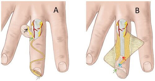
After the flap and broaden pedicle were designed, the skin was cut along the designed outline. The flap was cut at the superficial layer of the extensor tendon so as to protect the fascia and inner venous plexus. Postoperative tendon adhesion was also prevented and survival rate of the grafted skin was improved since the integrity of the aponeurosis was not destroyed upon separation at this level. Aiming to restore the sensory function of the amputated fingertip, digital nerves within the scope of the flap (, Black Arrow) were dissected and traversed in order to flip with the distal end of the flap, being anastomosed with the severed radial or ulnar proper digital nerve (, Gray Arrow) at the amputated finger end.
To form a superficial skin channel for creating and accommodating the nutrient stripe of the flap (, Blue Arrow), the whole skin layer from distal end of the flap to the proximal end of the defect was cut in a large zigzag incision and the skin incision placed laterally (ulnar or radial, according to the oblique direction of amputation) at the distal level of the distal interphalangeal joint in order to protect the nail bed and maximize the distal interphalangeal joint’s range of motion.
After the skin channel was cut and formed, the fascia tissue beneath the skin layer was dissected and 0.5–0.7 cm wide fascia tissue was reserved as the flap’s vascular nutrient stripe. Next, we rotated the flap in the pivot point by about 180° to cover the defect area. The pivot point (, Red Arrow) was designed at 0.5–0.8 cm proximal to the distal interphalangeal joint. The axis line refers to the finger dorsal midline, which includes the connecting line of dorsal midpoints of the distal and proximal interphalangeal joints and its extension line.
The transected digital nerve in the flap was then seamed to the stump of the radial or ulnar proper digital nerve (9-0 Prolene Polypropylene Suture; Ethicon US, LLC, Somerville, NJ) using the epineurium suture method. The flap was sutured with silk thread (5-0 non-absorbable suture, Ethicon US, LLC, Somerville, NJ) starting from the top of the flap (i.e. the opposite side of the broaden pedicle tip), followed by the two sides of the flap. The superficial skin channel was closed after completely suturing the skin flap. When suturing the skin edge near the pivot point, one should ensure that the tension at the skin edge is not too high and that the skin edge on both sides is slightly aligned to guarantee blood supply of the flap due to the existence of a double fascia layer around the pivot point. Finally, direct suture or free skin grafts can be used depending on size of the donor wound based on following principles: a) if the flap width was within 1.0 cm, the donor wound surface can be directly sutured, b) if the flap width is larger than 1.0 cm, free skin graft was appropriate. Forced suture should not be used on either. Typical surgical procedure was shown in .
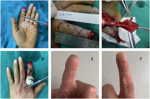
Figure 2. Typical case presentation. An Allen’s type 3 fingertip amputation is shown in (A). The exact location and width of flap and broaden pedicle is shown in (B). During operation, we dissected the digital nerve in the flap (C). Postoperative status was shown in (D). At 18 months follow-up, the index finger showed good contouring and appearance (E and F).
Postoperative management
Postoperative wound coverage was performed using a gauze to provide suitable pressure. Warmth was provided to the fingertip by light illumination. Antibiotics, anti-spastic, and anti-thrombotic treatments were given. Flap circulation was monitored for at least 2 days by visually inspecting tissue color and capillary refilling. Based on flap condition, sutures were removed in a timely manner to reduce wound tension and venous obstruction. Active range-of-motion exercises were initiated after the removal of sutures. All patients underwent formal hand motion exercises.
Outcome evaluation
Outcomes were evaluated by a hand surgeon at our department who did not attend the operation. The wounded finger’s range of motion (ROM) was assessed using the total active motion (TAM) scoring system of the American Society for Surgery of the Hand. The parameters were compared with the contralateral side (100% excellent; 75%–99% good; 50%–74% fair; <50% poor) [Citation6]. The sensibility of the flap and the donor site were evaluated using the Semmes Weinstein monofilament test with a five-piece hand kit (2.83: normal sensation, 3.61: residual texture sensation, 4.31: residual protective sensation, 4.56: loss of protective sensation, and 6.65: residual deep pressure sensation) [Citation7]. Static 2PD tests were rated using the modified American Society for Surgery of the Hand guidelines (<6 mm excellent; 6–10 mm good; 11–15 mm fair; >15 mm poor) [Citation8,Citation9]. The visual analogue scale (VAS) was used to evaluate residual pain in the flap and the donor site, ranging from 0 (no pain) to 10 (worst pain) [Citation10]. To quantify postoperative physical function and symptoms, all patients were evaluated using the Quick Disabilities of the Arm, Shoulder, and Hand score (quick DASH) [Citation11]. Scars were assessed using the Vancouver scar scale (VSS), ranging from 0 to 3 in 4 categories [Citation12,Citation13]. Follow-up assessments were completed by a hand surgeon at our department who did not participate in the operation or therapy.
Results
Patients
After rigorous screening, follow-up was available for 17 patients (14 males and 3 females, mean age: 44.7 ± 12.9 years) and 18 long fingers. The mean follow-up was 16.6 ± 4.0 months (range: 12–25 months). 13 fingers were on the left hand (72.2%) and 5 on the right hand (27.8%). The operations involved 6 index fingers (33.3%), 6 middle fingers (33.3%), 4 ring fingers (22.3%), and 2 little fingers (11.1%). The average size of fingertip defects was 1.11 × 1.13 cm (range: 0.6 × 1.0–2.0 × 1.8 cm). The mean flap size was 1.32 × 1.32 cm (range: 0.8 × 1.2–2.3 × 2.0 cm). Mean nutrient stripe length was 4.18 ± 0.34 cm (range: 3.4 − 4.6 cm). Mean broaden pedicle length was 0.44 ± 0.08 cm (range: 0.3 − 0.5 cm). Mean operating time was 123 ± 32 min (range: 90–185 min). The main etiology for fingertip wounds was crush amputations (55.6%) and clear-cut injuries (44.4%) ( and ). All flaps survived. One flap showed partial necrosis and two cases showed tension blister but healed with wound care. No wound infections were observed.
Table 1. Patients and results.
Table 2. Demographic and surgical details of the cases in this study.
Motor ability
At the final follow-up, the mean TAM for the injured fingers was 255.6°±8.0°, which was over 95% of the contralateral side (unaffected side: 268.4°±5.7°, p < 0.05), with all fingers scored as good grade. Detailed statistics are shown in .
Table 3. Outcomes at the final follow-up.
Neurological assessment
Mean Semmes Weinstein monofilament score in the flap was 3.89 ± 0.37 g (range: 3.6–4.56 g, contralateral side 3.18 ± 0.39 g, range: 2.83–3.61 g, p < 0.05). Mean Semmes Weinstein monofilament score at the donor site was 3.22 ± 0.40 g (range: 2.83–3.61 g) (contralateral side 3.00 ± 0.33 g, range 2.83–3.61 g, p < 0.05). Mean values for static two-point discrimination in the flap was 5.6 ± 1.2 mm (range: 4–8 mm), including 9 excellent and 9 good results (contralateral side: 3.7 ± 0.8 mm, range: 3–5 mm, p < 0.05). Mean values of the static two-point discrimination in the donor zone was 4.3 ± 0.6 mm (range: 4–6 mm), including 17 excellent and 1 good results (contralateral side. 3.6 ± 0.6 mm, range 3–5 mm, p > 0.05). Mean quick dash scores was 5.81 (range: 0–20). Mean VAS score in the flap and donor site was 0.7 (range: 0–4) and 0.2 (range: 0–2) ().
Appearance and satisfaction
Based on the 5-point Likert scale, aesthetic satisfaction was rated ‘sufficient’ by 3 patients, ‘good’ by 8 patients, and ‘excellent’ by 6 patients (). Vancouver scar scale results at the donor and recipient sites are shown in . Typical follow-up appearance was shown in . By the end of follow-up, all patients were satisfied with the curative effects and aesthetic appearance.
Table 4. Vancouver scar scale results at the donor and recipient sites.
Discussion
After ruling out post-traumatic infection, the main objective of fingertip defect treatment is to preserve the joints’ range of motion, restore sensory function, preserve the length of affected long fingers, and to improve post-treatment appearance as much as possible [Citation14]. In all Allen type I injuries and a part of Allen II type injuries, conservative treatment with a closed dressing covering the wound is acceptable [Citation15–17]. However, for patients with Allen II and III amputations, especially those with large defects and bone exposure, conservative treatment is not effective and urgent surgery is often needed [Citation18].
Currently, the treatment of fingertip defects mainly involves finger replantation, amputation, and flap reconstruction [Citation19–21]. For acute transverse injury, fingertip reconstruction is considered the best treatment for fingertip amputation. However, due to the harsh conditions, high technical requirements and high risk of failed replantation, it is difficult to carry out this technique in primary hospitals [Citation22]. Amputation surgery is simple and convenient for postoperative management, but due to loss of aesthetics and function, it is not performed routinely [Citation23]. Due to these factors, flap therapy is an ideal treatment strategy. Common surgical methods for repairing fingertip defects in long fingers include: 1) V–Y advancement flap, whereby the flap is mainly aimed at the dorsal or oblique defect, and can be usually be repaired in a small area but not a large area of defect. The most popular methods include Segmuller, extended Segmuller, Mouchet-Gilbert, and Venkataswami [Citation24,Citation25]; 2) cross-finger flap, whereby the flap is mainly used to repair the dorsal defect of the finger and can cover a large area of the defect. However, it requires a long time of two-finger fixation and a second operation to open the flap [Citation26]; 3) antegrade homodigital island flap, whereby the flap effectively covers digital abdomen defects. However, it is also not suitable for large defects and requires dissection of bilateral neurovascular bundles, which is difficult to conduct [Citation27]; 4) heterodigital island flap, which is a complementary option to the homodigital island flap. It can be considered when adjacent soft tissue is damaged [Citation28]; 5) Free flap, which is suited for small fingertip defects, or for patients with low sensory requirements, such as fingertip defects in the little finger in which sensation is much smaller than in other fingers [Citation25].
The fingertip is a special anatomical structure. Its main function is to grasp and feel objects. Tactile recognition is a key function of the hand, especially the fingertip. Achieving the best sensory function is the aim of fingertip reconstruction [Citation29,Citation30]. Sensory function can be assessed clinically using the 2PD and SWM tests. Kim and colleagues examined 8 patients with static 2PD, 12 months post-surgery and obtained a mean 2PD result of 5.0 mm [Citation31]. Dağhan Dağdelen and colleagues measured static 2PD at 7 months after repairing fingertip injury, with an average measurement value of 4.5 mm [Citation32]. Comparison of our findings with past reports revealed that the sensory function of the recipient flap in this study was acceptable, although the mean static 2PD and SWM scores in the flap differed significantly from the contralateral side
Another major function of the fingertips is gripping and resistance to mechanical forces [Citation33]. A major goal of therapy after fingertip amputation is to restore normal active motion without the development of flexion contracture. In this study, injured fingers had a mean TAM of 255.6°±8.0°, while contralateral fingers had a mean TAM of 268.4°±5.7°. The IS/CS rate was 95.16%, which corresponds to ‘good’ according to Kleiner [Citation34] – no significant limitation in active and passive activity was observed. Forced splints and cross-finger flaps require additional postoperative fixation and increase the risk of joint stiffness [Citation27]. Here, TAM results showed that the affected finger’s postoperative mobility recovered well and benefited from the avoidance of postoperative fixation of the affected finger and early postoperative joint activity and rehabilitation exercise.
In addition to motor and sensory functions, aesthetic recovery is important aim when treating fingertip defects [Citation35]. VSS analysis indicated that our study had satisfactory aesthetic clinical outcomes. Quick DASH scores and VAS scores indicated that few patients suffered postoperative pain that affected their daily life and work, which is consistent with the results of all patients being satisfied.
HDNBPIF has several advantages in the repair of fingertip defects. First, the flap relies on the vascular network within the fascia to provide circulation support, without the need to dissect specific digital arteries, which reduces the operation’s difficulty. Secondly, the dorsal finger nerve is dissected in the flap and transplanted to the stump of digital proper nerves, leading to satisfying sensory function recovery in the long term. Third, the flap’s broaden pedicle design appropriately increases the width of the skin at the junction between the flap and the stump, which can reduce the suture tension and soft tissue pressure and ensure the blood supply of the distal flap and reduce the possibility of necrosis of the distal flap. Furthermore, compared with cross finger flaps, each long finger can be treated separately without affecting each other when multiple fingers are injured. Additionally, the flap is designed on the dorsal of the lacerated finger, which does not affect other fingers. In the case of multiple fingertip amputations, all wounded long fingers can receive HDNBPIF therapy simultaneously and in a timely manner. Besides, HDNBPIF avoids long-term splint fixation after surgery and second operation, allowing early functional exercise of the affected finger. Finally, the skin flap has similar texture and color as the surrounding tissue, as well as good blood supply, friction resistance, and cold resistance.
However, HDNBPIF also has some disadvantages: First, it requires dissection of the dorsal digital nerve and suture of the severed digital proper nerve. Secondly, as the blood supply source of the flap is not as steady as digital artery’s supply, mild ischemia and flap retraction, as well as blister formation may occur after surgery. After surgery, it is necessary to promptly aspirate effusion and release pedicle sutures. Finally, compared with palmar flaps, this method is more invasive and requires more soft tissue dissection.
This study also has some limitations. First, this is a single-center cohort study. Second, because the study lacked a control group, it could only be compared with past studies. Third, the study had a small sample size (only 17 patients were followed up). Finally, the follow-up time was short in some cases.
In this study, clinical data on the use of homodigital dorsal neurofascial broaden pedicle island flaps (HDNBPIF) to treat Allen type II and III fingertip injury were analyzed. The results show that HDNBPIF has several advantages including preservation of finger length, restoration of sensory function, good aesthetic appearance, and does not impair joint activity, all of which results in high patients’ satisfaction. In summary, HDNBPIF can be used to reliably repair fingertip defects in long fingers at primary clinics.
Ethical approval
The study was approved by ethical committee of Suzhou Yongding Hospital with an approve No. 201801.
Acknowledgement
The authors thanks all participants and hospital staff for their valuable contribution to this work. We also thank the education and health project of Wujiang District of Suzhou City for supporting this study.
Disclosure statement
No potential conflict of interest was reported by the authors.
Correction Statement
This article has been republished with minor changes. These changes do not impact the academic content of the article.
Additional information
Funding
References
- Harris AP, Goodman AD, Gil JA, et al. Incidence, timing, and risk factors for secondary revision after primary revision of traumatic digit amputations. J Hand Surg Am. 2018;43(11):1040.e1–1040.e11.
- Lim JX, K, C, Chung. VY advancement, thenar flap, and cross-finger flaps. Hand Clin. 2020;36(1):19–32.
- Sérane-Fresnel J, Lafosse T, Amsallem L, et al. Fingertip reconstruction by palmar bipedicular island flap in long fingers (modified neurovascular Tranquilli-Leali flap): a dual-center study. Hand Surg Rehabil. 2020;39(1):59–64.
- Loréa P, Chahidi N, Marchesi S, et al. Reconstruction of fingertip defects with the neurovascular Tranquilli-Leali flap. J Hand Surg Br. 2006;31(3):280–284.
- Lee DH, Mignemi ME, Crosby SN. Fingertip injuries: an update on management. J Am Acad Orthop Surg. 2013;21(12):756–766.
- Libberecht K, Lafaire C, Van Hee R. Evaluation and functional assessment of flexor tendon repair in the hand. Acta Chir Belg. 2006;106(5):560–565.
- Feng Y, Schlösser FJ, Sumpio BE. The Semmes Weinstein monofilament examination as a screening tool for diabetic peripheral neuropathy. J Vasc Surg. 2009;50(3):675–682, 682.e1.
- Crosby PM, Dellon AL. Comparison of two-point discrimination testing devices. Microsurgery. 1989;10(2):134–137.
- Dellon AL, Kallman CH. Evaluation of functional sensation in the hand. J Hand Surg Am. 1983;8(6):865–870.
- Reed MD, Van Nostran W. Assessing pain intensity with the visual analog scale: a plea for uniformity. J Clin Pharmacol. 2014;54(3):241–244.
- Beaton DE, Wright JG, Katz JN; Upper Extremity Collaborative Group. Development of the QuickDASH: comparison of three item-reduction approaches. J Bone Joint Surg Am. 2005;87(5):1038–1046.,
- Brusselaers N, Pirayesh A, Hoeksema H, et al. Burn scar assessment: a systematic review of different scar scales. J Surg Res. 2010;164(1):e115-23–e123.
- Brunetti B, Salzillo R, Tenna S, et al. Perforator-based flap reconstruction after melanoma resection: evaluation of oncological, aesthetic, and functional outcomes. J Reconstr Microsurg. 2022;38(7):555–562. doi:10.1055/s-0041-1740925
- Neustein TM, Payne SH Jr, Seiler JG 3rd. Treatment of fingertip injuries. JBJS Rev. 2020;8(4):e0182.
- Quadlbauer S, Pezzei C, Jurkowitsch J, et al. The semi-occlusive dressing in treating Allen III and IV fingertip injuries as an alternative to local skin flaps. Unfallchirurg. 2017;120(11):961–968.
- Lasserre G, Bakkouch S, Pauchot J, et al. Fingertip reconstruction with occlusive dressing: clinical results and biological analysis of the dressing content’s. Chir Main. 2010;29(5):315–320.
- Boudard J, Loisel F, El Rifaï S, et al. Fingertip amputations treated with occlusive dressings. Hand Surg Rehabil. 2019;38(4):257–261.
- Krauss EM, Lalonde DH. Secondary healing of fingertip amputations: a review. Hand (N Y). 2014;9(3):282–288.
- Harenberg PS, Jakubietz RG, Jakubietz MG, et al. Reconstruction of the thumb tip using palmar neurovascular flaps. Oper Orthop Traumatol. 2012;24(2):116–121.
- Unglaub F, Langer MF, Unglaub JM, et al. Defect coverage of fingers and thumb: indications and treatment. Unfallchirurg. 2018;121(4):321–334.
- Zhou X, Wang L, Mi J, et al. Thumb fingertip reconstruction with palmar V-Y flaps combined with bone and nail bed grafts following amputation. Arch Orthop Trauma Surg. 2015;135(4):589–594.
- Barbary S, Dap F, Dautel G. Finger replantation: surgical technique and indications. Chir Main. 2013;32(6):363–372.
- Yang J, Yu C, Jia X. A new surgical method to reconstruct the fingertip. Ann Plast Surg. 2019;83(6):647–649.
- Atasoy E, Ioakimidis E, Kasdan ML, et al. Reconstruction of the amputated finger tip with a triangular volar flap. A new surgical procedure. J Bone Joint Surg Am. 1970;52(5):921–926.
- Tang JB. Fingertip repair methods: choices for different fingers and sides emphasizing sensation. J Hand Surg Eur Vol. 2019;44(10):1109–1111.
- Koch H, Kielnhofer A, Hubmer M, et al. Donor site morbidity in cross-finger flaps. Br J Plast Surg. 2005;58(8):1131–1135.
- Katz RD. The anterograde homodigital neurovascular island flap. J Hand Surg Am. 2013;38(6):1226–1233.
- Pham DT, Netscher DT. Vascularized heterodigital island flap for fingertip and dorsal finger reconstruction. J Hand Surg Am. 2015;40(12):2458–2464.
- Dellon ES, Mourey R, Dellon AL. Human pressure perception values for constant and moving one- and two-point discrimination. Plast Reconstr Surg. 1992;90(1):112–117.
- Sano K, Ozeki S, Kimura K, et al. Relationship between sensory recovery and advancement distance of oblique triangular flap for fingertip reconstruction. J Hand Surg Am. 2008;33(7):1088–1092.
- Kim KS, Kim ES, Hwang JH, et al. Fingertip reconstruction using the hypothenar perforator free flap. J Plast Reconstr Aesthet Surg. 2013;66(9):1263–1270.
- Dağdelen D, Aksoy A. Evaluation of neurotized hypothenar free perforator flaps used for fingertip reconstruction. Ann Plast Surg. 2020;84(2):e1–e6.
- Hwang K, Han JY, Chung IH. Hypothenar flap based on a cutaneous perforator branch of the ulnar artery: an anatomic study. J Reconstr Microsurg. 2005;21(5):297–301.
- Kleinert HE, Verdan C. Report of the committee on tendon injuries (International Federation of Societies for Surgery of the Hand). J Hand Surg Am. 1983;8(5):794–798.
- Erba P, Ogawa R, Vyas R, et al. The reconstructive matrix: a new paradigm in reconstructive plastic surgery. Plast Reconstr Surg. 2010;126(2):492–498.