Abstract
Human parvovirus B19 (B19V) is a human pathogen known to be associated with many non-erythroid diseases, including hepatitis. Although B19V VP1-unique region (B19-VP1u) has crucial roles in the pathogenesis of B19V infection, the influence of B19-VP1u proteins on hepatic injury is still obscure. This study investigated the effect and possible inflammatory signaling of B19-VP1u in livers from BALB/c mice that were subcutaneously inoculated with VP1u-expressing COS-7 cells. The in vivo effects of B19-VP1u were analyzed by using live animal imaging system (IVIS), Haematoxylin-Eosin staining, gel zymography, and immunoblotting after inoculation. Markedly hepatocyte disarray and lymphocyte infiltration, enhanced matrix metalloproteinase (MMP)-9 activity and increased phosphorylation of p38, ERK, IKK-α, IκB and NF-κB (p-p65) proteins were observed in livers from BALB/c mice receiving COS-7 cells expressing B19-VP1u as well as the significantly increased CRP, IL-1β and IL-6. Notably, IFN-γ and phosphorylated STAT1, but not STAT3, were also significantly increased in the livers of BALB/c mice that were subcutaneously inoculated with VP1u-expressing COS-7 cells. These findings revealed the effects of B19-VP1u on liver injury and suggested that B19-VP1u may have a role as mediators of inflammation in B19V infection.
Introduction
Human parvovirus B19, generally referred to as B19V or erythrovirus B19, is a major human pathogen.Citation1 Infection with B19V is associated with many different pathological processes and clinical manifestations, including erythema infectiosum, cardiovasculitis, thrombocytopenia, neurologic disorders, hepatitis, arthropathy and autoimmune disorders.Citation2-4 B19 virus can be found in blood, bone marrow, liver, immune cells, synovial membranes, and various tissues.Citation5 In non-erythroid tissues, including heart, liver, lymphoid, synovial, skin, colon, and thyroid cells, expression of B19 virus was also associated with specific changes in inflammatory genes such as TNF-α and IL-6, indicating persistent B19 virus expression can impact the cellular microenvironment.Citation5
The B19V is a small non-enveloped virus enclosing a single-stranded linear 5.6-kb DNA genome. The icosahedral capsid of B19V consists of a nonstructural protein (NS1) and 2 capsid proteins, VP1 and VP2, which are identical except for 227 amino acids at the amino-terminal end of the VP1-protein, the so called VP1-unique region (VP1u).Citation6 Although VP2 proteins predominate in the capsid, VP1 is critical for eliciting appropriate immune responses in both humans and animals.Citation7-9 The peptide from residues 60 to 100 in B19-VP1u is also known to induce prolonged immune responses in humans.Citation10 Additionally, B19-VP1u has been demonstrated to have phospholipase A2 (PLA2) activity, which is essential for its infectivity.Citation11-12 B19-VP1u protein alone could induce pathological changes in the synovial compartment and myocardium.Citation13-14 Antibodies against B19-VP1u also induce inflammatory responses on vascular endothelia cells and accelerated cardiac and liver injury in NZB/W F1 mice.Citation15-16 Additionally, in naïve mice, antibodies against B19-VP1u induce antiphospholipid antibodies and antiphospholipid syndrome (APS)-like autoimmunity.Citation17 Accordingly, serum from patients with acute B19 infection has high recognition of CL and β2GPI, and the phospholipase domain observed in the B19-VP1u may contribute to antiphospholipid antibodies (aPL) production.Citation18-19
Evidences have reported that B19V could cause acute hepatitis, chronic hepatitis and even fulminant hepatic failure.Citation20-23 However, the precise role of B19-VP1u on induction of hepatic inflammation is still unclear. Therefore, this study investigated the effect of B19-VP1u in BALB/c mice subcutaneously inoculated with COS-7 cells expressing B19-VP1u to determine whether B19-VP1u participates in the induction of inflammation.
Materials and Methods
Ethics
Fifteen female BALB/c mice at age of 6 weeks were purchased from National Laboratory Animal Center, Taiwan and divided into 3 groups (5 mice /group) and housed under supervision of the Institutional Animal Care and Use Committee at Chung Shan Medical University, Taichung, Taiwan. All procedures and protocols were approved by the Institutional Animal Care and Use Committee at Chung Shan Medical University (Affidavit of Approval of Animal Use Protocol, No. 1318) and followed the Guide for the Care and Use of Laboratory Animals published by the United States National Institutes of Health.
Plasmids and transfection
Plasmid pTurboFP650-C1 was purchased from Evrogen (Evrogen, Moscow, Russia). pTurboFP650-C is a mammalian expression vector encoding near-infrared fluorescent protein TurboFP650. TurboFP650-tagged fusions retain fluorescent properties of the native protein allowing fusion localization in vivo.Citation24 The VP1u gene in the pET32a vectorCitation18 was extracted using restriction endonuclease digestion at the Bgl II site and Sal I sites prior to ligation into the pTurboFP650-C1 expression vector. The ligatant, so called pTurboFP650-VP1u was then transformed into Escherichia Coli DH5α competent cells, which were purchased from Life Technologies (Carlsbad, California, USA). Restriction enzyme digestion and DNA sequencing analysis were used to verify the plasmid. The purified pTurboFP650-VP1u ligatants were then stored in −20°C before use. COS-7 cells were originally obtained from American type culture collection (ATCC) (Manassas, Va, USA) and were grown in Dulbecco's modified Eagle medium (DMEM) supplemented with 10% fetal bovine serum (FBS) (GIBCO-BRL, Carlsbad, California, USA) at 37°C and 5% CO2 incubator. A total of 1×10⁶ cells were grown to 70% confluent in 100 mm2 culture plates before transfection. The transfection reaction was performed by using Lipofectamine plus reagents (Invitrogene, California, USA) with 2μg of each plasmid, pTurboFP650-C1 or the pTurboFP650-VP1u constructant according to the manufacture's instruction. The cells were then cultured in serum-free DMEM at 37°C in a 5% CO2 humidified incubator for 12hr and subsequently in DMEM with 10% FBS. After transfection, both pTurboFP650-C1 and pTurboFP650-VP1u transfectants were treated with G418 antibiotics (600 μg/ml), and the stable clones were obtained after this selection process for 3 months.
Cell preparation and animal groups
The viability of cells was performed prior to IVIS experiments. No difference in viability was detected among control cells and the stable clones (Fig S1). The cells were suspended in 200ul of sterile phosphate buffer saline (PBS) in a syringe that was kept on ice before use. Aliquots containing 1 × 10⁶ cells of control group (COS-7 cells) with PBS only, TurboFP650-C1 or TurboFP650-VP1u cells were injected subcutaneously of the BALB/c mice at the age of 8 weeks and was monitored by a live animal imaging system (IVIS) (The Caliper IVIS Lumina II Imaging System, Lincolnshire, UK). The mice were then sacrificed at day 28 by CO2 asphyxiation. The brain, kidneys and livers tissues were collected and stored at −80°C until use.
Haematoxylin-eosin staining
The liver samples of animals were excised and soaked in formalin and covered with wax. Slides were prepared by deparaffinization and dehydration. They were passed through a series of graded alcohols (100%, 95% and 75%), for 15 min. each. The slides were then dyed with haematoxylin. After gently rinsing with water, each slide was then soaked with 85% alcohol, 100% alcohol I and II for 15 min. each. At the end, they were soaked with Xylene I and Xylene II. Photomicrography was performed with Zeiss Axiophot microscopes and photomicrographs were obtained. For quantification, the number of infiltrated lymphocytes was counted in 5 randomly selected fields of a section slide. All measurements were made using at least 4 section slides from 3 independent animals.
Gel zymography
Matrix metalloproteinase (MMP)-9 and MMP-2 activities were analyzed by gelatin zymography. Ten microliters of 10× diluted serum or 25 μg protein lysate of tissues from BALB/c mice was separated by an 8% sodium dodecyl sulfate–polyacrylamide gel electrophoresis (SDS-PAGE) gel polymerized with 1 mg/ml gelatin. Gels were washed once for 30 mins in 2.5% Triton X-100 to remove the SDS and then soaked in the reaction buffer containing 50 mM Tris-HCl, 200 mM NaCl, 10 mM CaCl2, and 0.02% (w/v) Brij 35 (Sigma, St. Louis, MO; pH 7.5) for 30 mins. The reaction buffer was changed to a fresh one, and the gels were incubated at 37°C for 24 hrs. Gelatinolytic activity was visualized by staining the gels with 0.5% Coomassie brillant blue and quantified by densitometry (Appraise; Beckman-Coulter, Brea, CA).
Western blot
The loading sample for each lane of Western blot was a pool of liver homogenized lysates from 5 random selected mice of a same group. Protein samples were separated in 10% or 12.5% SDS-PAGE and electrophoretically transferred to nitrocellulose membrane (Amersham Biosciences, Piscataway, NJ, USA) as described in our previous study.Citation26 After blocking with 5% non-fat dry milk in PBS, antibodies against CRP, IL-1 β, IL-6, IFN-γ, p-P38, P38, p-ERK, ERK, p-P65, Ikk-α, IκB, p-STAT-1, STAT-1, p-STAT-3, STAT-3 (Santa Cruz Biotechnology, CA, USA), RFP (Applied Biological Materials Inc., BC, Canada), B19-VP1uCitation15-17 and β-actin (Upstates, Charlottesville, VA, USA) were diluted in PBS with 2.5% BSA and incubated for 1.5 h with gentle agitation at room temperature. The membranes were washed twice with PBS-Tween for 1 h and secondary antibody conjugated with horseradish peroxidase (HRP) (Santa Cruz Biotechnology, Santa Cruz, CA, USA) was added. Immobilon Western Chemiluminescent HRP Substrate (Millipore, MA, USA) was used to detect antigen–antibody complexes. Quantified results were performed by densitometry (Appraise, Beckman-Coulter, Brea, CA, USA).
Statistical analysis
All of the statistical analyses were performed using GraphPad Prism 5 software (GraphPad Software, CA) by one-way analysis of variance (One-way ANOVA) followed by Tukey multiple-comparisons test. Data were represented as mean ± SEM and verified at least 3 independent experiments. A value of P < 0.05 was considered statistically significant. The significant differences were stressed with symbols as shown in figures.
Results
Expression of pTurboFP650-VP1u in BALB/c mice
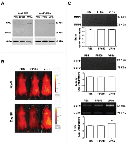
To verify the expressions of FP650 and FP650-VP1u recombinant proteins, antibodies against FP650 and B19-VP1u were used for Western Blots analysis. Expressions of FP650 and FP650-VP1u recombinant proteins were detected in stable clone of COS-7 cells transfected with pTurboFP650 and pTurboFP650-VP1u, respectively (). The in vivo effects of B19-VP1u were investigated by using IVIS to detect BALB/c mice subcutaneously inoculated with COS-7 cells, COS-7 cells transfected with pTurboFP650, or COS-7 cells transfected with pTurboFP650-VP1u, respectively, on 0 and 28 d after inoculation. shows that the animals that had received COS-7 showed no detectable fluorescence on 0 or 28 d after the inoculations. However, on day 28 after inoculation, only BALB/c mice receiving COS-7 cells transfected with pTurboFP650-VP1u revealed markedly increased fluorescence ().
Figure 1. Detection of FP650 and FP650-VP1u by Western Blots and IVIS, and activity of MMP-9 and MMP-2. (A) Expression of FP650 and FP650-VP1u recombinant proteins in COS-7 cells, COS-7 cells transfected with pTurboFP650 or pTurboFP650-VP1u were detected by Western Blots. (B) Fluorescence detection for BALB/c mice subcutaneously receiving COS-7 cells, COS-7 cells transfected with pTurboFP650, or COS-7 cells transfected with pTurboFP650-VP1u was performed with IVIS systems. (C) The MMP-9 and MMP-2 activity were analyzed by gel zymography after the treatments. Densitometric analysis results are shown in the lower panel. Similar results were observed in 3 independent experiments, and * indicates the significant difference, P < 0.05.
Liver inflammation induced by B19 VP1u
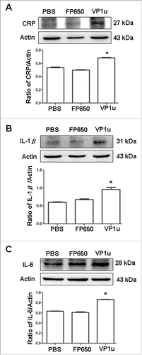
To identify the locations affected by B19-VP1u after inoculation with COS-7 cells transfected with pTurboFP650-VP1u, various organs such as the brain, kidneys and livers of BALB/c mice were analyzed by zymography (). MMP9/MMP2 ratio detected in brains and kidneys from mice receiving COS-7 cells did not significantly differ between those transfected with pTurboFP650 and those transfected with pTurboFP650-VP1u (). However, the MMP9/MMP2 ratios in livers of mice receiving COS-7 cells transfected with pTurboFP650-VP1u were significantly higher compared to those that receiving COS-7 cells without transfection or with pTurboFP650 transfection (). To confirm the effects of B19-VP1u on liver in BALB/c mice, a histopathological analysis was performed in liver tissue stained with hematoxylin and eosin. shows that lymphocyte infiltration was markedly higher in the livers of BALB/c mice that received COS-7 cells transfected with pTurboFP650-VP1u as compared to those mice that received COS-7 cells without transfection or with pTurboFP650 transfection. Quantified results were shown in . In addition, various inflammatory cytokines such as CRP, IL-1β and IL-6 were also detected. Accordingly, expressions of CRP, IL-1β and IL-6 in the liver were significantly higher in BALB/c mice receiving COS-7 cells transfected with pTurboFP650-VP1u ().
Figure 2. Histopathological analysis of hepatic tissue sections with hematoxylin and eosin staining. Livers from BALB/c mice receiving COS-7 cells without tranfection, COS-7 cells transfected with pTurboFP650, and COS-7 cells transfected with pTurboFP650-VP1u are shown were analyzed with hematoxylin and eosin staining. These images of hepatic sections were magnified by 200 times. Amplified images were shown in the right panel and the lymphocyte infiltration was indicated by an arrow. Similar results were observed in 3 independent experiments.
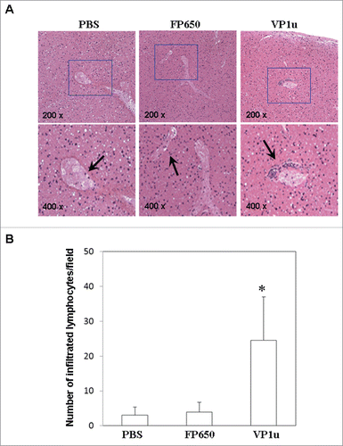
Figure 3. Expression of CRP, IL1-β and IL-6. Liver lysates obtained from BALB/c mice receiving COS-7 cells without tranfection, COS-7 cells transfected with pTurboFP650, and COS-7 cells transfected with pTurboFP650-VP1u are shown after the treatments were probed with antibodies against (A) CRP, (B) IL1-β and (C) IL-6. Bars represent the relative protein quantification on the basis of actin. Similar results were observed in 3 independent experiments, and * indicates the significant difference, P < 0.05.
Signaling involved in B19 VP1u induced liver inflammation
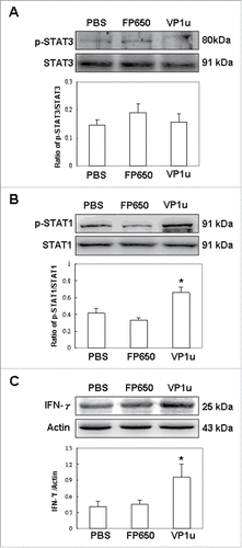
To verify that the signaling involved in B19-VP1u induced inflammatory responses in the liver of BALB/c mice, phosphorylated P-38 (p-P38), phosphorylated ERK (p-ERK), IKKα, IκB and phosphorylated (p-P65) were detected. Ratios of p-P38/p38 and p-ERK/ERK in the liver were significantly higher in BALB/c mice receiving COS-7 cells transfected with pTurboFP650-VP1u compared to mice that received COS-7 cells without transfection or with pTurboFP650 transfection (). Accordingly, expressions of IKKα, IκB and p-P65 in livers from BALB/c mice receiving COS-7 cells transfected with pTurboFP650-VP1u on the basis of actin were significantly higher than those in mice receiving COS-7 cells without transfection or with pTurboFP650 transfection (). The potential effects of B19-VP1u were also investigated by STAT3, STAT1, and their phosphorylated forms (p-STAT3, p-STAT1, respectively). The p-STAT3 detected in livers from BALB/c mice receiving COS-7 cells transfected with pTurboFP650-VP1u on the basis of STAT3 did not significantly differ from those in mice that received COS-7 cells without transfection or with pTurboFP650 transfection (). Interestingly, the ratio of p-STAT1/STAT1 detected in livers from BALB/c mice receiving COS-7 cells transfected with pTurboFP650-VP1u was significantly higher than that in mice receiving COS-7 cells without transfection or with pTurboFP650 transfection (). Meanwhile, IFN-γ in liver was significantly higher in BALB/c mice receiving COS-7 cells transfected with pTurboFP650-VP1u than in mice receiving COS-7 cells without transfection or with pTurboFP650 transfection ().
Figure 4. Expression of p-P38, P38, p-ERK, and ERK. Liver lysates obtained from BALB/c mice receiving COS-7 cells without tranfection, COS-7 cells transfected with pTurboFP650, and COS-7 cells transfected with pTurboFP650-VP1u are shown after the treatments were probed with antibodies against (A) p-P38 and P38, (B) p-ERK and ERK. The ratios of p-P38/P38 and p-ERK/ERK were shown in the lower panel, respectively. Similar results were observed in 3 independent experiments, and * indicates the significant difference, P < 0.05.
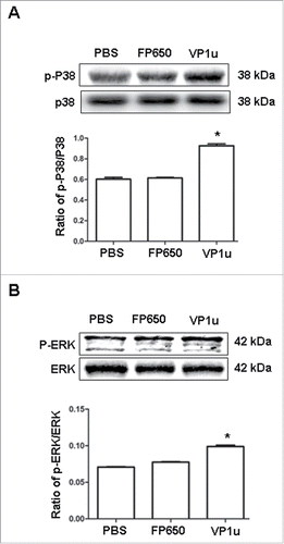
Figure 5. Expression of IKK-α, IκB, and p-P65. Liver lysates obtained from BALB/c mice receiving COS-7 cells without tranfection, COS-7 cells transfected with pTurboFP650, and COS-7 cells transfected with pTurboFP650-VP1u are shown after the treatments were probed with antibodies against (A) IKK-α, (B) IκB, and (C) p-P65. Bars represent the relative protein quantification on the basis of actin. Similar results were observed in 3 independent experiments, and * indicates the significant difference, P < 0.05.
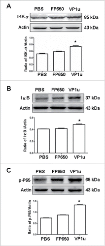
Figure 6. Expression of p-STAT3, STAT3, p-STAT1, STAT1 and IFN-γ. Liver lysates obtained from BALB/c mice receiving COS-7 cells without tranfection, COS-7 cells transfected with pTurboFP650, and COS-7 cells transfected with pTurboFP650-VP1u are shown after the treatments were probed with antibodies against (A) p-STAT3 and STAT3, (B) p-STAT1 and STAT1, and (C) IFN-γ. The ratios of p-STAT3/STAT3, p-STAT1/STAT1 and IFN-γ/actin were shown in the lower panel, respectively. Similar results were observed in 3 independent experiments, and * indicates the significant difference, P < 0.05.
Discussion
Although B19V infection is often described as a cause or trigger of various non-erythroid diseases, including hepatitis, the effects of B19-VP1u proteins on hepatic injury is still obscure. The possible role of B19-VP1u on induction of inflammation was further verified by using an IVIS system to observe and investigate the effect of B19-VP1u in naïve mice inoculated with COS-7 cells expressing B19-VP1u. Our experimental results revealed inflammatory phenomena and signaling, including enhanced MMP-9 activity and increased phosphorylation of p38, ERK, IKK-α, IκB and NF-κB (p65) proteins in the livers of BALB/c mice receiving COS-7 cells expressing B19-VP1u. Significant increases in CRP, IL-1β and IL-6 proteins were also noted. Meanwhile, a significant induction of phosphorylated STAT1, a crucial host-defense related molecule, was observed in livers from BALB/c mice receiving COS-7 cells expressing B19-VP1u. A significant induction of IFN-γ was also noted. However, induction of phosphorylated STAT3 was not statistically significant. These findings not only indicated the role of B19-VP1u on induction of inflammatory phenomena in naïve mice, but also indicated the signaling of anti-viral immunity through p-STAT1 activation.
Liver diseases caused by B19V infection have been demonstrated, which range from elevation of transaminases to acute hepatitis to fulminant liver failure and even chronic hepatitis.Citation20-23,26 In fact, spectrum of liver diseases may occur in 4.1% of patients infected by human parvovirus B19.Citation27 However, the mechanism by which parvovirus B19 infection may result in hepatic injury is exactly not clear. Two possible routes for B19 virus induced-liver injury have been reported, including direct cytopathic effect and indirect immunological effect.Citation28-30 B19V could enter hepatocytes through globoside and establishes a restricted infection with the production of NS1, leading to G1 arrest and apoptosis.Citation28 Alternatively, indirect immunological effect caused by B19V infection is thought to be due to increased circulating CD8+ cytotoxic T cells and elevated expressions of IFN-γ and TNF-α.Citation29 Our previous study has indicated that B19-VP1u induces inflammatory responses in macrophages.Citation30 In current study, significantly increased inflammatory associated proteins such as CRP, IL-1β, IL-6 and IFN-γ were detected in livers of naïve mice that were inocoulated with B19-VP1u expressing COS-7 cells. Therefore, herein we made a rational assumption that B19-VP1u may induce liver damage via indirect immunological effect. However, further investigations are needed to verify this assumption.
Signal transducers and activators of transcription (STATs) have well-known roles in the initiation, duration and intensity of immune responses. The essential roles of STAT members in the differentiation of Th1 and Th17 subsets have been demonstrated in mice with targeted deletion of STATs in T cells.Citation31 In fact, unbridled activation of STATs may also contribute to pathogenic autoimmunity.Citation32 Mice with targeted deletion of STAT3 in CD4+ T-cells (STAT3KO) reportedly cannot generate Th17 and do not develop experimental autoimmune uveoretinitis (EAU) or experimental allergic encephalomyelitis (EAE).Citation33 Another study indicated that STAT3 inhibitor reduces IL-17-induced TLR3 expression in fibroblast-like synoviocytes (FLS) from patients with rheumatoid arthritis (RA).Citation34 Additionally, experimental animal models of lupus and studies of patients with SLE reveal that activation of STAT1 and IFNs are closely associated with the pathogenesis of SLE.Citation35-36 Indeed, over-expressed and abnormal activated p-STAT1 promotes expression of apoptosis-related genes and production of auto-antibodies in lupus animal models.Citation37-38 Accordingly, suppression of cytokine signaling 1 (SOCS1) deficiency-induced high STAT1 expression leads to anti-DNA antibodies production and causes glomerulonephritis with glomerular IgG deposition.Citation39 Conversely, diminished production of p-STAT1 in NZB/W F1 mice by administration of suppressive synthetic oligodeoxynucleotides (ODN) significantly reduces the severity of lupus nephritis, as evidenced by decreased incidence of glomerulonephritis and reduced proteinuria formation.Citation40 Similar findings have been reported in human studies. The percentage of patients who have diffuse proliferative lupus nephritis (DPLN) with worse renal injury and who show STAT1 expression in glomeruli is higher than that in patients without STAT1 expression.Citation41 Significant higher level of STAT1 was also found in tissue from the swollen knees of untreated patients with SLE than that in rheumatoid arthritis (RA) patients and osteoarthritis (OA) patients.Citation42 Collectively, these data indicate that STAT1 and STAT3 play crucial roles in development of autoimmunity. This study firstly report that BALB/c mice receiving COS-7 cells expressing B19-VP1u have significantly increased IFN-γ and p-STAT1 but not p-STAT3 in the liver. Since STAT1 can bind to STAT3 target genes and directly suppress transcription by recruiting transcriptional repressors,Citation43 activation of p-STAT1 but not p-STAT3 was expected. Therefore, these findings may also explain the role of B19-VP1u in induction of inflammation and autoimmunity through activation of IFN-γ/p-STAT1 signaling.
Our previous studies implied that B19-VP1u contributes to autoimmunity by enhancing MMP9 activity and elevating inflammation-related proteins.Citation15-16 This study further revealed the induction of inflammatory responses and IFN-γ/STAT-1 signaling on hepatic injury in naïve mice subcutaneously inoculated with COS-7 cells transfected with pTurboFP650-VP1u. These findings revealed the effects of B19-VP1u on liver injury and suggest that B19-VP1u may have a role as mediators of inflammation in B19 infection.
Disclosure and Potential Conflict of Interest
No potential conflicts of interests were disclosed.
Supplemental Material
Supplemental data for this article can be accessed on the publisher's website.
KVIR_A_1122165_Supp.zip
Download Zip (2.7 MB)Acknowledgments
The authors thank Ted Knoy for his editorial assistance.
Funding
This study was supported by grants from National Science Council, Taiwan (NSC 101–2314-B-040–008), and Chung Shan Medical University Hospital (CSH-2014-C-009), Taichung, Taiwan.
References
- Servey JT, Reamy BV, Hodge J. Clinical presentations of parvovirus B19 infection. Am Fam Physician 2007; 75:373-6; PMID:17304869
- Bock CT, Klingel K, Aberle S, Duechting A, Lupescu A, Lang F, Kandolf R. Human parvovirus B19: A new emerging pathogen of inflammatory cardiomyopathy. J Vet Med B Infect Dis Vet Public Health 2005; 52:340-3; PMID:16316397; http://dx.doi.org/10.1111/j.1439-0450.2005.00867.x
- Servant-Delmas A, Lefrére JJ, Morinet F, Pillet S. Advances in human B19 erythrovirus biology. J Virol 2010; 84:9658-65; PMID:20631151; http://dx.doi.org/10.1128/JVI.00684-10
- Hatakka A, Klein J, He R, Piper J, Tam E, Walkty A. Acute hepatitis as a manifestation of parvovirus B19 infection. J Clin Microbiol 2011; 49:3422-4; PMID:21734024; http://dx.doi.org/10.1128/JCM.00575-11
- Adamson-Small LA, Ignatovich IV, Laemmerhirt MG, Hobbs JA. Persistent parvovirus B19 infection in non-erythroid tissues: possible role in the inflammatory and disease process. Virus Res 2014; 190:8-16; PMID:24998884; http://dx.doi.org/10.1016/j.virusres.2014.06.017
- Ozawa K, Ayub J, Hao YS, Kurtzman G, Shimada T, Young N. Novel transcription map for the B19 (human) pathogenic parvovirus. J Virol 1987; 61:2395-406; PMID:3599180
- Schwarz TF, Roggendorf M, Deinhardt F. Human parvovirus B19: ELISA and immunoblot assays. J Virol Methods 1988; 20:155-68; PMID:2843558; http://dx.doi.org/10.1016/0166-0934(88)90149-8
- Kurtzman GJ, Cohen BJ, Field AM, Oseas R, Blaese RM, Young NS. Immune response to B19 parvovirus and an antibody defect in persistent viral infection. J Clin Invest 1989; 84:1114-23; PMID:2551923; http://dx.doi.org/10.1172/JCI114274
- Anderson S, Momoeda M, Kawase M, Kajigaya S, Young NS. Peptides derived from the unique region of B19 parvovirus minor capsid protein elicit neutralizing antibodies in rabbits. Virology 1995; 206:626-32; PMID:7530397; http://dx.doi.org/10.1016/S0042-6822(95)80079-4
- Zuffi E, Manaresi E, Gallinella G, Gentilomi GA, Venturoli S, Zerbini M, Musiani M. Identification of an immunodominant peptide in the parvovirus B19 VP1 unique region able to elicit a long-lasting immune response in humans. Viral Immunol 2001; 14:151-8; PMID:11398810; http://dx.doi.org/10.1089/088282401750234529
- Filippone C, Zhi N, Wong S, Lu J, Kajigaya S, Gallinella G, Kakkola L, Söderlund-Venermo M, Young NS, Brown KE. VP1u phospholipase activity is critical for infectivity of full-length parvovirus B19 genomic clones. Virology 2008; 374:444-52; PMID:18252260; http://dx.doi.org/10.1016/j.virol.2008.01.002
- Leisi R, Ruprecht N, Kempf C, Ros C. Parvovirus B19 Uptake Is a Highly Selective Process Controlled by VP1u, a Novel Determinant of Viral Tropism. J Virol 2013; 87:13161-7; PMID:24067971; http://dx.doi.org/10.1128/JVI.02548-13
- Lu J, Zhi N, Wong S, Brown KE. Activation of synoviocytes by the secreted phospholipase A2 motif in the VP1-unique region of parvovirus B19 minor capsid protein. J Infect Dis 2006; 193:582-90; PMID:16425138; http://dx.doi.org/10.1086/499599
- Nie X, Zhang G, Xu D, Sun X, Li Z, Li X, Zhang X, He F, Li Y. The VP1-unique region of parvovirus B19 induces myocardial injury in mice. Scand J Infect Dis 2010; 42:121-8; PMID:19883162; http://dx.doi.org/10.3109/00365540903321580
- Tzang BS, Tsai CC, Chiu CC, Shi JY, Hsu TC. Up-regulation of adhesion molecule expression and induction of TNF-α on vascular endothelial cells by antibody against human parvovirus B19 VP1 unique region protein. Clin Chim Acta 2008; 395:77-83; PMID:18538665; http://dx.doi.org/10.1016/j.cca.2008.05.012
- Tsai CC, Tzang BS, Chiang SY, Hsu GJ, Hsu TC. Increased expression of Matrix Metalloproteinase 9 in liver from NZB/W F1 mice received antibody against human parvovirus B19 VP1 unique region protein. J Biomed Sci 2009; 16:14; PMID:19272186; http://dx.doi.org/10.1186/1423-0127-16-14
- Tzang BS, Lin TM, Tsai CC, Hsu JD, Yang LC, Hsu TC. Increased cardiac injury in NZB/W F1 mice received antibody against human parvovirus B19 VP1 unique region protein. Mol Immunol 2011; 48:1518-24; PMID:21555155; http://dx.doi.org/10.1016/j.molimm.2011.04.013
- Tzang BS, Lee YJ, Yang TP, Tsay GJ, Shi JY, Tsai CC, Hsu TC. Induction of antiphospholipid antibodies and antiphospholipid syndrome-like autoimmunity in naive mice with antibody against human parvovirus B19 VP1 unique region protein. Clin Chim Acta 2007; 382:31-6; PMID:17451664; http://dx.doi.org/10.1016/j.cca.2007.03.014
- Chen DY, Tzang BS, Chen YM, Lan JL, Tsai CC, Hsu TC. The association of anti-parvovirus B19-VP1 unique region antibodies with antiphospholipid antibodies in patients with antiphospholipid syndrome. Clin Chim Acta 2010; 411:1084-9; PMID:20385113; http://dx.doi.org/10.1016/j.cca.2010.04.004
- Kim BJ, Yoo KH, Li K, Kim MN. Parvovirus B19 infection associated with acute hepatitis in infant. Pediatr Infect Dis J 2009; 28:667; PMID:19561433; http://dx.doi.org/10.1097/INF.0b013e3181a645bd
- Krygier DS, Steinbrecher UP, Petric M, Erb SR, Chung SW, Scudamore CH, Buczkowski AK, Yoshida EM. Parvovirus B19 induced hepatic failure in an adult requiring liver transplantation. World J Gastroenterol 2009; 15:4067-9; PMID:19705505; http://dx.doi.org/10.3748/wjg.15.4067
- Wang C, Heim A, Schlaphoff V, Suneetha PV, Stegmann KA, Jiang H, Krueger M, Fytili P, Schulz T, Cornberg M, et al. Intrahepatic longterm persistence of parvovirus B19 and its role in chronic viral hepatitis. J Med Virol 2009; 81:2079-88; PMID:19856479; http://dx.doi.org/10.1002/jmv.21638
- Mogensen TH, Jensen JMB, Hamilton-Dutoit S, Larsen CS. Chronic hepatitis caused by persistent parvovirus B19 infection. BMC Infect Dis 2010; 10:246; PMID:20727151; http://dx.doi.org/10.1186/1471-2334-10-246
- Shcherbo D, Shemiakina II, Ryabova AV, Luker KE, Schmidt BT, Souslova EA, Gorodnicheva TV, Strukova L, Shidlovskiy KM, Britanova OV, et al. Near-infrared fluorescent proteins. Nat Methods 2010; 7:827-9; PMID:20818379; http://dx.doi.org/10.1038/nmeth.1501
- Hsu TC, Tsai CC, Chiu CC, Hsu JD, Tzang BS. Exacerbating effects of human parvovirus B19 NS1 on liver fibrosis in NZB/W F1 mice. PLoS One 2013; 8:e68393; http://dx.doi.org/10.1371/journal.pone.0068393
- Bihari C, Rastogi A, Saxena P, Rangegowda D, Chowdhury A, Gupta N, Sarin SK. Parvovirus B19 associated hepatitis. Hepat Res Treat 2013; 2013:472027; PMID:24232179
- Mihály I, Trethon A, Arányi Z, Lukács A, Kolozsi T, Prinz G, Marosi A, Lovas N, Dobner IS, Prinz G, et al. Observations on human parvovirus B19 infection diagnosed in 2011. Orvosi Hetilap 2012; 153:1948-57; http://dx.doi.org/10.1556/OH.2012.29447
- Cooling LL, Koerner TA, Naides SJ. Multiple glycosphingolipids determine the tissue tropism of parvovirus B19. J Infect Dis 1995; 172:1198-205; PMID:7594654; http://dx.doi.org/10.1093/infdis/172.5.1198
- Rauff B, Idrees M, Shah SA, Butt S, Butt AM, Ali L, Hussain A, Irshad-Ur-Rehman, Ali M. Hepatitis associated aplastic anemia: a review. Virol J 2011; 8:87; PMID:21352606; http://dx.doi.org/10.1186/1743-422X-8-87
- Tzang BS, Chiu CC, Tsai CC, Lee YJ, Lu IJ, Shi JY, Hsu TC. Effects of human parvovirus B19 VP1 unique region protein on macrophage responses. J Biomed Sci 2009; 16:13; PMID:19272185; http://dx.doi.org/10.1186/1423-0127-16-13
- Zhu J, Yamane H, Paul WE. Differentiation of effector CD4 T cell populations. Annu Rev Immunol 2010; 28:445-89; http://dx.doi.org/10.1146/annurev-immunol-030409-101212
- Egwuagu CE, Larkin Iii J. Therapeutic targeting of STAT pathways in CNS autoimmune diseases. JAKSTAT 2013; 2:e24134; PMID:24058800
- Liu X, Lee YS, Yu CR, Egwuagu CE. Loss of STAT3 in CD4+ T cells prevents development of experimental autoimmune diseases. J Immunol 2008; 180:6070-6; PMID:18424728; http://dx.doi.org/10.4049/jimmunol.180.9.6070
- Lee SY, Yoon BY, Kim JI, Heo YM, Woo YJ, Park SH, Kim HY, Kim SI, Cho ML. Interleukin-17 increases the expression of Toll-like receptor 3 via the STAT3 pathway in rheumatoid arthritis fibroblast-like synoviocytes. Immunology 2014; 141:353-61; PMID:24708416; http://dx.doi.org/10.1111/imm.12196
- Karonitsch T, Feierl E, Steiner CW, Karonitsch T, Feierl E, Steiner CW. Activation of the interferon-gamma signaling pathway in systemic lupus erythematosus peripheral blood mononuclear cells. Arthritis Rheum 2009; 60:1463-71; PMID:19404947; http://dx.doi.org/10.1002/art.24449
- Liang Y, Xu WD, Yang XK, Fang XY, Liu YY, Ni J, Qiu LJ, Hui P, Cen H, Leng RX, et al. Association of signaling transducers and activators of transcription 1 and systemic lupus erythematosus. Autoimmunity 2014; 47:141-5; PMID:24437638; http://dx.doi.org/10.3109/08916934.2013.873415
- Battle TE, Frank DA. The role of STATs in apoptosis. Curr Mol Med 2002; 2:381-92; PMID:12108949; http://dx.doi.org/10.2174/1566524023362456
- Hückel M, Schurigt U, Wagner AH, Stöckigt R, Petrow PK, Thoss K, Gajda M, Henzgen S, Hecker M, Bräuer R. Attenuation of murine antigen-induced arthritis by treatment with a decoy oligodeoxynucleotide inhibiting signal transducer and activator of transcription-1 (STAT-1). Arthritis Res Ther 2006; 8:R17; http://dx.doi.org/10.1186/ar1869
- Fujimoto M, Tsutsui H, Xinshou O, Tokumoto M, Watanabe D, Shima Y, Yoshimoto T, Hirakata H, Kawase I, Nakanishi K, et al. Inadequate induction of suppressor of cytokine signaling-1 causes systemic autoimmune diseases. Int Immunol 2004; 16:303-14; PMID:14734616; http://dx.doi.org/10.1093/intimm/dxh030
- Klinman DM, Tross D, Klaschik S, Shirota H, Sato T. Therapeutic applications and mechanisms underlying the activity of immunosuppressive oligonucleotides. Ann NY Acad Sci 2009; 1175:80-8; PMID:19796080; http://dx.doi.org/10.1111/j.1749-6632.2009.04970.x
- Martinez-Lostao L, Ordi-Ros J, Balada E, Segarra-Medrano A, Majó-Masferrer J, Labrador-Horrillo M, Vilardell-Tarrés M. Activation of the signal transducer and activator of transcription-1 in diffuse proliferative lupus nephritis. Lupus 2007; 16:483-8; PMID:17670846; http://dx.doi.org/10.1177/0961203307079618
- Nzeusseu Toukap A, Galant C, Theate I, Maudoux AL, Lories RJ, Houssiau FA, Lauwerys BR. Identification of distinct gene expression profiles in the synovium of patients with systemic lupus erythematosus. Arthritis Rheum 2007; 56:1579-88; PMID:17469140; http://dx.doi.org/10.1002/art.22578
- Hu X, Ivashkiv LB. Cross-regulation of signaling pathways by interferon-gamma: implications for immune responses and autoimmune diseases. Immunity 2009; 31:539-50; PMID:19833085; http://dx.doi.org/10.1016/j.immuni.2009.09.002
