
ABSTRACT
it was to explore effect of isosorbide dinitrate combined with exercise training and rehabilitation on endothelial progenitor cells (EPCs) in coronary heart disease. EPCs were isolated and cultured from peripheral blood of coronary heart disease patients, and morphology and surface markers were detected. Then, 116 patients were rolled into treatment group (isosorbide dinitrate + exercise rehabilitation training) and control group (isosorbide dinitrate). Characteristics of EPCs cells after treatment were compared. The mononuclear cells were round and small in size and were not evenly distributed in the culture plate. EPCs cells grew as colonies after 8d-culture, and the surrounding cells grew outward in a germinating manner with colonies as the center, forming multiple cell populations. Positive rates of EPCs surface markers CD133, CD34, and vascular endothelial growth factor receptor (KDR) were 11.25 ± 3.07%, 48.18 ± 9.13%, and 76.36 ± 8.27%, respectively. Proliferation activity of EPCs in the treatment group was dramatically higher versus controls at day three, five, and seven (P < 0.05). Adhesion ability of EPCs in treatment group was dramatically higher than controls at day three, five, and seven (P < 0.05). Migration ability of EPCs in treatment group was dramatically higher versus control group at day three, five, and seven (P < 0.05). In short, isosorbide dinitrate plus exercise rehabilitation greatly enhanced the proliferation activity, adhesion ability, and migration ability of EPCs cells, which also played a beneficial role in the repair of endothelial injury, with notable effects.
Abbreviation list Abbreviation: Full name; EPCs: endothelial progenitor cells; RPMI: roswell Park Memorial Institute;VEGF: vascular endothelial growth factor; BFGF: basic fibroblast growth factor; DiL-acLDL: DiL-labeled acetyl low density lipoprotein; PBS: phosphate buffer; FITC-UEA-I: FITC-labeled Vitex agglutinin I; EDTA: ethylenediamine tetra-acetic acid; MTT: (3-(4,5-dimethylthiazol-2-yl)-2,5-diphenyltetrazolium bromide) tetrazolium.
1. Introduction
Coronary heart disease is short for coronary atherosclerosis. When a coronary artery narrows, it reduces the amount of oxygenated blood the heart can supply. Mild people generally have no obvious symptoms or only physical discomfort after strenuous activities. The most common symptom is chest pain and discomfort, mainly in the upper part of the sternum body, which can be left shoulder, left upper arm medial, neck and other radiation, the nature of the pain is compression or pressing. Some people may have chest tightness but no chest pain, which will be relieved after a few minutes [Citation1–3]. In addition, chest pain can be accompanied by symptoms ranging from shortness of breath, tooth pain, cold sweat, dizziness, nausea, vomiting, or indigestion. Strictly speaking, coronary heart disease can’t be completely cured, requiring long-term medication and long-term follow-up [Citation4,Citation5]. When normal people are with no symptoms of coronary heart disease or with no coronary artery manifestations, there may be some risk factors. Primary prevention is required when the patient is old, male, smoking, and other risk factors exist. Secondary prevention, which mainly includes the use of antiplatelet drugs, anticoagulant drugs and lipid-lowering drugs, should be given when patients have coronary heart disease and have been clearly diagnosed [Citation6,Citation7].
Endothelial progenitor cell (EPC) is a precursor of endothelial cells, also called angioblast. Stimulated by physiological or pathological factors, it can participate in the repair of damaged vessels from bone marrow mobilization to peripheral blood [Citation8]. In 1997, Asahara et al. proved for the first time that there were precursor cells that could differentiate into vascular endothelial cells in circulating peripheral blood, and named them vascular EPCs. Studies suggested that EPCs play an important role in cardiovascular and cerebrovascular diseases, peripheral vascular diseases, tumor angiogenesis and wound healing [Citation9,Citation10], which provides new ideas for the research and treatment of ischemic diseases. The research on the biological characteristics and therapeutic effects of EPCs has become a new hotspot in this field. Unfortunately, no specific markers of EPCs have been found. A certain type of cells can only be regarded as EPCs based on the fact that they can differentiate into mature vascular endothelial cells and have the function of vascular endothelial cells. Therefore, it is still quite difficult to separate and purify EPCs [Citation11–13]. Isosorbide dinitrate, also known as slow-release isosorbide dinitrate, is a sustained-release tablet. Its main ingredient is isosorbide nitrate, which is used for the prevention and treatment of angina pectoris. Its functions and indications include long-term treatment of coronary heart disease and adjuvant treatment of myocardial infarction with persistent angina symptoms, as well as chronic pulmonale and chronic congestive heart failure [Citation14,Citation15]. Therefore, isosorbide dinitrate combined with exercise rehabilitation training was applied in the nursing treatment of EPCs in coronary heart disease in this research, to provide help for the clinical diagnosis and treatment of coronary heart disease.
In summary, EPCs may be involved in the treatment of coronary heart disease, and the combined treatment mode of drug therapy plus external exercise training may have a better effect on endothelial progenitor cells than single drugs. Therefore, a comprehensive program of isosorbide dinitrate plus exercise training rehabilitation was adopted to treat patients with coronary heart disease. EPCs were isolated and cultured from peripheral blood of patients, and their morphology, surface markers, and activity were detected. The specific participation mechanism of peripheral blood EPCs in the treatment of coronary heart disease and the clinical efficacy of isosorbide dinitrate combined with exercise training and rehabilitation were explored.
2. Materials and Methods
2.1 Research samples
A total of 116 patients with coronary heart disease treated in X Hospital from 1 January 2020 to 8 May 2021 were selected as the research subjects, including 72 males and 44 females. This study had been approved by the medical ethics committee of the hospital. Patients and their families had been informed of this study and had signed the informed consent.
Inclusion criteria: (i) patients with complete basic data; (ii) patients who had signed the informed consent; (iii) patients with no formal exercise history; (iv) patients with stable clinical symptoms and signs for more than one month.
Exclusion criteria: (i) patients with unstable angina pectoris or acute myocardial infarction; (ii) patients with malignant arrhythmia or high atrioventricular block; (iii) patients with fluid dynamics instability; (iv) patients with severe valvular heart disease; (v) patients with unstable diseases of lower limbs; (vi) patients who were unwilling to cooperate with the training.
2.2 Treatment plans
The patients were divided into treatment group and control group. The patients in treatment group were treated with isosorbide dinitrate combined with exercise rehabilitation training, while those in control group were treated with single drug isosorbide dinitrate.
The patients in control group were given isosorbide dinitrate orally, one tablet each time, twice a day.
In addition to regular oral medication, physiologic ischemia training was also required in the treatment group. The scheme was as follows.
The maximal autonomous isometric clenched motion was adopted to produce physiological ischemia of skeletal muscle. During the training, patients kept clenching their fists with one hand grip force device and timed it with their subjective best effort. Each time lasted for one minute, and then they relaxed for one minute. One group exercise was repeated ten times. Then, the other side of the limb clenched fist to do one group. There were two times a day in the morning and afternoon, with a total of four groups. The training lasted for five days a week, training for three months. During exercise, patients were asked to breathe naturally and avoid holding their breath.
2.3 Isolation and culture of endothelial progenitor cells
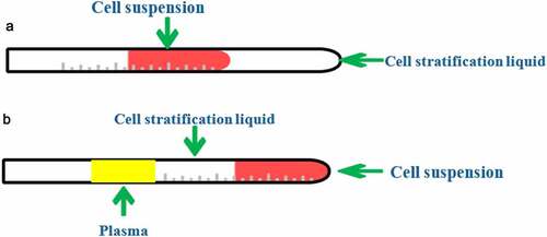
Cell separation process was as follows [Citation16]. (i) First, 20 mL peripheral blood of the patient was collected aseptically in the morning and injected into a 50 mL anticoagulant centrifuge tube with 500 U low molecular weight heparin added. After being covered, the blood was shaken gently immediately to fully anticoagulate, and the specimen was processed within four hours. (ii) The collected peripheral blood was diluted equifold (Hank’s solution of the same volume was added) on the ultra-clean workbench. (iii) 15 mL of lymphocyte separation solution was placed in a 50 mL centrifuge tube, and the diluted blood was slowly added to the stratified solution along the wall of the test tube at 1 cm from the interface of the stratified solution. (iv) The centrifuge tube was put into a horizontal centrifuge, centrifuged at 25 degrees Celsius for half an hour (2,500 r/min). After centrifugation, the tube was divided into four layers, namely the upper layer of plasma, blood diluent, and most of the platelets. The lowest layer was composed of erythrocytes and granulocytes, and the middle layer was composed of cellular stratified fluid. The cloudy gray layer at the junction between the stratified fluid and plasma was the mononuclear cell layer (). (v) The upper plasma and blood diluent were sucked out with a sterile pipette and discarded. Then, the mononuclear cell layer was carefully absorbed along the tube wall and moved into another sterile centrifuge tube. It was important to absorb all mononuclear cells, while avoiding excessive absorption of stratified fluid and plasma to avoid mixing with other cellular components. (vi) The mononuclear cell suspension was washed twice with Hank’s solution and centrifuged at 2,000 r/min and 1,500 r/min, respectively, at 25°C for 10 minutes to remove the mixed platelet and lymphocyte separation solution.
Figure 1. Display of the layering results of the cytocentrifuge tube. (a) the cell layering before centrifugation; (b) the cell layering after centrifugation).
Cell culture process was as follows [Citation17]. (i) The supernatant was absorbed with a pipette, and the cells were suspended in the culture medium containing 10% bovine serum (Roswell Park Memorial Institute). The mononuclear cells were suspended at a density of 2–3 × 10⁶/mL in a 6-well plate coated with 2ug/ mL fibronectin. (ii) 2 mL RPMI-1640 (Roswell Park Memorial Institute) medium containing 10% fetal bovine serum, 10 ng/mL vascular endothelial growth factor (VEGF)1, and 20 ng/mL basic fibroblast growth factor (BFGF) were added to each well, and cultured in an incubator with saturated humidity and 5% CO2 and 37°C. (iii) On day four and day seven, the medium was replaced and VEGF and BFGF were added again. According to the purpose of the experiment, the cells were collected at different time points for experimental application.
2.4 Cell analysis experiments
I. Observation of cell morphology. The cells at the 1st, 3rd, 5th, 7th, and 11th days of culture were taken and observed under an inverted microscope. The morphological changes of cells were recorded.
II. Cell staining and identification. i) The cells cultured for eight days were added with 2.5 mL DiL-labeled acetyl low density lipoprotein (DiL-acLDL) and incubated in 5% CO2 at 37°C for twelve hours. ii) The cells were fixed with 2% paraformaldehyde for half an hour, soaked with phosphate buffer (PBS) twice, then added with 10 μg/mL FITC-labeled Vitex agglutinin I (FITC-UEA-I), and cultured for another one hour. iii) After the cells were taken out, they were washed once with PBS, and then continuously washed three times, each time for five minutes. Finally, the PBS was washed off with running water and dried for inspection. iv) A drop of 0.03 mol/L glycerin was added to the specimen, identified with fluorescence microscope, and the cells of each hole were counted under microscope.
III. Cell proliferation activity. i) Cells cultured for eight days were washed in Hank’s solution, and 0.5% trypsin + ethylenediamine tetra-acetic acid (EDTA) was added. ii) Cells were inoculated in 96-well plates with 4 × 10⁵ cells, 200 μL cell suspension was added to each well, and three multiple wells were set and cultured for seven days. iii) Cell proliferation activity was detected at day 0, 3, 5, and 7. iv) Then, 20 μL of (3-(4,5-dimethylthiazol-2-yl)-2,5-diphenyltetrazolium bromide) tetrazolium (MTT) was added to each well, cultured for four hours under the same conditions. After the supernatant was discarded, 150 μL of dimethylsulfoxide was added in these wells one by one, shaken for ten minutes. v) The absorbance at 490 nm was detected by colorimetry, and the cell survival curve was plotted.
IV. Cell adhesion ability. i) 0.25% trypsin was added into EPCs, and a little serum-containing medium was added after 2–3 min digestion to stop digestion. ii) The straw absorbed the culture medium in the hole of the culture plate, and blew the adherent cells repeatedly. The blowing should be carried out in order, the action should be gentle, and the foam should not appear as far as possible. Cell suspension was formed after cell separation from the wall. iii) The cell suspension was collected in a centrifuge tube and centrifuged at 800–1,000 r/min for 3–5 min. The supernatant was discarded, and the culture medium was added for cell counting. iv) The same amount of EPCs were spread on the culture plate coated with human fibronectin and cultured at 37°C for 30 min. v) Three high magnification fields (×200) were randomly selected under an inverted microscope to count adherents, and the average was taken. The adhesion ability of adherent cells with more adherent cells was stronger.
V. Cell migration. i) 0.25% trypsin was added into EPCs, and a little serum-containing medium was added after 2–3 min digestion to stop digestion. ii) Bend pipepipe absorbed the culture medium in the bottle and repeatedly blew adherent cells, blowing should be carried out in order, the action should be gentle as far as possible not to appear foam, and cells detached from the bottle wall to form a cell suspension. iii) The cell suspension was collected in a centrifuge tube and centrifuged at 800–1,000 r/min for 3–5 min. The supernatant was discarded, and the culture medium was added for cell counting. iv) The lower chamber of the modified Boyden chamber was added with 20 μL of medium and 50 ng/mL of VEGF, and then the upper chamber of the chemochemoic chamber was covered with a filter membrane (with the bright side down of the filter membrane). v) 2 × 10⁴ EPCs were placed in 50 μL culture medium and injected into the upper chamber. Glass slides were placed on the cover to cover all holes. Bubbles could not be produced during the operation, for bubbles would hinder the movement of cells. vi) The cells were cultured at 37°C in 5% CO2 for 24 h. The unmoved cells were scraped off the filter membrane, fixed with methanol for 3–5 min, stained with Giemsa for 15–30 min, rinsed with distilled water, and examined under microscope after drying. Three high-power visual fields (×200) were randomly selected to count the cells migrating to the bottom, and the average was taken.
VI. Flow cytometry [Citation18]. i) After seven days of cell growth factor induction and differentiation culture, suspension cells were sucked out. After the cells were washed with Hank’s solution, the attached cells were digested with 0.5% pancreatin for 10 minutes, and the digestion was terminated with RPMI-1640 medium supplemented with 10% calf serum. ii) The cells were centrifuged at 1,000rpm for five minutes, then the cell density was adjusted to 1 × 10⁶ with 1640 medium, and the cells were fixed with 4% paraformaldehyde for 40 minutes. iii) After centrifugation at 1,000 rpm for 5 minutes, cells were washed with PBS, and then centrifuged at 1,000 rpm for 5 minutes and resuspend in Kang tube A and B, respectively. Tube A was added with 10 μL FITC-labeled CD34 mouse anti-human monoclonal IgG1 antibody (10 μL), PE-labeled CD133 mouse anti-human monoclonal IgG1 antibody (10 μL), and FITC-labeled KDR mouse anti-human monoclonal IgG1 antibody (10 μL). Tube B was added with FITC-labeled mouse IgG1 antibody (10 μL), PE-labeled mouse IgG1 antibody (10 μL), and FITC-labeled mouse IgG1 antibody (10 μL), as isotype controls. After mixing, they were incubated at 2–8°C for 30 minutes in the dark or 15 minutes at room temperature. Then, 500 μL of fixative was added to each tube, which were mixed and incubated in the dark for ten minutes. iv) PBS was added into the tubes, mixed well, centrifuged at 2,000rpm for five minutes, and the unbound antibodies were washed off. The supernatant was discarded, and the cells were resuspended in PBS. 1% paraformaldehyde was added to fix the cells, which were then sent to flow cytometry for detection.
2.5 Observation indexes
Clinical data of the patients were recorded, including basic data (age and ratio of male to female), cardiac function (systolic blood pressure, diastolic blood pressure, and left ventricular ejection fraction), medical history (hypertension, diabetes, and hyperlipidemia), and medication (aspirin, clopidogrel, and calcium antagonist). The echocardiographic imaging data of the patients were collected.
2.6 Statistical Methods
SPSS 19.0 was employed for data statistics and analysis. Mean ± standard deviation (͞x ± s) was how measurement data were expressed, and percentage (%) was how count data were expressed. The pairwise comparison was performed by one factor analysis of variance. The difference was statistically considerable with P < 0.05.
3. Results
This study aimed to analyze the influence mechanism of isosorbide dinitrate combined with exercise training and rehabilitation on EPCs in patients with coronary heart disease. It was assumed that endothelial progenitor cells may be involved in the treatment of coronary heart disease. A total of 116 patients were divided into treatment group (isosorbide dinitrate combined with exercise rehabilitation training) and control group (isosorbide dinitrate alone). Then, EPCs were isolated and cultured from peripheral blood of patients by density gradient method. The morphology, surface markers, and activity of EPCs were detected by flow cytometry, so as to comprehensively evaluate the specific participation mechanism of EPCs in the treatment of coronary heart disease. It was hoped to provide reference for clinical drug treatment and rehabilitation training treatment of coronary heart disease.
3.1 Cell observation results
3.1.1. Observation of cell morphology
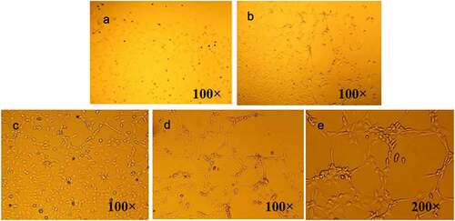
In below, the mononuclear cells preliminarily isolated were round and small in volume and not evenly distributed in the culture plate. After three days of culture, EPCs cells became larger in volume, increased in transparency, and spindle cells began to appear. After five days of culture, EPCs cells increased in number, and small rod cells began to appear. After eight days of culture, EPCs cells began to grow like colonies, and circular cells were located in the center of the cell mass. Adherant spindle cells grew outward from around the cell mass to form clones. The surrounding cells, centered on the colony, grew outward in a germinating manner, forming multiple cell groups.
Figure 2. Observation of cell morphology under microscope. (a) preliminarily isolated mononuclear cell; (b) EPCs cell after three days of culture; (c) EPCs cells cultured for five days; (d) EPCs cells cultured for eight days (100×); (e) EPCs cells cultured for eight days (200×).
3.1.2 Cell staining observation
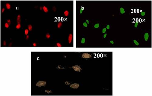
showed the results of cell fluorescence double staining. The cells stained by Dil-acLDL were red under the microscope, the cells stained by FITC-UEA-I were green under the microscope, and the double-stained cells were yellow under the microscope.
Figure 3. Cell fluorescence double staining observation. (A: Dil-acLDL staining; B: FITC-UEA-I staining; C: double staining).
3.1.3. Observation of cell surface markers
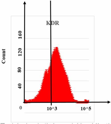
, , nd showed the detection results of EPCs surface markers CD133, CD34, and KDR, respectively. The positive rates of CD133, CD34, and KDR in cell expression were 11.25 ± 3.07%, 48.18 ± 9.13%, and 76.36 ± 8.27%, respectively. This result can well confirm that the isolated and cultured cells were EPCs.
Figure 4. Flow cytometry detection result of EPCs surface marker CD133.
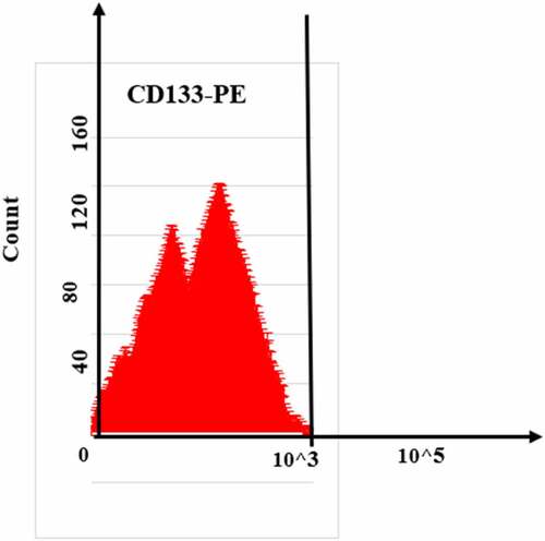
Figure 5. Flow cytometry detection result of EPCs surface marker CD34.
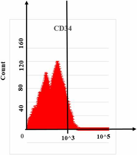
Figure 6. Flow cytometry detection result of EPCs surface marker KDR.
3.2 Comparison of basic data of the two groups of patients
showed the comparison of the basic data of the two groups of patients. For basic information of the two groups of patients (age and male to female ratio), cardiac function (systolic blood pressure (SBP) and diastolic blood pressure (DBP), left ventricular ejection fraction (LVEF)), medical history ratio (history of hypertension, diabetes, and hyperlipidemia), and medication (aspirin, clopidogrel, and calcium antagonist), the difference between the pairwise comparisons was not considerable (P > 0.05).
Figure 7. Comparison of basic data of the two groups of patients.
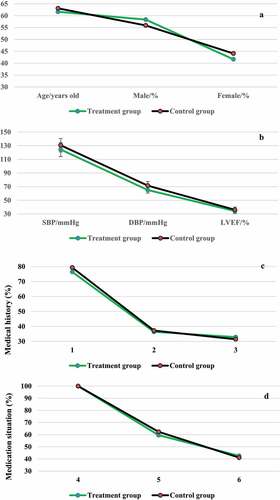
(1, 2, and 3 indicated history of hypertension, diabetes, and hyperlipidemia, respectively; 4, 5, and 6 indicated patients’ aspirin, clopidogrel, and calcium antagonist, respectively; A: the patient’s age and male to female ratio; B: the patient’s systolic blood pressure, diastolic blood pressure, and left ventricular ejection fraction; C: the patient’s medical history ratio; D: the patient’s medication status)
3.3 Echocardiographic analysis of some patients
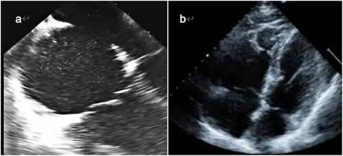
was an echocardiogram of a 25-year-old man. The patient came in with sudden chest pain, reported a heart murmur since childhood. Echocardiography showed a large atrial septal defect with bidirectional shunt between the right and left atria. was an echocardiogram of a 40-year-old female patient with clinically suspected right ventricular dysplasia and sudden death early in life in the family. The cardiogram showed enlargement of the right atrium and right ventricular lumen, right ventricle flowing into the tract muscles and apical trabeculae of the apex, and thin right ventricular wall.
Figure 8. Partial echocardiographic analysis of patients. (A was a sample of a 25-year-old male patient; B was a sample of a 40-year-old female patient).
3.4 EPCs cell proliferation activity
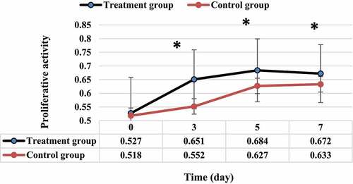
In below, the EPCs cell proliferation activity of the two groups of patients showed a trend that first increased and then stabilized with the increase of treatment time. EPCs cell proliferation activity of the treatment group was dramatically greater than that of the control group at three, five, and seven days, and the difference was considerable (P < 0.05).
Figure 9. Comparison of cell proliferation activity between the two groups of EPCs.
3.5 EPCs cell adhesion ability
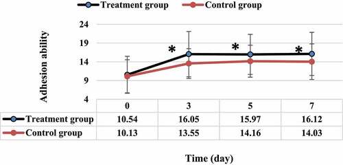
In below, the EPCs cell adhesion ability of the two groups of patients increased first and then stabilized with the increase of treatment time. The EPCs cell adhesion ability of the treatment group was dramatically greater than that of the control group at three, five, and seven days, and the difference was considerable (P < 0.05).
Figure 10. Comparison of cell adhesion ability between the two groups of EPCs.
3.6 EPCs cell migration ability
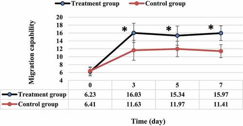
In below, the EPCs cell migration ability of the two groups of patients increased first and then stabilized with the increase of treatment time. The EPCs cell migration ability of the treatment group was dramatically greater than that of the control group at three, five, and seven days, and the difference was considerable (P < 0.05).
Figure 11. Comparison of cell migration ability between the two groups of EPCs.
4. Discussion
EPCs, as the precursor cells of vascular endothelium, are often called vascular cells. Studies revealed that EPCs are not only found in peripheral blood, but also in cord blood and bone marrow. Under the action of certain stimuli, EPCs were released into the blood, but no specific markers of EPCs have been found. It can only be inferred that a certain type of isolated and cultured cells can differentiate into mature vascular endothelial cells and have the function of vascular endothelial cells. Therefore, in this work, the gradient density method was innovatively used to separate EPCs from the peripheral blood of patients with coronary heart disease after treatment, and the information of cell morphology and surface markers was detected. It was found that the preliminarily separated mononuclear cells were round, relatively small in size, and unevenly distributed on the culture plate. EPCs cells cultured for about eight days began to grow in colony-like cell clusters. The surrounding cells took the colony as the center and grew outward in a sprouting manner, forming multiple cell clusters. This manifestation is consistent with the research of many domestic and foreign scholars, and it was preliminarily confirmed that the cells isolated and cultured in this work were EPCs [Citation19]. From the results of double fluorescent staining, the cells stained by Dil-acLDL were red under the microscope, and the cells stained by FITC-UEA-I were green under the microscope. The double-stained cells showed yellow under the microscope. Further flow cytometry showed that the positive rates of EPCs surface markers CD133, CD34, and KDR in cell expression were 11.25 ± 3.07%, 48.18 ± 9.13%, and 76.36 ± 8.27%, respectively. This was similar to the research results of Sun et al. (2020) [Citation20], indicating that the cells isolated and cultured in this study were EPCs. From the above results, under certain conditions, such as through the action of growth factors, the mononuclear cells extracted from peripheral blood by density gradient centrifugation not only promoted the differentiation of EPCs, but also strengthened the proliferation ability of EPCs and increased the number of EPCs.
The proliferation activity of EPCs cells isolated and cultured in the two groups were compared, and the proliferation activity of EPCs cells in the two groups showed a trend of first increasing and then stabilizing with the increase of treatment time. The proliferation activity of EPCs cells in the treatment group was dramatically higher than that in the control group at day three, five, and seven, and the difference was considerable (P < 0.05). Cell proliferation is one of the important physiological functions of living cells, an important life characteristic of organisms, and the basis of organism growth, development, reproduction, and genetics, which can effectively reflect the growth state and activity of cells [Citation21]. The result shows that isosorbide dinitrate combined with exercise rehabilitation training can promote the proliferation activity of EPCs cells in patients with coronary heart disease better than single drug [Citation22]. The adhesion ability of EPCs cells in both groups was firstly increased and then stabilized with the increase of treatment time. The adhesion ability of EPCs cells in the treatment group was dramatically higher than that in the control group at three, five, and seven days, and the difference was considerable (P < 0.05), which indicated that isosorbide dinitrate combined with exercise rehabilitation training had a greater impact on the adhesion ability of EPCs cells than single drug [Citation23]. In addition, the migration ability of EPCs cells in both groups was firstly increased and then stabilized with the increase of treatment time. The migration ability of EPCs cells in the treatment group was dramatically higher than that in the control group at three, five, and seven days (P < 0.05). These results indicated that isosorbide dinitrate combined with exercise rehabilitation training can dramatically enhance the migration ability of EPCs cells, and the effect was better than that of single drug [Citation24]. In addition, this study is biased in the selection of patient samples. The sample size is small, and the number of male patients is significantly higher than that of females, which need to be adjusted in subsequent studies.
5. Conclusion
In this research, EPCs were firstly isolated and cultured from peripheral blood of patients with coronary heart disease, and their morphology and surface markers were detected. Then, 116 patients were divided into treatment group (isosorbide dinitrate combined with exercise rehabilitation training) and control group (single drug isosorbide dinitrate), and the characteristics of EPCs cells in the two groups after treatment were compared. The results showed that isosorbide dinitrate combined with exercise rehabilitation training could dramatically enhance the proliferation activity, adhesion ability, and migration ability of EPCs cells. In addition, it also played a beneficial role in the repair of endothelial injury, and the effect was obviously better than that of single drug. However, there are still some problems to be improved in this study, such as a small number of patient samples were selected, and the method of isolation and culture of peripheral blood EPCs cells was rough. Subsequent large sample and multicenter analysis is necessary. In conclusion, the results of this study provide a scientific theoretical basis for the treatment of EPCs in patients with coronary heart disease.
Acknowledgements
Ruozhu Dai, Study on the mechanism of mobilization of endothelial progenitor cells in patients with coronary heart disease by isosudine combined with exercise training rehabilitation (Project No.: 2018J01202)
Disclosure statement
No potential conflict of interest was reported by the author(s).
Additional information
Funding
References
- Zhu Y, Yang T, Duan J, et al. MALAT1/miR-15b-5p/MAPK1 mediates endothelial progenitor cells autophagy and affects coronary atherosclerotic heart disease via mTOR signaling pathway. Aging (Albany NY). 2019Feb21;11(4):1089–1109. PMID: 30787203; PMCID: PMC6402525.
- Yubero-Serrano EM, Fernandez-Gandara C, Garcia-Rios A, et al. Mediterranean diet and endothelial function in patients with coronary heart disease: an analysis of the CORDIOPREV randomized controlled trial. PLoS Med. 2020Sep9;17(9):e1003282. PMID: 32903262; PMCID: PMC7480872.
- Madonna R, Renna FV, Lanuti P, et al. The acute impact of high-dose lipid-lowering treatment on endothelial progenitor cells in patients with coronary artery disease-The REMEDY-EPC early substudy. PLoS One. 2017Apr10;12(4):e0172800. PMID: 28394933; PMCID: PMC5386268.
- Hammer Y, Soudry A, Levi A, et al. Effect of vitamin D on endothelial progenitor cells function. PLoS One. 2017May17;12(5):e0178057. PMID: 28545072; PMCID: PMC5435351.
- Zhou H, Tu Q, Zhang Y, et al. Shear stress improves the endothelial progenitor cell function via the CXCR7/ERK pathway axis in the coronary artery disease cases. BMC Cardiovasc Disord. 2020Sep7;20(1):403. PMID: 32894067; PMCID: PMC7487552.
- Cavalcante SL, Lopes S, Bohn L, et al., Effects of exercise on endothelial progenitor cells in patients with cardiovascular disease: a systematic review and meta-analysis of randomized controlled trials. Rev Port Cardiol (Engl Ed). 2019 Nov;38(11):817–827. English, Portuguese. Epub 2020 Feb 7. PMID: 32037059
- Pelliccia F, Pasceri V, Moretti A, et al. Endothelial progenitor cells predict long-term outcome in patients with coronary artery disease: ten-year follow-up of the PROCREATION extended study. Int J Cardiol. 2020Nov1;318:123–125. Epub 2020 Jun 6. PMID: 32522679.
- Sheng ZQ, Li YF, Zheng KL, et al. The relationship between number and function of EPCs and concentration of VEGF165 and SDF-1 in coronary artery spasm. Eur Rev Med Pharmacol Sci. 2018May;22(9):2767–2777. PMID: 29771429.
- Poh KK, Lee PSS, Djohan AH, et al. Transplantation of endothelial progenitor cells in obese diabetic rats following myocardial infarction: role of thymosin Beta-4. Cells. 2020Apr12;9(4):949. PMID: 32290541; PMCID: PMC7226991.
- Zhou E, Zou Y, Mao C, et al. MicroRNA-221 inhibits the transition of endothelial progenitor cells to mesenchymal cells via the PTEN/FoxO3a signaling pathway. Adv Clin Exp Med. 2021 Oct 5. Epub ahead of print. PMID: 34610220. 10.17219/acem/141446
- Li YP, Fan ZX, Gao J, et al. Influencing factors of vascular endothelial function in patients with non-obstructive coronary atherosclerosis: a 1-year observational study. BMC Cardiovasc Disord. 2020Jan30;20(1):40. PMID: 32000667; PMCID: PMC6993456.
- Zhang S, Chen L, Zhou Z, et al. Effects of puerarin on clinical parameters, vascular endothelial function, and inflammatory factors in patients with coronary artery disease. Med Sci Monit. 2019Jan14;25:402–408. PMID: 30636768; PMCID: PMC6342064.
- Chan YH, Ngai MC, Chen Y, et al. Cumulative rheumatic inflammation modulates the bone-vascular axis and risk of coronary calcification. J Am Heart Assoc. 2019Jun4;8(11):e011540. PMID: 31130038; PMCID: PMC6585350.
- Morrone D, Felice F, Scatena C, et al. Role of circulating endothelial progenitor cells in the reparative mechanisms of stable ischemic myocardium. Int J Cardiol. 2018Apr15;257:243–246. Epub 2017 Sep 14. PMID: 28918896.
- Neumüller J, Neumüller-Guber SE, Lipovac M, et al. Immunological and ultrastructural characterization of endothelial cell cultures differentiated from human cord blood derived endothelial progenitor cells. Histochem Cell Biol. 2006Dec;126(6):649–664. Epub 2006 Jun 10. PMID: 16767408.
- Hu Z, Wang H, Fan G, et al. Danhong injection mobilizes endothelial progenitor cells to repair vascular endothelium injury via upregulating the expression of Akt, eNOS and MMP-9. Phytomedicine. 2019Aug; 61: 152850 Epub 2019 Jan 29. PMID: 31035054
- Lam YT, Hsu CJ, Simpson PJL, et al. Androgens stimulate EPC-mediated neovascularization and are associated with increased coronary collateralization. Endocrinology. 2020May1;161(5):bqaa043. PMID: 32157309.
- Denollet J, van Felius RA, Lodder P, et al. Predictive value of Type D personality for impaired endothelial function in patients with coronary artery disease. Int J Cardiol. 2018May15;259:205–210. Epub 2018 Feb 20. PMID: 29477262.
- Ghem C, Dias LD, Sant’Anna RT, et al. Combined analysis of endothelial, hematopoietic, and mesenchymal stem cell compartments shows simultaneous but independent effects of age and heart disease. Stem Cells Int. 2017;2017:5237634. Epub 2017 Jul 27. PMID: 28819363; PMCID: PMC5551513.
- Sun L, Zhang Y, Zhang J, et al. Atorvastatin improves the proliferation and migration of endothelial progenitor cells via the miR-221/VEGFA axis. Biosci Rep. 2020Nov27;40(11):BSR20193053. PMID: 32936287; PMCID: PMC7689653.
- Suárez-Cuenca JA, Robledo-Nolasco R, Alcántara-Meléndez MA, et al. Coronary circulating mononuclear progenitor cells and soluble biomarkers in the cardiovascular prognosis after coronary angioplasty. J Cell Mol Med. 2019Jul;23(7):4844–4849. Epub 2019 May 8. PMID: 31069956; PMCID: PMC6584722.
- Yi M, Wu Y, Long J, et al. Exosomes secreted from osteocalcin-overexpressing endothelial progenitor cells promote endothelial cell angiogenesis. Am J Physiol Cell Physiol. 2019Nov1;317(5):C932–C941. Epub 2019 Aug 14. PMID: 31411920.
- Wang H, Huang H, Ding SF. Sphingosine-1-phosphate promotes the proliferation and attenuates apoptosis of endothelial progenitor cells via S1PR1/S1PR3/PI3K/Akt pathway. Cell Biol Int. 2018Nov;42(11):1492–1502. Epub 2018 Jul 8. PMID: 29790626.
- Kawamura M, Paulsen MJ, Goldstone AB, et al. Tissue-engineered smooth muscle cell and endothelial progenitor cell bi-level cell sheets prevent progression of cardiac dysfunction, microvascular dysfunction, and interstitial fibrosis in a rodent model of type 1 diabetes-induced cardiomyopathy. Cardiovasc Diabetol. 2017Nov2;16(1):142. PMID: 29096622; PMCID: PMC5668999.