
Abstract
Objective: Primary lateral sclerosis (PLS) is a rare form of motor neuron disease characterised by UMN degeneration leading to slowly progressive spasticity. Whether it is a separate disease or a subtype of ALS has been debated. In ALS comorbid frontotemporal dementia (FTD) is frequently seen (±15%). However, cognitive and behavioural changes are generally not considered to be a part of PLS. Methods: To report the clinical findings and frequency of PLS patients that developed FTD in a referral-based cohort and provide an overview of the literature. Results: In our cohort six out of 181 (3.3%) PLS patients developed FTD. In the literature a few cases of PLS with FTD have been reported and only a limited number of small studies have investigated cognition in PLS. However, when these studies are summarised a pattern emerges with FTD diagnoses in ±2% and frontotemporal impairment in 22% of patients. Conclusions: These findings suggest that PLS is part of the FTD-MND continuum and would favour viewing it as a subtype of ALS. It is, however, not a restricted (isolated UMN involvement) phenotype.
Introduction
Primary lateral sclerosis (PLS) is a rare form of motor neuron disease (MND), characterised by the degeneration of upper motor neurons (UMN) leading to slow progressive spasticity. Whether it is a separate disease or a subtype of ALS has been debated. In ALS comorbid frontotemporal dementia (FTD) is frequently seen (±15%) (Citation1) and up to 50% show signs of frontotemporal dysfunction without fulfilling formal diagnostic criteria (Citation2). Parkinsonian features, sensory abnormalities and mild frontal lobe dysfunction have been reported in ±10% of PLS patients (termed PLS-plus) (Citation3), but frank (frontotemporal) dementia is not considered as part of PLS. Here we describe a case series identified through the review of electronic medical records of PLS patients that subsequently developed FTD, report the approximate frequency of PLS-FTD in a referral-based cohort and provide an overview of the literature.
Material and methods
Retrospective case series
The Academic Medical Centre (AMC Amsterdam) and University Medical Centre Utrecht (UMCU) form the national referral centre for MND and together we see 80% of MND patients in the Netherlands. Patients were seen between 2006 and 2016 at outpatient clinics and diagnosed with PLS by neurologists with extensive experience of MND. Patients were considered to have PLS when they fulfilled the diagnostic criteria for clinically pure PLS or PLS-plus according to the diagnostic criteria proposed by Gordon et al. (Citation4), which in brief require evident and progressive upper motor neuron signs without focal muscle atrophy or visible fasciculations and no denervation on EMG four years from onset. The age at onset must be >40 years. Secondary and mimicking conditions must have been excluded, which in our cases was done through an extensive work-up (MRI imaging of the neuraxis, laboratory investigations (including metabolic disorders), serology (HIV, syphilis, Borrelia and HTLV1 and -2), CSF (if appropriate), genetic testing for hereditary spastic paraplegias (HSPs) and/or ALS and needle EMGs (to exclude LMN involvement)). Particular emphasis is placed on ruling out (Citation1) UMN-predominant ALS by repeating EMGs, (Citation2) HSP by taking detailed family histories, genetic testing and following patients over time (Citation5,Citation6) and (Citation3) progressive supranuclear palsy; vertical gaze palsy, frequent falling within first year, proximal more than distal symmetrical akinesia or rigidity, abnormal neck posture (retrocollis) and early onset of cognitive impairment (Citation7–9).
All patients suspected of having PLS were followed at one of our centres until a disease duration of four years (longer in the vast majority of cases), after which patients were referred to the nearest multidisciplinary specialised MND care team, which in some cases was not one of our centres.
During follow-up some patients were referred back because they had developed severe behavioural and/or cognitive changes. We had also observed this in some patients that had remained in our own care. In all of these patients we performed an additional work-up, which included imaging, laboratory investigations, serology, neuropsychological assessment, genetic testing and EMG. In all cases, it was the opinion of the treating physician that the behavioural/cognitive changes could be attributed to FTD.
We considered the observation that multiple patients had appeared to have developed FTD intriguing, as frank FTD is not listed in any of the proposed diagnostic criteria and multiple studies even report cognition and behaviour to be unaffected in PLS. We therefore retrospectively reviewed the electronic medical records of our patients with the objective of identifying all PLS patients that developed FTD and re-evaluated these cases against the current diagnostic criteria for bvFTD and primary progressive aphasia (PPA) (Citation10,Citation11). Review of medical records was carried out with the approval of the relevant medical ethics committees.
Literature overview
After having identified multiple cases of PLS with FTD, we performed a search of the literature () with the following objectives:
Providing an approximate frequency of published PLS cases with concomitant FTD or cognitive/behavioural changes. Based on the available data in the publications we evaluated the reported patients against current criteria for bvFTD or PPA (Citation10,Citation11) or classified them as PLS with cognitive impairment (ci), behavioural impairment (bi) or without cognitive impairment according to the Strong criteria (Citation12).
Characterizing the cognitive profile of PLS by listing the affected cognitive domains as reported as well as by performing a meta-analysis. To be included in the meta-analysis studies needed to have a case-control design (matched for age and education or by applying age- and education-corrected standard scores) and report data from at least one validated neuropsychological test as mean and SD or raw or standardised test scores for both cases and controls. Studies had to report on unique cohorts and in the case of overlap the largest study was included. Data were categorised in 10 cognitive domains (ACE-R was included as a global measure of cognition), as described before (Citation13), and extracted by two authors (BSdV, LMMR) in order to reduce extraction errors. Using Review Manager 5.3 we calculated the effect size of each cognitive domain per study expressed as Hedges’ g. When multiple neuropsychological tests for one cognitive domain were used in a study, we calculated an average effect size so that each study only added one effect size to the final analysis. A random effects model was applied to obtain an average weighted effect size across the studies.
Summarizing imaging and neuropathology findings in PLS patients with comorbid dementia.
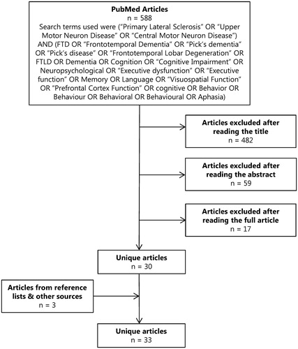
Figure 1. Summary of the results from our literature search. The search was performed using MEDLINE (via PubMed) on articles in English from July 1963 to January 2017 reporting on cognition and/or behaviour in PLS. Titles and abstracts were screened and relevant full text articles were retrieved. References were screened for additional studies and publications were also identified from the authors’ literature collections. When overlap between studies was expected, corresponding authors were contacted for further details. Since the inclusion of the search term “PLS” led to a high amount of irrelevant hits (e.g. partial least squares, post ligation syndrome, parathyroid lesions, etc.) it was not included in our list of search terms. We included studies on PLS in which dementia, cognition or behaviour was mentioned.
Results
Retrospective case series
Between 2006 and 2016, 181 patients were diagnosed with PLS. Interrogation of their medical records showed that concomitant FTD had been diagnosed in six (3.3%). All of these patients had developed severe behavioural changes or language deficits after years of having progressive, but isolated UMN signs. As frank FTD is not generally considered to be a feature of PLS an additional work-up was performed to rule out other causes (such as comorbid Alzheimer’s disease, delirium, metabolic disorders, mass lesions, etc.) through neuropsychological evaluation (behaviour, cognition), MRI imaging and laboratory investigations.
In five patients severe and progressive deterioration of behaviour (apathy, binge eating, loss of decorum, perseverative/stereotypical behaviour, diminished social interest or personal warmth) was the most prominent feature (bvFTD). In one patient progressive language deficits were the most prominent feature with impairment in confrontation naming, word comprehension, object recognition and emotion perception and empathy, while memory and visual spatial functions remained relatively spared. This patient was considered to have semantic dementia.
Repeated imaging in four out of these six cases showed progressive (asymmetrical) frontal and/or temporal atrophy (). A summary of the clinical characteristics and full case descriptions are provided in and Supplementary material.
Figure 2. The initial axial T2 weighted MRI (A) showed left temporal lobe atrophy. The second scan made 2 years later (B) demonstrated bilateral temporal lobe and hippocampus atrophy (axial and coronal T2 weighted MRI), but more severe on the left side with a knife blade aspect of the temporal gyri while the general cerebral atrophy was limited (far right: sagittal T1 weighted MRI).
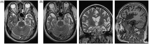
Table 1. Clinical characteristics of PLS-FTD patients.
Because we considered these cases unusual, we also re-evaluated the initial PLS diagnosis. None of the cases showed clinical or electrophysiological evidence of LMN involvement at the moment of re-evaluation (four to nine years after onset) excluding conversion to ALS-FTD. Moreover, genetic testing for C9orf72 was negative. In the lower limb-onset patients we considered (complex) HSP to be highly unlikely because genetic testing was negative, onset was late, asymmetrical and all of them had developed bulbar involvement within a relatively short time-frame. Saccadic eye movements, tremor or slight cogwheel rigidity was seen in three patients, but Parkinsonism was not a prominent feature in any of the cases and none had vertical gaze palsy. We therefore also considered PSP or other forms Parkinsonism to be unlikely.
We did not detect evidence for another non-degenerative nervous system, medical or psychiatric disorder as cause for the observed cognitive and behavioural changes. Findings on neuropsychological examination were not considered to be consistent with Alzheimer’s disease or any other type of dementia. Cases 1 and 3– 6 fulfil the current criteria for bvFTD while case 2 meets criteria for semantic dementia (Citation10,Citation11).
Overview of the literature
We found 33 publications on cognition/behaviour in PLS reporting on a total of 307 patients, of which seven had concomitant FTD (2.3%). In 68 cases cognitive or behavioural changes were present, but which did not meet criteria for FTD (22.1%) ().
Table 2. Studies on PLS patients that interrogated cognition and/or behaviour.
Cognitive profile: We identified a total of 19 studies in which some form of neuropsychological evaluation was performed in PLS patients. There were considerable differences with regard to the test batteries used, which in some studies was limited to simple screening instruments (e.g. MMSE), whereas others performed full neuropsychological examinations. Executive dysfunction was the most commonly observed impairment (reported in 12 out of 19 studies). Language deficits, memory impairment and behavioural change were also observed. The frequency of cognitive deficits was highest in studies that administered a full neuropsychological examination (Citation14–23), which may suggest under-reporting in studies that only applied simple screening instruments ().
A total of four studies fulfilled the inclusion criteria for the meta-analysis encompassing 10 cognitive domains. Most cognitive domains were only assessed in a single study and the maximum number of studies per cognitive domain was 2. Large effect sizes (Hedges’ g > 0.8) were observed for executive function, delayed verbal memory, psychomotor speed and fluency. A large effect was also seen for the ACE-R, included as a global measure of cognition. The other domains did not demonstrate any significant effects. It must be noted that there is considerable heterogeneity and therefore these results should be interpreted with caution ().
Table 3. Pooled weighted effect sizes and heterogeneity statistics of cognitive domains.
Imaging: Diagnostic criteria for PLS include that brain imaging should be without structural abnormalities, although atrophy of the precentral gyrus is allowed (Citation24). The corticospinal tracts may also appear hyper-intense on T2-weighted images, thought to be reflective of Wallerian degeneration. More advanced imaging techniques also demonstrate abnormalities of motor cortex and the corticospinal tracts. For instance, 18FDG-PET imaging may show a stripe of hypometabolism in the precentral gyrus (Citation25) and diffusion tensor imaging (DTI) studies show reduced fractional anisotropy within the corticospinal tracts and mid-body of the corpus callosum (Citation15,Citation26,Citation27). The number of imaging studies that also included PLS patients with cognitive disturbances is unfortunately limited (). Interestingly, these studies show that there may be more widespread involvement of the brain in PLS. In particular, DTI studies demonstrate white matter tract damage may be quite extensive affecting the basal ganglia, corpus callosum, frontal and temporal lobes (Citation15,Citation27). SPECT, CT-perfusion and fMRI studies confirm involvement of these non-motor regions (Citation14,Citation18,Citation21). PLS patients with cognitive impairment predominantly had executive dysfunction and in some studies the severity of cognitive dysfunction correlated with the extent of frontal involvement (Citation14,Citation15). Memory impairment and/or hippocampal atrophy was not reported in these studies.
Table 4. Overview of neuroimaging findings in PLS.
Neuropathology: In pure PLS there is prominent loss of pyramidal neurons, often complete absence of Betz cells, in the precentral gyrus with severe degeneration of the corticospinal tract, but with preservation of the motor nuclei in the brainstem and anterior horn cells in the spinal cord. Neurofibrillary tangles, senile plaques and Lewy bodies are absent and the observed protein aggregates are ubiquitin-positive, but tau-negative. As most of these studies predated the discovery of TDP-43 as the main component of aggregates within the MND-FTD spectrum, it is unclear whether these aggregates stain for TDP-43. However, Kosaka et al. report TDP-43-positive pathology in the motor cortex, frontal and temporal lobe in a PLS patient with frontotemporal lobar degeneration (Citation28).
In the autopsies of PLS patients with comorbid dementia the hippocampus appears to be unaffected. The most severe pathology outside of the motor system is seen in the frontal and temporal regions (gliosis, white matter damage) which may be accompanied by neuronal loss (Citation28–31). To a lesser degree atrophy of the basal ganglia (thalamus and striatum, but not substantia nigra) may also be seen (Citation28–31). Again, without evidence for tauopathies, including AD or α–synucleinopathies ().
Table 5. Overview of neuropathological findings in PLS.
The only exception to these findings is the case reported by Engel et al. where there is only mild loss of neurons in the precentral gyrus that is not considered to be disproportionate to neuronal loss elsewhere in the brain (Citation22). It appears UMN signs resulted from a prominent loss of myelinated axons in the spinal cord, which would be more consistent with HSP than PLS. The authors acknowledge this, but favour a diagnosis of PLS based on the late and asymmetrical onset combined with a negative family history. Similarly, the authors struggle to classify the cognitive changes in this patient. Clinically it was considered to be a frontal syndrome, but neuropathology shows amyloid-positive diffuse plaques and neurofibrillary tangles, which would suggest Alzheimer’s disease. The most prominent pathology is, however, not seen in the hippocampus region, but rather in the orbitofrontal cortex and they even consider the possibility that the observed amyloid plaques could be attributed to normal aging. After consideration of all these observations, the authors consider this a disorder within the PLS-HSP spectrum with neuropathological features of AD. In this case there is no definitive clinical, genetic or pathological diagnosis and it is not clear whether we should consider this as PLS with dementia or perhaps HSP with comorbid AD.
Discussion
Studying PLS is challenging for many reasons. First it is a very rare disorder (2–5% of patients in adult neuromuscular clinics). Secondly, there is no diagnostic test that definitively demonstrates PLS, and this is further complicated by the fact that different sets of diagnostic criteria have been proposed. Knowledge of the disease is therefore based on small and possibly heterogeneous cohorts. Despite these issues, PLS is a relatively well-defined clinical syndrome characterised by the progressive loss of UMNs. In over half of cases, there is an insidious onset of symptoms in the legs, which slowly and relatively symmetrically ascends to the cervical and bulbar regions. In the remaining patients the disease is characterised by prominent bulbar symptoms with a patchier pattern of progression. Perhaps the most relevant debate with regard to PLS is whether it is a subtype of ALS. The most recent version of the El Escorial criteria considers PLS to be a restricted phenotype of ALS, meaning it is characterised by ‘UMN deficits existing in isolation’ (Citation32). Keeping it as a subcategory was, however, considered desirable as patients with clinically pure UMN dysfunction have a more benign prognosis (survival >10 years to normal life expectancy). Seemingly contradicting the view of PLS as a pure UMN disorder is the fact that Parkinsonian features, sensory abnormalities and mild frontal lobe dysfunction are seen in ±10% of patients (PLS-plus) (Citation4,Citation20,Citation33). This suggests more widespread involvement of the nervous system, at least in some cases.
Here, we report a case series of PLS patients that went on to develop FTD over the course of the disease. We considered this to be an intriguing finding, given that the commonly used Pringle criteria list cognition and behaviour to be normal in PLS and the Gordon criteria allow mild frontal impairment, but none lists frank FTD (or dementia) as a disease feature (Citation4,Citation26).
We therefore aimed to provide an estimate of the frequency of dementia as well as to classify the type of dementia observed in PLS. Based on our own retrospective case series and the literature; we found that comorbid dementia occurs in 3% of PLS patients, which is clinically considered to be FTD by most authors. Our review of the literature indicates 22% of patients show cognitive deficits within the spectrum of FTD without fulfilling the formal diagnostic criteria.
We must however emphasise that this is a rough estimate that could be an overestimation as well as underestimation for several reasons: this is a retrospective study based on medical records (potentially incomplete, non-standardised follow-up, varying disease durations), referral bias (leading to more unusual cases presenting at academic centres) and publication bias (lack of publications on normal cognition in PLS).
In our meta-analysis on neuropsychological studies in PLS we could only include four studies and for many cognitive domains data were only available from one study (with a maximum of two studies for a single domain). The meta-analysis shows considerable heterogeneity and large confidence intervals indicating that these results should be interpreted with caution. It is, however, interesting to note that the meta-analysis shows a profile with deficits in the same domains as in the other studies (not included in the meta-analysis) when one simply lists the affected domains. Moreover, these results are also similar to the neuropsychological findings in the patients from our case series, suggesting consistent findings in the cognitive profile of PLS with deficits in executive function, delayed verbal memory, psychomotor speed and fluency.
It is of note that memory was never affected in isolation, principally or as the most prominent feature, suggesting this is not comorbid Alzheimer’s disease. This seems to be reinforced by findings from imaging and neuropathological studies. Imaging studies report abnormalities outside of the motor system, in particular in frontotemporal brain regions, characterised by atrophy, white matter tract damage, decreased connectivity, metabolism and perfusion. Neuropathological studies report atrophy of frontal and temporal lobes, white matter damage in frontal regions, with preservation of the hippocampus. Intra-neuronal ubiquitin-positive inclusions are observed, in one case also staining for TDP-43, without evidence for amyloid, tau or α-synuclein pathology. Based on these findings it is our opinion comorbid FTD may develop in PLS. This places PLS within the MND-FTD continuum and perhaps provides further evidence for a link to ALS, but also demonstrates that PLS is not a disorder characterised by UMN deficits existing in isolation (restricted phenotype).
These observations also have consequences for the clinical management as it has been shown in ALS that cognitive/behavioural impairment is associated with non-compliance with treatment recommendations, negatively impacts survival and significantly increases care-giver burden (Citation34). We would therefore recommend that PLS patients undergo cognitive screening at regular intervals. In the reported cases, cognitive and behavioural changes developed after years of isolated UMN signs. As very few natural history studies have been performed in PLS and nearly all published studies are cross-sectional, it seems likely that cognitive involvement in PLS is under-reported.
From a scientific point of view characterising PLS as a subtype of ALS may also be very relevant. Survival in ALS is highly variable, ranging from a few months to >15 years, and therefore many studies looking for (genetic) modifiers have been undertaken. In these studies, fast progressing ALS patients are compared to those with slow disease progression. Perhaps including PLS patients in these studies would increase power to find such associations, as these are the slowest progressors. Further study of PLS may also provide insight into the mechanisms underlying the spread of disease as the relatively slow rate of progression might facilitate longitudinal (imaging, biomarker, other) studies, which are often complicated in ALS due to the severity of the disease. Finally, in three of our cases we observed years of slow motor decline that remained slow although cognitive deterioration was very rapid and severe. That motor and cognitive decline occurred at a very different pace may suggest that these are different pathophysiological processes.
In this study we aimed to characterise the nature of cognitive and behavioural changes in PLS by re-evaluating our own and published cases against current diagnostic criteria for dementia, which show that roughly 3% of PLS patients fulfil criteria for FTD and a further 22% show some degree of frontotemporal impairment. One may, however, also argue that attempting to label these cases within existing classification systems is overly rigid. Perhaps the take home message of this paper is that pathology in PLS first seems to spread within the motor cortex and subsequently to functionally related areas of the brain (frontal and temporal lobes). These observations are similar to findings in ALS, where TDP-43 pathology appears to be propagated along axonal pathways with more frontotemporal involvement in advanced stages (Citation35). Similarly, FTD patients may develop clinical or histopathological evidence of UMN involvement later in the disease, which has been labelled as FTLD-PLS (Citation36–41). Therefore it has been proposed that ALS and FTD should be considered the phenotypic extremes of a spectrum, the MND-FTD continuum. Here, we provide evidence that PLS should also be viewed as part of the MND-FTD continuum with highly similar neuropsychological, imaging and histopathological findings to ALS, but that PLS is not a restricted phenotype (limited to upper motor neurons). Further studies of PLS are relevant to diagnosis, management and prognosis of PLS and may further provide insight into mechanisms underpinning the pathophysiology of the FTD-MND continuum.
Supplementary material available online
IAFD_Michael_et_al_Supplemental_content.zip
Download Zip (92.1 KB)Disclosure statement
MAvE serves on the UK Motor Neurone Disease Association biomedical research advisory panel, has consulted for Biogen and has received travel grants from Shire (formerly Baxalta).
Funding
MAvE receives funding from the Netherlands Organisation for Health Research and Development (Veni scheme), The Thierry Latran Foundation and the ALS Foundation Netherlands.
Related Research Data
References
- Phukan J, Elamin M, Bede P, Jordan N, Gallagher L, Byrne S, et al. The syndrome of cognitive impairment in amyotrophic lateral sclerosis: a population-based study. J Neurol Neurosurg Psychiatry. 2012;83:102–8.
- Montuschi A, Iazzolino B, Calvo A, Moglia C, Lopiano L, Restagno G, et al. Cognitive correlates in amyotrophic lateral sclerosis: a population-based study in Italy. J Neurol Neurosurg Psychiatry. 2015;86:168–73.
- Singer MA, Statland JM, Wolfe GI, Barohn RJ. Primary lateral sclerosis. Muscle Nerve. 2007;35:291–302.
- Gordon PH, Cheng B, Katz IB, Pinto M, Hays AP, Mitsumoto H, et al. The natural history of primary lateral sclerosis. Neurology. 2006;66:647–53.
- Vázquez-Costa JF, Bataller L, Vílchez JJ. Primary lateral sclerosis and hereditary spastic paraplegia in sporadic patients. An important distinction in descriptive studies. Ann Neurol. 2016;80:169–70.
- Yang Y, Zhang L, Lynch DR, Lukas T, Ahmeti K, Sleiman PMA, et al. Compound heterozygote mutations in SPG7 in a family with adult-onset primary lateral sclerosis. Neurol Genet. 2016;2:e60.
- King A, Curran O, Al-Sarraj S. Atypical progressive supranuclear palsy presenting as primary lateral sclerosis. J Neurol Sci. 2013;329:69.
- Nagao S, Yokota O, Ikeda C, Terada S, Ihara Y, Uchitomi Y. Progressive supranuclear palsy presenting as primary lateral sclerosis. J Neurol Sci. 2013;329:70–1.
- McFarland NR. Diagnostic Approach to Atypical Parkinsonian Syndromes. Continuum (Minneap Minn). 2016;22:1117–42.
- Rascovsky K, Hodges JR, Kipps CM, Johnson JK, Seeley WW, Mendez MF, et al. Diagnostic criteria for the behavioral variant of frontotemporal dementia (bvFTD): current limitations and future directions. Alzheimer Dis Assoc Disord. 2007;21:S14–S8.
- Gorno-Tempini ML, Hillis AE, Weintraub S, Kertesz A, Mendez M, Cappa SF, et al. Classification of primary progressive aphasia and its variants. Neurology. 2011;76:1006–14.
- Strong MJ, Grace GM, Freedman M, Lomen-Hoerth C, Woolley S, Goldstein LH, et al. Consensus criteria for the diagnosis of frontotemporal cognitive and behavioural syndromes in amyotrophic lateral sclerosis. Amyotroph Lateral Scler. 2009;10:131–46.
- Beeldman E, Raaphorst J, Klein Twennaar M, de Visser M, Schmand BA, de Haan RJ. The cognitive profile of ALS: a systematic review and meta-analysis update. J Neurol Neurosurg Psychiatry. 2016;87:611–9.
- Agosta F, Canu E, Inuggi A, Chiò A, Riva N, Silani V, et al. Resting state functional connectivity alterations in primary lateral sclerosis. Neurobiol Aging. 2014;35:916–25.
- Canu E, Agosta F, Galantucci S, Chiò A, Riva N, Silani V, et al. Extramotor damage is associated with cognition in primary lateral sclerosis patients. PLoS One. 2013;8:e82017.
- Caselli RJ, Smith BE, Osborne D. Primary lateral sclerosis: a neuropsychological study. Neurology 1995;45:2005–9.
- Grace GM, Orange JB, Rowe A, Findlater K, Freedman M, Strong MJ. Neuropsychological functioning in PLS: a comparison with ALS. Can J Neurol Sci. 2011;38:88–97.
- Murphy MJ, Grace GM, Tartaglia MC, Orange JB, Chen X, Rowe A, et al. Cerebral haemodynamic changes accompanying cognitive impairment in primary lateral sclerosis. Amyotroph Lateral Scler. 2008;9:359–68.
- Piquard A, Le Forestier N, Baudoin-Madec V, Delgadillo D, Salachas F, Pradat P-F, et al. Neuropsychological changes in patients with primary lateral sclerosis. Amyotroph Lateral Scler 2006;7:150–60.
- Le Forestier N, Maisonobe T, Piquard A, Rivaud S, Crevier BL, Salachas F, et al. Does primary lateral sclerosis exist? A study of 20 patients and a review of the literature. Brain. 2001;124:1989–99.
- de Koning I, van Doorn PA, van Dongen HR. Slowly progressive isolated dysarthria: longitudinal course, speech features, and neuropsychological deficits. J Neurol. 1997;244:664–6.
- Engel PA, Grunnet M. Atypical dementia and spastic paraplegia in a patient with primary lateral sclerosis and numerous necortical beta amyloid plaques: new disorder or Alzheimer’s disease variant? J Geriatr Psychiatry Neurol. 2000;13:60–4.
- Lobo PP, Pinto S, Rocha L, Reimão S, de Carvalho M. Orofacial apraxia in motor neuron disease. Case Rep Neurol. 2013;5:47–51.
- Statland JM, Barohn RJ, Dimachkie MM, Floeter MK, Mitsumoto H. Primary Lateral Sclerosis. Neurol Clin. 2015;33:749–60.
- Cosgrove J, Jamieson S, Chowdhury FU. Teaching NeuroImages: Hypometabolism of the primary motor cortex in primary lateral sclerosis: the stripe sign. Neurology. 2015;84:e206.
- Pringle CE, Hudson AJ, Munoz DG, Kiernan JA, Brown WF, Ebers GC. Primary lateral sclerosis. Clinical features, neuropathology and diagnostic criteria. Brain. 1992;115: 495–520.
- Coon EA, Whitwell JL, Jack CR, Josephs KA. Primary lateral sclerosis as progressive supranuclear palsy: diagnosis by diffusion tensor imaging. Mov Disord. 2012;27:903–6.
- Kosaka T, Fu Y-J, Shiga A, Ishidaira H, Tan C-F, Tani T, et al. Primary lateral sclerosis: upper-motor-predominant amyotrophic lateral sclerosis with frontotemporal lobar degeneration–immunohistochemical and biochemical analyses of TDP-43. Neuropathology. 2012;32:373–84.
- Sugihara H, Horiuchi M, Kamo T, Fujisawa K, Abe M, Sakiyama T, et al. A case of primary lateral sclerosis taking a prolonged clinical course with dementia and having an unusual dendritic ballooning. Neuropathology. 1999;19:77–84.
- Tan C-F, Kakita A, Piao Y-S, Kikugawa K, Endo K, Tanaka M, et al. Primary lateral sclerosis: a rare upper-motor-predominant form of amyotrophic lateral sclerosis often accompanied by frontotemporal lobar degeneration with ubiquitinated neuronal inclusions? Report of an autopsy case and a review of the literature. Acta Neuropathol. 2003;105:615–20.
- Mochizuki A, Komatsuzaki Y, Iwamoto H, Shoji S. Frontotemporal dementia with ubiquitinated neuronal inclusions presenting with primary lateral sclerosis and parkinsonism: clinicopathological report of an autopsy case. Acta Neuropathol. 2004;107:377–80.
- Ludolph A, Drory V, Hardiman O, Nakano I, Ravits J, Robberecht W, et al. A revision of the El Escorial criteria - 2015. Amyotroph Lateral Scler Frontotemporal Degener. 2015;16:291–2.
- Norlinah IM, Bhatia KP, Ostergaard K, Howard R, Arabia G, Quinn NP. Primary lateral sclerosis mimicking atypical parkinsonism. Mov Disord. 2007;22:2057–62.
- Chiò A, Vignola A, Mastro E, Giudici AD, Iazzolino B, Calvo A, et al. Neurobehavioral symptoms in ALS are negatively related to caregivers’ burden and quality of life. Eur J Neurol. 2010;17:1298–303.
- Brettschneider J, Del Tredici K, Toledo JB, Robinson JL, Irwin DJ, Grossman M, et al. Stages of pTDP-43 pathology in amyotrophic lateral sclerosis. Ann Neurol. 2013;74:20–38.
- Hu WT, Seelaar H, Josephs KA, Knopman DS, Boeve BF, Sorenson EJ, et al. Survival profiles of patients with frontotemporal dementia and motor neuron disease. Arch Neurol. 2009;66:1359–64.
- Dickson DW, Josephs KA, Amador-Ortiz C. TDP-43 in differential diagnosis of motor neuron disorders. Acta Neuropathol. 2007;114:71–9.
- Josephs KA, Dickson DW. Frontotemporal lobar degeneration with upper motor neuron disease/primary lateral sclerosis. Neurology. 2007;69:1800–1.
- Kobayashi Z, Tsuchiya K, Arai T, Yokota O, Yoshida M, Shimomura Y, et al. Clinicopathological characteristics of FTLD-TDP showing corticospinal tract degeneration but lacking lower motor neuron loss. J Neurol Sci. 2010;298:70–7.
- Doran M, Enevoldson TP, Ghadiali EJ, Larner AJ. Mills syndrome with dementia: broadening the phenotype of FTD/MND. J Neurol. 2005;252:846–7.
- Yokota O, Tsuchiya K, Arai T, Yagishita S, Matsubara O, Mochizuki A, et al. Clinicopathological characterization of Pick's disease versus frontotemporal lobar degeneration with ubiquitin/TDP-43-positive inclusions. Acta Neuropathol. 2009;117:429–44.
- Russo LS. Clinical and electrophysiological studies in primary lateral sclerosis. Arch Neurol. 1982;39:662–4.
- Sotaniemi KA, Myllylä VV. Primary lateral sclerosis; a debated entity. Acta Neurol Scand. 1985;71:334–6.
- Kiernan JA, Hudson AJ. Frontal lobe atrophy in motor neuron diseases. Brain. 1994;117: 747–57.
- Yoshida M. Amyotrophic lateral sclerosis with dementia: the clinicopathological spectrum. Neuropathology. 2004;24:87–102.
- Tartaglia MC, Rowe A, Findlater K, Orange JB, Grace G, Strong MJ. Differentiation between primary lateral sclerosis and amyotrophic lateral sclerosis: examination of symptoms and signs at disease onset and during follow-up. Arch Neurol. 2007;64:232–6.
- Tomik B, Zur KA, Szczudlik A. Pure primary lateral sclerosis-Case reports. Clin Neurol Neurosurg. 2008;110:387–91.
- Zago S, Poletti B, Corbo M, Adobbati L, Silani V. Dysgraphia in patients with primary lateral sclerosis: a speech-based rehearsal deficit?. Behav Neurol. 2008;19:169–75.
- Huey ED, Koppel J, Armstrong N, Grafman J, Floeter MK. A pilot study of the prevalence of psychiatric disorders in PLS and ALS. Amyotroph Lateral Scler. 2010;11:293–7.
- Gilbert RMW, Fahn S, Mitsumoto H, Rowland LP. Parkinsonism and motor neuron diseases: twenty-seven patients with diverse overlap syndromes. Mov Disord. 2010;25:1868–75.
- Amato N, Riva N, Cursi M, Martins-Silva A, Martinelli V, Comola M, et al. Different Frontal Involvement in ALS and PLS Revealed by Stroop Event-Related Potentials and Reaction Times. Front Aging Neurosci. 2013;5:82.
- Kolind S, Sharma R, Knight S, Johansen-Berg H, Talbot K, Turner MR. Myelin imaging in amyotrophic and primary lateral sclerosis. Amyotroph Lateral Scler Frontotemporal Degener. 2013;14:562–73.
- Meoded A, Kwan JY, Peters TL, Huey ED, Danielian LE, Wiggs E, et al. Imaging findings associated with cognitive performance in primary lateral sclerosis and amyotrophic lateral sclerosis. Dement Geriatr Cogn Dis Extra. 2013;3:233–50.
- Mitsumoto H, Nagy PL, Gennings C, Murphy J, Andrews H, Goetz R, et al. Phenotypic and molecular analyses of primary lateral sclerosis. Neurol Genet. 2015;1:e3.
- Proudfoot M, Menke RAL, Sharma R, Berna CM, Hicks SL, Kennard C, et al. Eye-tracking in amyotrophic lateral sclerosis: a longitudinal study of saccadic and cognitive tasks. Amyotroph Lateral Scler Frontotemporal Degener. 2015;17:101–11.
- Wais V, Rosenbohm A, Petri S, Kollewe K, Hermann A, Storch A, et al. The concept and diagnostic criteria of primary lateral sclerosis. Acta Neurol Scand. 2016. [Epub ahead of print]. doi:10.1111/ane.12713
- Higgins JP, Green S, editors. Cochrane Handbook for Systematic Reviews of Interventions. Chichester, UK: John Wiley & Sons, Ltd; 2008.