
Abstract
The Lantana camara Linn root extract derived gold nanoparticles (Au NPs) were characterized by Ultraviolet-Visible spectroscopy, X-ray diffraction, fourier transform-infrared, high resolution transmission electron microscopy, selected area electron diffraction pattern and energy dispersive X-ray analyses. In DPPH assay, the inhibitory concentration (IC50) of Au NPs and gallic acid was 24.17 and 5.39 μg/ml, whereas, for cytotoxicity assay, the IC50 of Au NPs was 17.72 and 32.98 μg/ml on MBA-MB-231 and Vero cells, respectively. Thus, the Au NPs possess significant in vitro antioxidant and cytotoxic properties which could be considered as potential alternate for the development of anticancer drug in future.
Keywords:
Introduction
Nanobiotechnology is a dynamic field of research in materials science. Nanotechnology has contributed in several applications, such as therapeutics (Liu et al. Citation2010), catalysis (Shin et al. Citation2009), biosensing devices (Zhou et al. Citation2011), biomedicine due to its ready manufacturing process (Ragaseema et al. Citation2012), quality control, biocompatibility, and indeed its use in wound healing and antibacterial applications are already part of clinical practice (Fong and Wood Citation2006). Of late, applications of nanoparticles are improving due to their enriched properties on specific characteristics viz., size, distribution, and morphology. The metal oxide nanoparticles have been applied for cancer therapy, hyperthermia, drug delivery, tissue repair, cell labeling, targeting, and immune assays, detoxification of biological fluids, magnetic resonance imaging, and magnetically responsive drug delivery therapy (Huang et al. Citation2007, Iv et al. Citation2015, Khlebtsov and Dykman Citation2011). Among all metals, gold nanoparticles (Au NPs) are being used for tumor detection, angiogenesis, genetic disease and genetic disorder diagnosis, photo-imaging, and photo-thermal therapy (Kumar et al. Citation2016a). Also, gold has lower toxicity than other metallic nanoparticles, in which, problems curtailing toward the specificity of targeting the tumor cells must be fixed, prior the introduction of Au NPs directly to the clinic. Plant-mediated NPs synthesis has advantages, including biocompatibility, scalability, and the medical applicability of synthesizing NPs using the universal solvent (water), as a reducing medium (Noruzi Citation2015). Plants are readily available with nontoxic nature that could replenish the demand raised for NPs with suitable applications in the biomedical systems (Velusamy et al. Citation2016). Lantana camara Linn is an aromatic shrub belonging to the family Verbenaceae, and is native to tropical America, which was introduced as an ornamental and hedge plant in India (Sharma & Sharma 1989). Traditionally, it was used to treat bronchitis and arterial hypertension (Rastogi and Mehrotra Citation1995), cold, headache, uterine hemorrhage, chicken pox, eye injuries, whooping cough, and asthma (Ross Citation1999). The roots are used for the treatment of malaria, rheumatism, and skin rashes (Taoubi et al. Citation1997). The powdered root of L. camara in milk was given to children for stomach-ache, as a vermifuge (Kalita et al. Citation2012) and also for the treatment of tooth-ache (Kirtikar and Basu Citation2006) antipyretic, antispasmodic (Saxena et al. Citation2012), and antimalarial agent (Weenen et al. Citation1990). Lantana oil is used for the treatment of skin itches, antiseptic for wounds, leprosy, and scabies (Ashish Saraf et al. Citation2011). The characteristic features of the root are sweet and bitter tasting, refrigerant, antifebrile (Kumarasamyraja et al. Citation2012). Also, several triterpenoids, naphthoquinones, flavonoids, alkaloids, and glycosides isolated from this plant are known to exert diverse biological activities (Ghodake et al. Citation2013). Further, it was considered as an effective biological agent with multiple potentials. Cancer is the most important cause of mortality in the world. Many cancers initially respond to chemotherapy, but later develop resistance (Johnston Citation1997). Currently, available chemo-preventives and chemotherapeutic agents cause undesirable side effects (Brown Citation2002) therefore, developing a biocompatible and cost-effective method of treatment for cancer is indispensable. Breast cancer is the second leading cause of cancer death among women in the USA. Every year, the Times of India estimates 1, 00, 000–1, 25, 000 new breast cancer cases in India (12 October 2012), and this statistical survey is projected to an increase in double by 2025. In 2016, an estimated 1,685 210 new cases of cancer will be diagnosed in the USA and 595 690 people will die from the disease (https://www.cancer.gov/about-cancer/understanding/statistics). And also the estimated deaths and new cases among different age groups of women were 138, 370, and 739 980, respectively (American Cancer Society Citation2016). The mortality rate of patients and the must for cancer therapy coerces the need for technological breakthrough in terms of easy availability, cost effectiveness, and safety in terms of side effects. The existing cytotoxic agents used for the breast cancer treatment are found to be expensive and inefficient because they induce severe side effects due to their toxicity in non-cancerous tissues (Kim et al. Citation2007, Yeruva et al. Citation2008). Therefore, it is of urgent need to develop novel therapeutic agents that are biocompatible and cost-effective (Franco-Molina et al. Citation2010). Though, there have been plenty of reports on synthesis of nanoparticles from L. camara Linn by several researchers (Ajitha et al. Citation2015; Dash et al. Citation2015; Kumar et al. Citation2016a, Citationb, Citationc; Kamarasamyraja and Jaganathan 2013; Mavukkandy et al. Citation2016; Sivakumar et al. Citation2012; Thirumurugan et al. Citation2011), this is the first attempt to synthesize Au NPs using the root aqueous extract of L. camara Linn to evaluate its antioxidant as well as cytotoxic effect on human breast cancer cell line (MDA-MB-231) by MTT assay, and the possible mechanism for cell death was addressed through acridine orange and ethidium bromide (AO/EB) dual staining and DNA fragmentation assays.
Materials and methods
Chloroauric acid (HAuCl4) was procured from ACROS, Belgium, and 3–(4, 5-dimethylthiazol-2-yl)-2, 5-diphenyl-2H-tetrazolium bromide (MTT), a yellow tetrazole, was obtained from Invitrogen, St. Louis, MO. The acridine orange, ethidium bromide, and all other fine chemicals were obtained from Sigma–Aldrich, St. Louis, USA. L. camara Linn with healthy roots were collected from the Periyar University campus (11.7186° N, 78.0772° E), Salem, India. And the roots were subjected to nanoparticle synthesis.
Preparation of root extract for nanoparticles biosynthesis
The roots of the collected healthy plants of L. camara were excised and washed under running tap water followed by distilled water until no debris remained. Further, the roots were chopped and surface sterilized using 70% ethanol, 0.1% mercuric chloride, and Tween 20 of each at 5 min of interval followed by a wash with distilled water. The roots were shadow dried at room temperature and annihilated with a sterile electrical blender in order to obtain a powdered form. The powdered samples were stored in a brown bottle and protected from exposure to sunlight until further use.
Lantana camara root aqueous broth preparation and synthesis of Au NPs
Two gram of root fine powder was mixed with 100 ml of deionized water and boiled for 30 min, cooled, and filtered through Whatman filter paper No. 1. From this, 40 ml of L. camara root aqueous broth was supplemented with 60 ml of 1 mM HAuCl4 solution, and the mixture was placed in orbital shaker at room temperature, for the bioreduction of Au3+ to Au0 (Singh et al. Citation2016a). The dark violet color formed after the addition of HAuCl4 was characterized in the range 200–700 nm using Cyberlab UV-100, Spectrophotometer operated at a resolution of 1 nm. The bio-reduction of Au0 was monitored periodically by measuring the absorbance of the solutions. The overall optimized reaction condition was observed in 1 mM HAuCl4 solution with neutral pH. The Au NPs obtained from the reaction mixture were purified by repeated centrifugation at 2000 rpm for 10 min followed by dispersion of the pellet thrice in deionized water to remove the water-soluble biomolecules such as proteins and secondary metabolites. The water-suspended NPs were dried at 30 °C overnight and then kept under vacuum for 24 h to dry the NPs (Jayaseelan et al. Citation2013).
Characterization of gold nanoparticles (Au NPs)
The water-suspended NPs were subjected to centrifugation at 15 000 ×g for 10 min, and the resulted pellet was dissolved in deionized water and filtered through Millipore filter (0.45 mm). An aliquot of this filtrate containing Au NPs was used for X-Ray Diffraction (XRD), Fourier transform infrared (FTIR) spectroscopy, high resolution transmission electron microscopy (HRTEM), and energy dispersive X-ray Spectroscopy (EDX) analyses. Dried and powdered NPs were used for XRD analysis. The spectra were recorded under Rigaku Miniflex II, Japan advance X-ray diffractometer. Characterization involved FTIR analysis of the synthesized Au NPs by scanning in the range 400–4000 cm −1 at a resolution of 4 cm −1. These measurements were carried out on a Perkin Elmer spectrum in the diffuse reflectance. The surface morphology of NPs was examined with the JEOL JEM 2100 high-resolution transmission electron microscope. The purity and the chemical composition of the Au NPs were analyzed by EDX spectroscopy an inbuilt of JEOL JEM 2100 (Balasubramani et al. Citation2015a).
In vitro antioxidant assay
DPPH free radical scavenging assay
The DPPH free radical scavenging assay was performed by modifying the early prescribed method (Balasubramani et al. Citation2015b). One ml of 0.1 mM DPPH (in methanol) was added to different concentrations (20, 40, 60, 80, & 100 μg/ml) of gallic acid, Au NPs, and root aqueous extract of L. camara. The reaction mixtures were stirred well and incubated in the dark for 30 min. The absorbance at 517 nm was measured against a blank (methanol). Gallic acid was used as the standard, and the control was prepared as described above without a sample. The lower absorbance of the reaction mixture indicated a higher percentage of scavenging activity. The percentage (%) of inhibition or scavenging of free radicals was determined by the following formula: % inhibition = (Abscontrol − Abstest/Abscontrol) × 100. The inhibitory concentration (IC50) of the tested samples was calculated using the software Graph Pad Prism 6.
In vitro cytotoxicity assay
Cell culture
The MDA-MB-231 cells were purchased from National Centre for Cell Sciences (NCCS), Pune (India). The cells were stored in liquid nitrogen. The frozen vials were thawed by gentle agitation in a 37 °C water bath immediately after being taken out from the liquid nitrogen tank. Cells were grown in RPMI 1640 medium containing penicillin (110 IU/ml), streptomycin (100 μg/ml), and 2 mM glutamine and supplemented with 10% fetal bovine serum. The cells were routinely cultured in 50 cm2 plastic culture flasks at 37 °C in a humidified atmosphere with 5% CO2 and 95% air. The medium was refreshed every 24 h, and cells were trypsinized (0.1% trypsin) on reaching 80% confluency (Zhang et al. Citation2014).
MTT assay
To determine the cytotoxic effect of Au NPs, cell viability study was done with the conventional MTT-reduction assay with slight modifications (Kotha et al. Citation2006). Briefly, MDA-MB- 231 cells were seeded in a 96-well plate at the density of 5 × 10³ cells/well. The cells were allowed to attach and were grown in a 96-well plate for 24 h, in 200 ml of RPMI with 10% FBS. Further, media was removed and replaced with suspension of various concentrations of Au NPs viz., 0.01, 0.1, 1, 10, and 100 mg/ml (minimum three wells were seeded with each concentration). Equal concentrations of L. camara root extract were used as positive control, and the cells were incubated for 48 h. After the addition of MTT (10 ml, 5 mg/ml), the cells were incubated at 37 °C for another 4 h. Optical density of the formazan product was read at 495 nm using scanning multi well spectrophotometer. The results were given as mean of three independent experiments.
Acridine orange/ethidium bromide dual staining
Acridine orange/ethidium bromide (AO/EB) dual staining was carried out to detect the morphological evidence of apoptosis in Au NPs-treated cells. Twenty-five microliters of treated and untreated cell suspension (5 × 10⁶ cells/ml) was stained with 1 ml of acridine orange and ethidium bromide dye mix (100 mg/ml of acridine orange and ethidium bromide prepared in PBS separately) (Winnicka et al. Citation2007). Then the samples were examined under fluorescent microscopy (Nikon Eclipse TS 100).
Statistical analyses
The data analyses were done using the SPSS Statistical Software Package version 20.0. All data were expressed as mean ± SD. One-way analysis of variance (ANOVA) was performed with Tukey’s HSD test for the in vitro cytotoxic effects of Au NPs. Two-way ANOVA was performed with Bonferroni’s test by comparing the DPPH scavenging ability of Au NPs versus root extract and gallic acid. In all analyses, a probability level of P < 0.05 was used as the significance of differences between values. The difference between two groups was considered to be significant when the “P” value was less than 0.05 and highly significant when the “P” value was less than 0.01 or 0.001.
Results
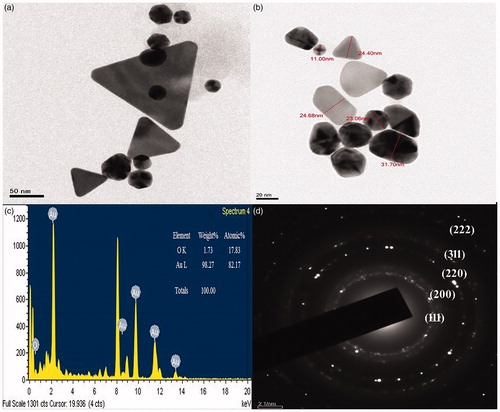
The boiled root of L. camara retained pale yellow color after 30 min. The reaction was rapid, when the colored root broth was mixed with 1 mM HAuCl4 and the mixture was turned into dark purple color within 10 min (). The span of overall bio-reduction reaction was within 2 h, in which the Au3+ reduced into Au° ions in optimum condition, and the formation of Au NPs was confirmed. The UV-visible spectra of synthesized Au NPs showed the strong surface plasmon resonance corresponds to the peak at 552 nm, during different time intervals of bio-reduction (). The exact nature and size of the Au NPs formed were studied through XRD analysis. shows strong and narrow diffraction peaks indicated that the product have well crystalline. The XRD peaks at 38°, 44°, 64°, and 77° can be indexed to the (1 1 1), (2 0 0), (2 2 0), and (3 1 1) Bragg’s reflections of cubic structure of metallic gold respectively and the crystallinity of the Au NPs was confirmed Joint Committee on Powder Diffraction Standards (JCPDS no. 04–0784). The broadening of Bragg’s peaks indicates the formation of NPs. Nearly monodispersed Au NPs with controllable size and uniform shape can be easily obtained in the simple aqueous reduction method. The mean size of Au NPs was calculated using the Debye–Scherrer’s equation by determining the full width half maxima of the (1 1 1) Bragg’s reflection. The intense IR bands are observed at 3408.49 cm −1 is due to N–H and O–H stretching. A peak was observed around 2920 and 2853 cm −1 that could be assigned to the C–H stretching vibration of alkyl and aldehyde groups. The bands observed at 1607 cm −1 assigned to the stretching vibrations of the C = C, respectively (). The band located at 1258 and 1063 cm −1 was due to C-N stretching of aromatic amines and aliphatic amines. The typical peak at 619 cm −1 was observed in functionalized Au NPs pattern close to 600 cm −1, which signify the presence of R–CH. The functional groups N–H and O–H and C = C, the biomolecule present in the plant extract, facilitated the reduction of Au ions to Au NPs. Thus FT-IR analysis clearly shows that bio-molecules present in plant’s root aqueous extract which could be responsible for the formation of Au NPs. Representative HRTEM images were recorded at a magnification of 10 000× revealed the size, shape, and distribution of synthesized NPs shown in (). The high-resolution study of the nanoparticles using HRTEM predominates with spherical morphologies with an average size of around 11–32 nm. Most of the nanoparticles were roughly circular with smooth edges and few were triangle with sharp edges. The nanoparticles obtained are highly crystalline as shown in by clear lattice fringes and the upper inset pattern of SAED confirmed the crystalline nature of synthesized Au NPs through bright circular Bragg’s rings corresponding to their reflections. shows the spot-profile EDX of Au NPs showed strong signals for gold atoms along with weak signals from carbon and oxygen. These weak signals could have been arisen from X-ray emission from macromolecules like proteins/enzymes bound to the NPs or in the vicinity of the particles.
Figure 1. L. camara Linn root extract treated with HAuCl4 resulted Au NPs (dark purple).
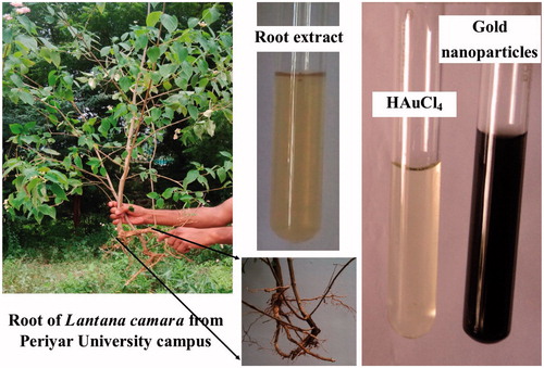
Figure 2. UV-visible spectra of Au NPs synthesized by L. camara root extract.

Figure 3. X-ray diffraction patterns of synthesized Au NPs.
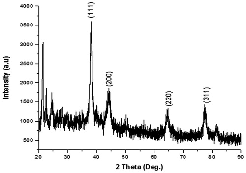
Figure 4. FT-IR spectrum of synthesized Au NPs of L. camara root.
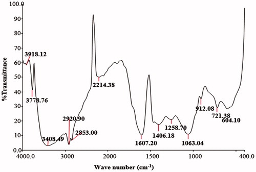
Figure 5. HR-TEM of Au NPs synthesized from root extract of L. camara. (a) and (b) the shapes and different sizes of Au NPs; (c) EDX analysis of Au NPs of L. camara; (d) SAED pattern showing that the nanoparticles are crystalline.
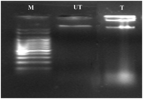
The DPPH radical scavenging activities of root aqueous extract and Au NPs increased gradually in a dose-dependent manner. The significant differences among the treated concentrations and the tested samples (Au NPs vs. root extract and gallic acid) were observed. The percentage inhibition of the aqueous root extract, Au NPs, and the control (Gallic acid) was represented in . The root aqueous extract, Au NPs, and gallic acid showed 67.06, 88.95, and 95.60% of scavenging effect at the sample concentration of 100 μg/ml, respectively. The inhibitory concentrations (IC50) of root aqueous extract, Au NPs, and Gallic acid were found to be: 60.58 ± 0.13, 24.17 ± 0.07, and 5.39 ± 0.16 μg/ml, respectively. Thus, the root extract-mediated Au NPs possess considerable scavenging activity, when compared to gallic acid. The cytotoxicity assay was carried out with different concentrations of Au NPs which resulted in the decreased percentage of growth on breast cancer cell line (MDA-MB-231) and normal cell line (Vero). The L. camara Au NPs exhibited significant inhibition in MDA-MB-231 and normal Vero cell line at a dose-dependent manner. The percentage growth of both the cells was gradually decreased in the increased concentration of Au NPs. The significant differences were found, among all different treated concentrations of Au NPs on MDA-MB-231 (F = 494.765; df =10, 22; P < 0.05) and Vero cells (F = 281.710; df =10, 22; P < 0.05), respectively (). The growth inhibitory rate (IC50) of synthesized Au NPs against human breast cancer (MDA-MB-231) cells and normal Vero cells was found to be 17.72 μg/ml and 32.98 μg/ml after 48 h of incubation. An Apoptotic morphological change caused by Au NPs was studied using acridine orange/ethidium bromide differential staining method. The stained cells were characterized to viable (light green), early apoptotic (bright green fluorescence and condensed chromatin), late apoptotic (orange fluorescence), and nonviable cells (red-colored fluorescence) (). The Au NPs-treated cells showed condensed nuclei, membrane blebbing, and apoptotic bodies. In contrast, the control cells showed intact nuclear architecture. However, very few apoptotic bodies were noticed in HAuCl4-treated cells. The biologically synthesized Au NPs induced cell death via apoptosis; DNA laddering assay was performed on agarose gel. A clear fragmented DNA ladder was observed in Au NPs-treated MDA-MB- 231 cells whereas HAuCl4-treated cells did not show such clear fragmented DNA ladder (). In addition, the untreated (control) cells did not show any prominent DNA ladder on the agarose gel. Therefore, the data obtained from this study confirm that Au NPs induced cell death through apoptosis.
Figure 6. The DPPH scavenging assay of root extract, Au NPs, and Gallic acid.
DPPH scavenging ability of Au NPs vs. root extract and ascorbic acid. Values are mean of independent determinations. Two-way ANOVA, significant different from Au NPs: “*” p < 0.001.
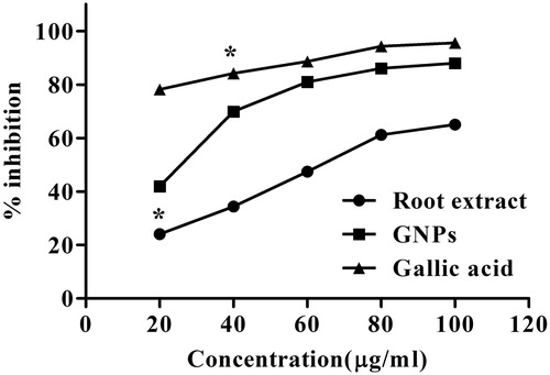
Figure 7. Percentage growth curve of MDA-MB-231 against the Au NPs.
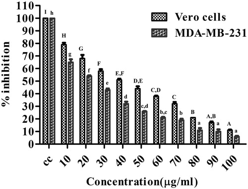
Figure 8. Cytotoxicity (a) Untreated, (b) treated MDA-MB-231 cells at 48 h, (c) Live cells (AO-Et-Br staining), and (d) damaged cells.
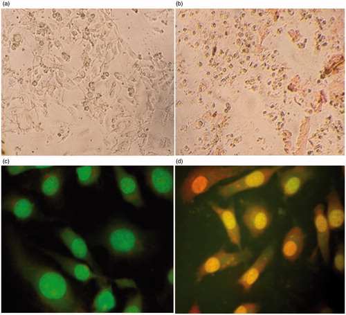
Figure 9. DNA fragmentation assay.
Where, M, Marker; UT, untreated cells at 48 h; T- MDA-MB-231 cells exposed with GNPs.
Discussion
The weed, L. camara Linn, has various medicinal applications which were contributed with earlier synthesized nanoparticles by several researchers (;;; Ajitha et al. Citation2015; Dash et al Citation2015; Kumar et al. Citation2016a, Citationb, Citationc; Mavukkandy et al. Citation2016; Kamarasamyraja and Jaganathan Citation2013; Sivakumar et al. Citation2012; Thirumurugan et al. Citation2011). Their results described about multi-potentials of L. camara which opened up its use in biomedical applications. Therefore, the present study focused on the in vitro antioxidant and cytotoxic effects of Au NPs synthesized from root aqueous extract. L. camara on human breast cancer (MDA-MB-231) and normal (Vero) cells. Recently, researchers have successfully synthesized the Au NPs using the root extract from the medicinal herbal plant Panax ginseng (Singh et al. Citation2015; Singh et al. Citation2016a) which suggested the use of medicinal plants as resources. Similarly, the present findings utilized the L. camara’s root aqueous extract for the Au NPs synthesis by their medicinal value inculcated earlier. The bio-reduction of Au3+ to Au0 was achieved within 10 min after adding HAuCl4 solution. Au NPs are known to exhibit a purplish color in the reaction mixture which is due to the excitation of the surface plasmon vibrations (SPR). The colloidal Au NPs showed intense color due to SPR arising from combined oscillation of electrons induced by an electromagnetic field (Toderas et al. Citation2009). Generally, the synthesized Au NPs are confirmed by the terminated ruby red within 10 min as reported earlier by Sheny et al. (Citation2011). In our present study, the optimum time required for the completion of reaction was rapid, i.e., in 8 min, which supports early finding (Jayaseelan et al. Citation2013) for the pre-confirmation of Au NPs. As the earlier report of Mittal et al. (Citation2013), the present study supports for the bio-reduction of metal ion into base metal which was quite rapid, readily, and easily scaled up. The spectrophotometric wavelength ranges of 500–550 nm are normally used in characterizing the Au NPs (Karuppaiya et al. Citation2013). The peak of absorption spectrum at 552 nm was quite clear like that of the earlier findings of several researchers, which could be attributed to the blue-shifted peak range of Au about 560 nm (Aromal and Philip Citation2012; Subramaniam et al. Citation2016). Au NPs synthesized from root extract of L. camara showed three prominent Bragg reflections viz., (111), (200), and (220) that were analogous on the basis of the face-centered cubic nature of Au (Punuri et al. Citation2012) with Joint Committee on Powder Diffraction Standards (JCPDS no. 04–0784), which indicates the formation of nanoparticles. From the result of selected area energy-dispersive study, the upper inset pattern of synthesized Au NPs accompanied the earlier findings of Philip (Citation2009) that endorses the crystalline nature of gold through circular rings analogous to the Bragg’s reflections. Plant extracts may act both as reducing agents and stabilizing agents during the synthesis of nanoparticles (Kumar and Yadav 2009). The findings of several researchers have demonstrated the functional groups that act in capping, bioreduction, and stabilization of NPs. Presently, the Au NPs synthesized from L. camara root extract containing biomolecules, which have been highlighted that the stabilization of inert surfaces, might be due to the electrostatic barriers on the surfaces of NPs as reported earlier by El-Batal et al. (Citation2013) and Murugan et al. (Citation2015). The different functional groups with their respective stretches obtained in the presently synthesized Au NPs are in line with the previous findings of Varshney et al. (Citation2010). Recent studies have shown that biomolecules such as protein, phenol, and flavonoids present in the plant extract play an important role in the reduction of metals ions and capping of the nanoparticles (Mittal et al. Citation2013). The size ranges (between 11 and 32 nm) of Au NPs obtained were similar to the earlier report of Ghosh et al. Citation2011 from Dioscorea bulbifera, and the observed differences in size of NPs could be due to the reduction of ions to gold being formed at different times (Rajasekhar et al. Citation2012). The size of the NPs may be smaller at higher concentration of extract owing to the phytochemicals (present in the extract) that can effectively stabilize the NPs (Paul et al. Citation2013). The different size and shape might occur due to the aggregates of leaf aqueous extract. The EDX attachment on the HRTEM provided elemental composition analysis in view as well as spot analyses of minute particles and thus confirms the presence of specific elements. is a representative plot of the spot EDX analysis in which 98.73% signals the gold (Au) and the rest is oxygen (O). The HRTEM-EDX analysis displayed the well-known signature spectra for gold and thus convincingly evidenced the presence of this noble metal gold (Au). These results are in accordance with other reports on the EDX analysis of gold structures synthesized using extracts derived from Diospyros kaki (Song et al. Citation2009). The SAED patterns revealed that the particles are crystalline in nature. The fringe spacing for the colloids are correspond to the spacing between the (111) plane of face-centered cubic (fcc) nature of gold. The SAED pattern shows the diffraction ring from inner to outer which can be indexed as (111), (200), and (220) reflections, respectively of fcc gold. The earlier report of Philip (Citation2009) has interestingly noted that the particles with deviated fringe spacing are not almost in contact and they are separated from each other with invariable distance as obtained in this present SAED pattern. In DPPH assays, lower IC50 values showed higher free radical scavenging activity. Au NPs showed significant free radical scavenging activity (IC50–24.17) against DPPH radicals, over the standard antioxidant gallic acid (IC50–5.39) which has higher inhibitory effects (95.60%). The root aqueous extract comprises a noticeable amount of hydrogen donor molecules, which might reduce the free radicals in DPPH scavenging assays as in agreement with earlier report (Murugan et al. Citation2016). Also, the scavenging ability might endorsed by the phenolic contents in plants probably due to their redox potentials, which could act as reducing agents and singlet oxygen quenchers (Al-Shmgani et al. Citation2016, Saravanakumar et al. Citation2016). The IC50 values obtained by the several researchers in earlier for the Au NPs on MDA-MB-231 cells were 50 and 56.65 μg/ml (Adeyemi and Whiteley Citation2015, Krishnaraj et al. Citation2014). In this line, the presently synthesized Au NPs revealed the IC50 value of 17.72 μg/mL on MDA-MB-231 cells which was 2-fold decrease in concentration. The cytotoxic effect of Au NPs could be exhibited due to the physical and chemical interactions of gold ions with functional groups of intracellular components viz., proteins, nitrogen bases, and phosphate groups (Moteriya and Chanda Citation2016). Of late, Singh et al. (Citation2016b) have been reported that the biologically synthesized nanoparticles are quite potent and highly preferable in terms of effectiveness as compared with physiochemically synthesized nanoparticles. Therefore, the L. camara-mediated Au NPs might also considered being a significant bio-controlling agent for cancer cells. Apoptosis of the Au NPs-treated cells was escorted by a fall in the percentage of cells in G0/G1 phase and a raise in the percentage of G2/M phase cells, representing cell cycle arrest at G2/M (Lee et al. Citation2011). The ROS can act as signal molecules promoting cell cycle progression and can induce oxidative DNA damage (Hu et al. Citation2009). DNA fragmentation is broadly considered as a characteristic feature of apoptosis (Allen et al. Citation1997). Induction of apoptosis can be confirmed by two factors such as irregular reduction in size of cells, in which the cells are reduced and shrunken, and lastly DNA fragmentation. The present study, therefore, illustrated the anticancer property of L. camara-mediated Au NPs, which may be employed as a drug in nanomedicine to treat breast cancer in future. In breast cancer, nanoparticle-based therapies are paving the way in diagnosis, imaging, screening, and anti-proliferation. But the challenges met in the field of pharmacology, toxicology, immunology, large-scale manufacturing, and regulatory issues were disadvantaged severely in translating such advances from the bench to the bedside. However the presently synthesized Au NPs might act as a considerable biological agent on breast cancer treatment.
Conclusions
To sum up the present investigations, the in vitro antioxidant and cytotoxic properties of Au NPs synthesized from root extract of L. camara were confirmed. Stable and spherical-shaped Au NPs have been achieved from a simple, economic, nontoxic, and efficient green method. The crystalline nature of NPs is confirmed from bright rings in the SAED pattern, clear lattice fringes in the HRTEM images and peaks in the XRD pattern. From FT-IR spectrum, it is found that biomolecules responsible for capping and stabilization of Au NPs are flavonoids. Au NPs-treated cells exhibited dose-dependent cell death and membrane leakage. From the MTT assay, the synthesized Au NPs exhibited preponderant activity on human breast cancer (MDA-MB-231) cells and normal Vero cells. Au NPs also document the induction of apoptosis and fragmentation of DNA. Finally, the chance of using Au NPs synthesized from root aqueous extract to inhibit the growth and their cytotoxicity for potential therapeutic treatments can bids a new way to combat cancer diseases.
Acknowledgements
The authors are grateful to their host institutes for providing necessary facilities to carry out this work.
Disclosure statement
All the authors declare that they have no conflict of interests including any financial, personal, or other relationships with other people or organizations.
References
- Adeyemi OS, Whiteley CG. 2015. Metal nanoparticles caused death of metastatic MDA-MB-231 cells. World Acad Sci Eng Technol Chem Mol Eng. 2:3.
- Ajitha B, Ashok Kumar Reddy Y, Sreedhara Reddy P. 2015. Green synthesis and characterization of silver nanoparticles using Lantana camara leaf extract. Mater Sci Eng C. 49:373–381.
- Allen RT, Hunter WJ 3rd, Agrawal DK. 1997. Morphological and biochemical characterization and analysis of apoptosis. J Pharmacol Toxicol Methods. 37:215–228.
- Al-Shmgani HS, Mohammed WH, Sulaiman GM, Saadoon AH. 2016. Biosynthesis of silver nanoparticles from Catharanthus roseus leaf extract and assessing their antioxidant, antimicrobial, and wound-healing activities. Artif Cells, Nanomed Biotechnol. [Epub ahead of print]. DOI:10.1080/21691401.2016.1220950.
- American Cancer Society (ACS). 2016. Cancer Facts & Figures 2016. Atlanta: American Cancer Society, pp. 1–64.
- Aromal SA, Philip D. 2012. Benincasa hispida seed mediated green synthesis of gold nanoparticles and its optical nonlinearity. Physica E. 44:1329–1334.
- Balasubramani G, Ramkumar R, Krishnaveni N, Pazhanimuthu A, Natarajan T, Sowmiya R, Perumal P. 2015b. Structural characterization, antioxidant and anticancer properties of gold nanoparticles synthesized from leaf extract (decoction) of Antigonon leptopus Hook. & Arn. J Trace Elem Med Biol. 30:83–89.
- Balasubramani G, Ramkumar R, Krishnaveni N, Sowmiya R, Deepak P, Arul D, Perumal P. 2015a. GC–MS analysis of bioactive components and synthesis of gold nanoparticle using Chloroxylon swietenia DC leaf extract and its larvicidal activity. J Photochem Photobiol B: Biol. 148:1–8.
- Brown K. 2002. Breast cancer chemoprevention: risk-benefit effects of the antioestrogen tamoxifen. Expert Opin Drug Saf. 1:253–267.
- Dash SS, Bag BG, Hota P. 2015. Lantana camara Linn leaf extract mediated green synthesis of gold nanoparticles and study of its catalytic activity. Appl Nanosci. 5:343–350.
- El-Batal AI, Hashem AM, Abdelbaky NM. 2013. Gamma radiation mediated green synthesis of AuNPs using fermented soybean-garlic aqueous extract and their antimicrobial activity. Springerplus. 2:129–138.
- Fong J, Wood F. 2006. Nanocrystalline silver dressings in wound management: a review. Int J Nanomed. 1:441–449.
- Franco-Molina MA, Mendoza-Gamboa E, Sierra-Rivera CA, Gómez-Flores RA, Zapata-Benavides P, Castillo-Tello P, et al. 2010. Antitumor activity of colloidal silver on MCF-7 human breast cancer cells. J Exp Clin Cancer Res. 29:1–7.
- Ghodake PP, Kulkarni AS, Aloorkar NH, Osmani RA, Bhosale RR, Harkare BR, Kale BB. 2013. In-vitro antispasmodic activity analysis of methanolic leaves extract of Lantana camara Linn. on Excised Rat Ileum. J Pharmacogn Phytochem. 2:66–71.
- Ghosh S, Patil S, Ahire M, Kitture R, Jabgunde A, Kale S, et al. 2011. Synthesis of gold nano-anisotrops using Dioscorea bulbifera tuber extract. J Nanomat. 2011:1–8.
- Hu R, Yong KT, Roy I, Ding H, He S, Prasad PN. 2009. Metallic nanostructures as localized plasmon resonance enhanced scattering probes for multiplex dark-field targeted imaging of cancer cells. J Physic Chem. 113:2676–2684.
- Huang X, Jain PK, El-Sayed IH, El-Sayed MA. 2007. Gold nanoparticles: interesting optical properties and recent applications in cancer diagnostics and therapy. Nanomedicine (Lond). 2:681–693.
- Iv M, Telischak N, Feng D, Holdsworth SJ, Yeom KW, Daldrup-Link HE. 2015. Clinical applications of iron oxide nanoparticles for magnetic resonance imaging of brain tumors. Nanomedicine (Lond). 10:993–1018.
- Jayaseelan C, Ramkumar R, Abdul Rahuman A, Perumal P. 2013. Green synthesis of gold nanoparticles using seed aqueous extract of Abelmoschus esculentus and its antifungal activity. Ind Crop Prod. 45:423–429.
- Johnston SR. 1997. Acquired tamoxifen resistance in human breast cancer – potential mechanisms and clinical implications. Anticanc Drug. 8:911–930.
- Kalita S, Kumar G, Karthik L, Rao KVB. 2012. A review on medicinal properties of Lantana camara Linn. Res J Pharm Technol. 5:711–715.
- Kamarasamyraja D, Jaganathan NS. 2013. Antimicrobial activity of biosynthesized silver nanoparticles prepared from the leaf extract of Lantana camara. Int Res J Pharm. 4:203–207.
- Karuppaiya P, Satheeshkumar E, Chao WT, Kao LY, Chen ECF, Tsay HS. 2013. Anti-metastatic activity of biologically synthesized gold nanoparticles on human fibrosarcoma cell line HT-1080. Colloids Surf., B. 110:163–170.
- Khlebtsov N, Dykman L. 2011. Biodistribution and toxicity of engineered gold nanoparticles: a review of in vitro and in vivo studies. Chem Soc Rev. 40:1647–1671.
- Kim DW, Hong GH, Lee HH, Choi SH, Chun BG, Won CK, Hwang IK, Won MH. 2007. Effect of colloidal silver against the cytotoxicity of hydrogen peroxide and naphthazarin on primary cultured cortical astrocytes. Neuroscience. 117:387–400.
- Kirtikar KR, Basu BD. 2006. Indian Medicinal Plants. New Delhi, India: Periodical Experts Book Agency.
- Kotha A, Sekharam M, Cilenti L, Siddiquee K, Khaled A, Zervos A, et al. 2006. Resveratrol inhibits Src and Stat3 signaling and induces the apoptosis of malignant cells containing activated Stat3 protein. Mol Cancer Therapeutic. 5:621–629.
- Krishnaraj C, Muthukumaran P, Ramachandran R, Balakumaran MD, Kalaichelvan PT. 2014. Acalypha indica Linn: biogenic synthesis of silver and gold nanoparticles and their cytotoxic effects against MDA-MB-231, human breast cancer cells. Biotechnol Rep. 4:42–49.
- Kumar B, Smita K, Cumbal L, Debut A. 2016a. Extracellular biofabrication of gold nanoparticles by using Lantana camara berry extract. Synth React Inor Met Org and Nano-Metal Chemistry. in press. DOI:10.1080/15533174.2016.1157817.
- Kumar B, Smita K, Cumbal L. 2016b. Biofabrication of nanogold from the flower extracts of Lantana camara. IET Nanobiotechnol. 10:154–157.
- Kumar B, Smita K, Cumbal L. 2016c. Biosynthesis of silver nanoparticles using Lantana camara flower extract and its application. J Sol-Gel Sci Technol. 78:285–292.
- Kumar V, Yadav SK. 2009. Plant‐mediated synthesis of silver and gold nanoparticles and their applications. J Chem Technol Biotechnol. 84:151–157.
- Kumarasamyraja D, Jeganathan NS, Manavalan R. 2012. Pharmacological review of Lantana camara. L review article. Int J Pharm Ind Res. 2:1–5.
- Lee YS, Kim DW, Lee YH, Oh JH, Yoon S, Choi MS, et al. 2011. Silver nanoparticles induce apoptosis and G2/M arrest via PKCζ-dependent signaling in A549 lung cells. Archiv Toxicol. 85:1529–1540.
- Liu H, Liu Y, Wang Z, He P. 2010. Facile synthesis of monodisperse, size-tunable SnS nanoparticles potentially for solar cell energy conversion. Nanotechnology. 21:105707.
- Mavukkandy MO, Chakraborty S, Abbasi T, Abbasi SA. 2016. A Clean-green synthesis of platinum nanoparticles utilizing a pernicious weed Lantana (Lantana camara). American J Eng Appl Sci. 9:84–90.
- Mittal AK, Chisti Y, Banerjee UC. 2013. Synthesis of metallic nanoparticles using plant extracts. Biotechnol Adv. 31:346–356.
- Moteriya P, Chanda S. 2016. Synthesis and characterization of silver nanoparticles using Caesalpinia pulcherrima flower extract and assessment of their in vitro antimicrobial, antioxidant, cytotoxic, and genotoxic activities. Artif Cells Nanomed Biotechnol. [Epub ahead of print]. DOI:10.1080/21691401.2016.1261871.
- Murugan K, Aruna P, Panneerselvam C, Madhiyazhagan P, Paulpandi M, Subramaniam J, et al. 2016. Fighting arboviral diseases: low toxicity on mammalian cells, dengue growth inhibition (in vitro), and mosquitocidal activity of Centroceras clavulatum-synthesized silver nanoparticles. Parasitol Res. 115:651–662.
- Murugan K, Benelli G, Panneerselvam C, Subramaniam J, Jeyalalitha T, Dinesh D, et al. 2015. Cymbopogon citratus-synthesized gold nanoparticles boost the predation efficiency of copepod Mesocyclops aspericornis against malaria and dengue mosquitoes. Exp Parasitol. 153:129–138.
- Noruzi M. 2015. Biosynthesis of gold nanoparticles using plant extracts. Bioprocess Biosyst Eng. 38:1–14.
- Paul K, Bag BG, Samanta K. 2013. Green coconut (Cocus nucifera Linn) shell extract mediated size controlled green synthesis of poly shaped gold nanoparticles and its application in catalysis. Appl Nanosci. 4:769–775.
- Philip D. 2009. Honey mediated green synthesis of gold nanoparticles. Spectrochim Acta A Mol Biomol Spectrosc. 73:650–653.
- Punuri JB, Sharma P, Sibyala S, Tamuli R, Bora U. 2012. Piper betle-mediated green synthesis of biocompatible gold nanoparticles. Int Nano Lett. 2:18–26.
- Ragaseema VM, Unnikrishnan S, Kalliyana Krishnan V, Krishnan LK. 2012. The antithrombotic and antimicrobial properties of PEG-protected silver nanoparticle coated surfaces. Biomaterials 33:3083–3092.
- Rajasekhar RG, Jayakumar C, Antony BM, Sreenivasn D, Nagendra GN. 2012. Green synthesis, characterization and in-vitro antibacterial activity of polyshaped gold nanoparticles by using Senna siamea (Lam.) Plant leaf extract. Int J Green Chem Bioproc. 2:1–5.
- Rastogi RP, Mehrotra BN. 1995. Compendium of Indian Medicinal Plants. Vol I. New Delhi: Publication and Information Directorate, CSIR, pp. 50–66.
- Ross IA. 1999. Medicinal Plants of the World. New Jersey (NJ): Humana Press, Totowa. 179–82.
- Saraf A, Quereshi S, Sharma K, Afshan Khan N. 2011. Antimicrobial activity of Lantana camara L. J Exp Sci. 2:50–54.
- Saravanakumar A, Peng MM, Ganesh M, Jayaprakash J, Mohankumar M, Jang HT. 2016. Low-cost and eco-friendly green synthesis of silver nanoparticles using Prunus japonica (Rosaceae) leaf extract and their antibacterial, antioxidant properties. Artif Cell Nanomed Biotechnol. [Epub ahead of print]. DOI:10.1080/21691401.2016.1203795.
- Saxena M, Saxena J, Khare S. 2012. A brief review on: therapeutical values of Lantana camara plant. Int J Pharm Life Sci. 3:1551–1554.
- Sharma OP, Sharma PD. 1989. Natural products of the Lantana plant — the present and prospects. J Sci Ind Res. 48:471–478.
- Sheny DS, Mathew J, Philip D. 2011. Phytosynthesis of Au, Ag and Au–Ag bimetallic nanoparticles using aqueous extract and dried leaf of Anacardium occidentale. Spectrochim Acta a: Mol Biomol Spectrosc. 79:254–262.
- Shin KS, Choi JY, Park CS, Jang HJ, Kim K. 2009. Facile synthesis and catalytic application of silver-deposited magnetic nanoparticles. Catal Lett. 133:1–7.
- Singh P, Kim YJ, Wang C, Mathiyalagan R, El-Agamy Farh M, Yang DC. 2016a. Biogenic silver and gold nanoparticles synthesized using red ginseng root extract, and their applications. Artif Cell Nanomed Biotechnol. 44:811–816.
- Singh P, Kim YJ, Wang C, Mathiyalagan R, Yang DC. 2015. The development of a green approach for the biosynthesis of silver and gold nanoparticles by using Panax ginseng root extract, and their biological applications. Artif Cell Nanomed Biotechnol. 44:1150–1157.
- Singh P, Singh H, Ahn S, Castro-Aceituno V, Jiménez Z, Simu SY, Kim YJ, Yang DC. 2016b. Pharmacological importance, characterization and applications of gold and silver nanoparticles synthesized by Panax ginseng fresh leaves. Artif Cells Nanomedicine Biotechnol. [Epub ahead pf print]. DOI:10.1080/21691401.2016.1243547.
- Sivakumar P, Nethradevi C, Renganathan S. 2012. Synthesis of silver nanoparticles using Lantana camara fruit extract and its effect on pathogens. Asian J Pharm Clin Res. 5:97–101.
- Song JY, Jang HK, Kim BS. 2009. Biological synthesis of gold nanoparticles using Magnolia Kobus and Diopyros kaki leaf extracts. Proc Biochem. 44:1133–1138.
- Subramaniam J, Murugan K, Panneerselvam C, Kovendan K, Madhiyazhagan P, Dinesh D, et al. 2016. Multipurpose effectiveness of Couroupita guianensis-synthesized gold nanoparticles: high antiplasmodial potential, field efficacy against malaria vectors and synergy with Aplocheilus lineatus predators. Environ Sci Pol Res. 23:7543–7558.
- Taoubi K, Fauvel MT, Gleye J, Moulis C. 1997. Phenylpropanoid glycosides from Lantana camara and Lippia multiflora. Plan Medica. 19:63–65.
- Thirumurugan A, Anns Tomy N, Priyanka Kumar H, Prakash P. 2011. Biological synthesis of silver nanoparticles by Lantana camara leaf extracts. Int J Nanomat Biostruc. 1:22–24.
- Toderas F, Iosin M, Astilean S. 2009. Luminescence properties of gold nanorods. Nucl Instr Meth Phys Res B. 267:400–402.
- Varshney R, Bhadauria S, Gaur MS. 2010. Biogenic synthesis of silver nanocubes and nanorods using sun dried Stevia rebaudiana leaves. Adv Mat Lett. 1:232–237.
- Velusamy P, Kumar GV, Jeyanthi V, Das J, Pachaiappan R. 2016. Bio-inspired green nanoparticles: synthesis, mechanism, and antibacterial application. Toxicol Res. 32:95.
- Weenen H, Nkunya MH, Bray DH. 1990. Antimalarial activity of Tanzanian medicinal plants. Planta Med. 56:368–370.
- Winnicka F, Bielawski K, Bielawska A, Milty W. 2007. Apoptosis-mediated cytotoxicity of ouabain, digoxin and proscillaridin A in the estrogen independent MDA-MB-231 breast cancer cells. Archiv Pharm Res. 30:1216–1224.
- Yeruva L, Elegbede JA, Carper SW. 2008. Methyl jasmonate decreases membrane fluidity and induces apoptosis via tumor necrosis factor receptor 1 in breast cancer cells. Anti-Canc Drug. 19:766–776.
- Zhang M, Liu S, Xu G, Guo Y, Fu J, Zhang D. 2014. Cytotoxicity and apoptosis induced by nanobacteria in human breast cancer cells. Int J Nanomedicine. 9:265–271.
- Zhou L, He X, He D, Wang K, Qin D. 2011. Biosensing technologies for Mycobacterium tuberculosis detection: status and new developments. Clin Develop Immunol. 2011:193963.