
Abstract
Cord blood (CB) haematopoietic stem cell (HSC) is an alternative source of HSC transplantation. The limited cell number greatly restricts their clinic-scale therapeutic applications. The objective of this study was an ex vivo expansion of CB HSCs in a new three-dimensional polycaprolactone nano-scaffold coated with fibronectin (FN). First, we isolated CB CD34+ cells and cultured 10 days in presence of growth factors. The evaluation was performed by qRT-PCR, flow cytometry and clonogenicity. 3D PCL nano-scaffold coated with FN produced significantly higher total nucleated cells and CD34+ cells (p < .05) and also had significantly higher homing and self-renewality genes than 2D cell culture and before expansion (p < .05). The expression of CXCR-4, VLA-4, VLA-5 and LFA-1, and also HOXB-4, HOXA-9, BMI-1 and hTERT genes was higher in 3D than 2D. The CD13, CD14, CD33, CD34 and CD45 markers were significantly higher and CD2, CD3 and CD19 markers were significantly lower in 3D scaffold than 2D cell culture (p < .05). The type and number of colonies in 2D culture were lower than 3D culture medium (p > .05). 3D PCL nano-scaffold coated with FN could better keep specifications homing and self renewality of CB HSCs after expansion.
Introduction
After first successful umbilical cord blood transplantation (UCBT) in 1988 [Citation1], cord blood (CB) attracted a lot of attention to a new alternative haematopoietic stem cell (HSC) transplantation source [Citation2]. CB is the efficacious cell source for a treat some life-threatening haematological and non-haematological and also immunodeficiency disorders in paediatric patients [Citation3]. However, despite all advantages of CB, the limited number of transplantable HSCs in clinic greatly restricts their clinic-scale therapeutic applications [Citation4]. A number of these cells is the most important factor for a satisfactory transplantation, hence, different approaches have been assessed for overcoming this barrier and further achieving the treatment for haematological disease [Citation5–10]. One such solution is the ex vivo expansion of HSCs by creation appropriate status in cell culture medium so as to generate sufficient HSC numbers, sustain the expansion of HSCs ex vivo and improve the consequence of HSCs transplantation [Citation7,Citation11,Citation12]. In the last decade, a lot of effort has been focused on the ex vivo expansion of HSCs; but due to the easy-loss of HSCs stemness and lack of homing in ex vivo expansion contrast to body natural systems, there are still no satisfactory for this strategy [Citation13]. Bone marrow has unique and specialized microenvironment for HSCs, namely niche, where HSCs are contacted with a complex network and distinct types of cells, such as stromal cells, cytokine, growth factors and extracellular matrix (ECM) and they can with effect on growth, expansion and self-renewal regulate HSCs fate [Citation14,Citation15]. In fact, the crosstalk between HSC and their niche modulates HSC function.
The conventional way to amplify HSCs is two-dimensional (2D) expansion approaches that significantly diminish HSC proliferation compared to 3D culture systems [Citation16]. Thus, applying 3D scaffold that mimics bone marrow microenvironment in in vitro by ECM may be an effective way to stimulate the ex vivo expansion of HSCs. Also, it is important that 3D scaffold, producing sufficient cell number, retains the capacity for self-renewal and maintains a proliferation of HSCs in cell culture medium [Citation17]. ECM has an important role in physical and chemical signals that are amplified. The ECM component has a crucial role in HSC niche. Adhesiveness or interaction between HSCs and cell adhesion molecule providing homing or retaining HSCs in bone marrow niche is provided by these elements [Citation14,Citation18].
According to aforementioned study and due to the importance of the number of HSCs in transplantation, in this study, we aimed to establish the new 3D culture system by using specific nano-fibre and polycaprolactone (PCL) coated with fibronectin (FN) for the proliferation of HSCs derived from UCB with maintaining homing and self-renewality.
Materials and methods
Cord blood collection
After obtaining informed consent according to institutional guidelines, the cord was clamped and cut at the end of term deliveries by a well-trained personnel, umbilical CB was collected by gravity into sterile 250 ml CB collection bags (JMS, Singapore) containing 35 ml anticoagulant CPD-A1.
Purification of CB CD34+ cells
Unseparated CB cells were diluted in 1:5 with hydroxyl ethyl starch solution (HES, Grifols, Barcelona, Spain), and mononuclear cells (MNCs) were separated by centrifugation. The CD34+ cells were separated with a CD34 progenitor cell isolation kit (Miltenyi Biotec, San Diego, CA) with high-gradient magnetic field and mini-magnetically activated cell sorting (MACS).
To assess the purity of the isolated cells, we stained a portion with CD34-fluorescein isothiocyanate (FITC) (Dako, Glostrup, Denmark) antibody by flow cytometry. The rest of the cells were used for culture or were frozen.
Preparation culture medium
Culture medium was prepared by Stem line II serum-free media (Sigma, Darmstadt, Germany) supplemented with recombinant human stem cell factor (SCF, 50 ng/ml, Peprotech, London, UK), recombinant human thrombopoietin (TPO, 50 ng/ml, Gibco, Gran Island, NY) and recombinant human FMS-like tyrosine kinase 3 (FLT-3, 50 ng/ml, Gibco, Gran Island, NY).
Preparation 3D scaffolds
Sterilization of PCL scaffolds was performed by 1 h immersion in 70% ethanol, washed with PBS and then dried overnight under sterile condition. Scaffolds were placed in 24-well polystyrene plates and coated with FN in 50 μg/ml concentration for 24 h at 40 °C. Then, we removed FN solution and seeded cells.
Ex vivo expansion of CD34+ CB cells
In each well, 1 × 10⁴ CD34+ cells suspended in the 250 μl culture medium was added to the top centre of the scaffold (24-well format), avoiding tip/surface contact. Seeded scaffolds were then incubated for 3 h at 37 °C (20% O2 and 5% CO2 humidified atmosphere) for 10 days. Half of the medium was exchanged every 48 h with fresh medium and cells were counted. Tissue culture polystyrene (TCPS) was used as a control condition. At designed time points, expanded cells were harvested from the culture by pipetting for analysis.
Immunophenotype analysis
To evaluate the ability of HSC expansion and differentiation, cells were removed from scaffold and then the morphology of cells was checked and stained with monoclonal antibody CD34-PE and CD45-FITC, lymphoid markers (CD2-FITC, CD3-FITC and CD19-FITC), myeloid markers (CD13-FITC and CD33-FITC) and monocyte (CD14-FITC) (Dako, Glostrup, Denmark) against human epitope. The tubes were incubated in 4 °C for 30 min. Isotype control was used to set compensation and confirm the specificity. Ten thousand events were acquired on a Partec PAS (Munster, Germany) flow cytometer. Flow Jo software was used for data analysis (Tree Star, Inc., Ashland, OR).
Colony forming unit assay (CFU-assay)
The freshly isolated CD34+ CB cells and their progeny expanded in culture on day 10 were incubated in 2 ml methylcellulose culture medium (H4435, Stem Cell Technology, Vancouver, Canada) plated in six-well dishes at a concentration of 1 × 10⁴. H4435 contains 1% methylcellulose in Iscove’s MDM (IMDM, Sigma, Darmstadt, Germany). All cultures were performed in triplicate, maintained at 37 °C and 5% CO2 humidified atmosphere in the air, and scored on day 14 for granulocyte–erythrocyte–monocyte–megakaryocyte colony-forming units (CFU-GEMM), granulocyte-macrophage colony-forming units (CFU-GM) and erythroid burst-forming units (BFU-E) being counted.
RNA extraction and cDNA synthesis
RNA from purified progenitor cells and ex vivo expanded cells was isolated using Trizol (Invitrogen, Carlsbad, CA) according to the instructions of the supplier and then reverse-transcribed into first-strand cDNA using random hexamer primers and the Superscript II reverse transcriptase kit (Invitrogen, Carlsbad, CA) according to manufacturer’s instructions.
Real-time quantitative reverse transcription–polymerase chain reaction (qRT–PCR)
The expression levels of homing (CXCR4, VLA-4, VLA-5 and LFA-1), self renewality (BMI-1, hTERT, HOXA9 and HOXB-4) and the GAPDH reference gene were determined by SYBR Green I real-time PCR. Briefly, PCR was performed in a 12 μl total volume containing 1 μl of cDNA, 6.25 μl of 2× SYBR Green mix (Amplicon, Aalborg, Denmark), and 10 μmol/l primer pairs. After an initial denaturation at 95 °C for 15 min, 40 cycles consisting of the following procedure were performed using ABI step one PCR cycler (ABI, Vernon, CA): 20 s at 95 °C, 60 s at 60 °C for GAPDH, LFA-1, VLA-4, VLA-5, hTERT and BMI, and 62 °C for HOX-A9, and 64 °C for CXCR-4 and HOX-B4. The data are presented as the relative expression of the genes of interest relative to the internal control gene as determined by the 2(−ΔΔCT) method. Additionally, the specific amplification of the PCR products was analysed by melting curve analysis and agarose gel electrophoresis. The primers used for real-time PCR for all gene amplifications are shown in .
Table 1. Primer sequences used for real-time PCR (5′–3′).
Statistical analysis
All experiments were performed in triplicate (n = 3), an independent three samples, and each value represents the mean ± standard error. The parametric ANOVA test was used in order to compare the number and expansion of the total nucleated cells, the percentage and expansion of CD34+ cells as well as the number of colonies in the four studied groups. GraphPad Prism 6.0 software with a level of significance p < .05, p < .01 or p < .001 is used for analysis (GraphPad Software, La Jolla, CA).
Results
Expansion of CD34+ CB cells through the 3D culturing method
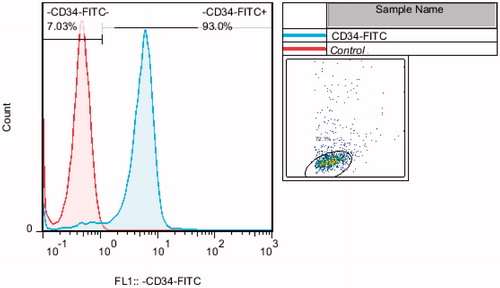
The number of isolated CD34+ cells obtained with the mini-MACS system was 7 × 10⁵/ml. The purity of isolated CD34+ cells was 85.4% ± 7.6% (). In general, the vast majority of cells were spherical cells that were non-adherent to plastic.
Figure 1. Flowgram of CD34+ cord blood cells purity percentage after extraction with MACS.
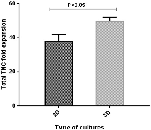
shows the effect of FN on the expansion of CD34+ cells. Average fold increases of the total nucleated cells after use of the conventional static (2D) and PCL coated with FN (3D) culture systems for 10 days were 38.3 ± 3.3 and 58 ± 1.63, respectively ().
Figure 2. Scanning electron micrograph of polycaprolactone nano-fibre (original magnification A × 100, B × 1000). CD34+ cord blood cells culture in 2D medium (original magnification C × 400) were growing in PCL nano-fibre coated with fibronectin after 10 days (original magnification D × 1000).
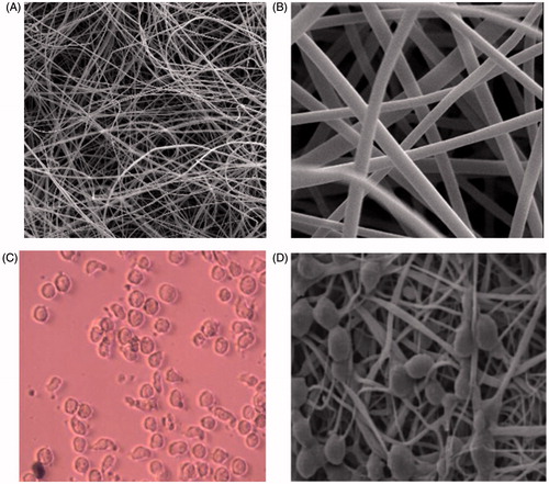
Figure 3. TNC fold expansion between 2D and 3D cell culture systems after 10 days culture.
Phenotypic changes of CD34+ CB cells during ex vivo expansion
The unexpanded cells start with 85.4%±7.6%. CD34+ decreased to 5.75%±1.25% after 10 days of culture in TCPS wells (p = .001, ). Cells expanded in FN-coated PCL scaffold could maintain the percentages of CD34+ cells at 65.65%±5.35%. These values were statistically significant (p = .001, ). Whereas nucleated cells proliferated more than 38 ± 3.3 and 58 ± 1.63 times during the 2D and 3D culture periods, respectively, the absolute number of CD34+ cells in the 2D and 3D culture systems increased continuously up to approximately 2.66 and 38 times the initial cell number, respectively (p = .001) ().
Figure 4. Total CD34+ cells fold expansion in 2D and 3D cell culture systems after 10 days culture.
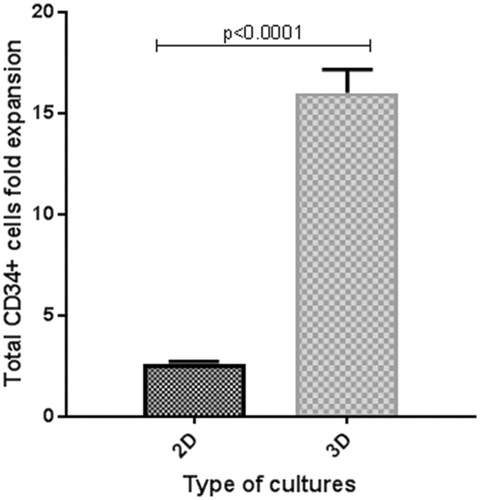
Table 2. Expression of surface markers on cells before and after expansion under different conditions.
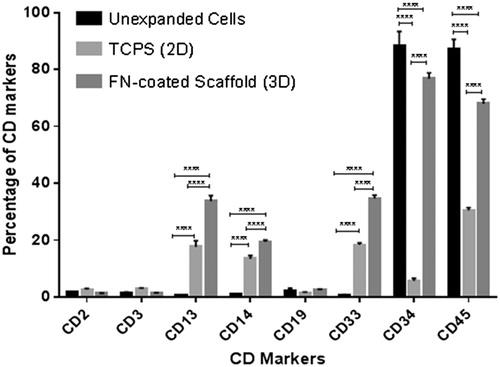
Compared with cells expanded in TCPS wells, cells expanded in FN-coated scaffold exhibited lower levels of surface markers for lymphoid markers (CD2+ and CD3+ for T cells and CD19+ for B cells) and mature megakaryocyte marker (CD41+). Interestingly, more cells which expanded on FN-scaffold (p>.05) expressed committed granulocyte–monocyte progenitors (CD13+ and CD33+) and mature monocytes (CD14) and CD45+ marker ( and ).
Figure 5. Haematopoietic stem cell (CD34+), granulocytes (CD13 and CD33), lymphocytes (CD2, CD3 and CD19) and monocyte (CD14) markers in unexpanded cells immediately after extraction, 2D and 3D cell culture systems after 10 days expansion.
CFU assays on expanded cells
To constitute the clonogenic haematopoietic progenitors among the expanded cells, CFU assays were conducted on cells expanded (). Compared with that cultured in the 2D cell culture system, cells expanded in FN-coated PCL scaffold showed a 2.1-fold increase of CFU-GEMM number, a 1.5-fold increase of BFU-E/CFU-E number, a 1.1-fold increase of CFU-GM number and a 1.3-fold increase in total CFU number.
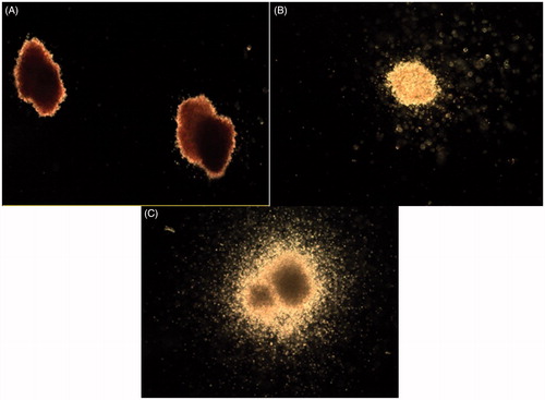
Figure 6. Image of BFU-E/CFU-E (A), CFU-GM (B) and CFU-GEMM (C) based on total CFU analysed for cells expanded in different culture system for 10 days.
The proportions of CFU-GM, BFU-E/CFU-E, CFU-GM and CFU-GEMM in total CFU number are plotted in and . Compared with unexpanded CD34+ cells, FN-coated scaffold maintained a similar percentage of CFU-GEMM (5% vs. 4%) for unexpanded cells (p > .05), a 8% increase of BFUE/CFU-E (57% vs. 49%) (p = .001) and a 7% increase of CFU-GM (43% vs. 36%) (p = .001).
Figure 7. Number of different colony in unexpanded CD34+ cells (primary), 2D and 3D cell culture systems after 10 days culture.
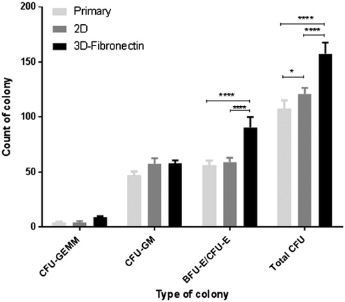
Table 3. Number of colony before and after expansion under different conditions.
Assessment of HSC homing and self renewality
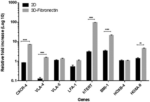
Evaluation of homing and self renewality measured the level of expression of CXCR-4, LFA-1, VLA-4, VLA-5 and BMI-1, hTERT, HOXA9 and HOXB4. Compared with unmanipulated cells, cells expanded in FN-coated PCL scaffold exhibited higher levels of homing genes, but only an expression of CXCR-4 (7.2 fold) was statically significant (p = .0001). Compared with that cultured in the 2D cell culture system, cells expanded in FN-coated PCL scaffold showed increased expression of homing genes, that CXCR-4 (8.7-fold) and VLA-4 (12.1-fold) were statically significant (p = .0001). Expanded cells in the 2D cell culture system decreased expression of homing genes compared to before expansion that declines expression of VLA-4 which was statically significant (p = .0001) and only VLA-5 had slightly increased.
Expression of self renewality genes in 3D culture system compared to unexpanded cells had increased and this increase for BMI-1 (20.5-fold) and hTERT (94.5-fold) was statically significant (p = .0001). The 2D cell culture system had increased in self renewality genes compared to before expansion which was not statically significant. 3D scaffold comparisons to 2D cell culture system showed increased expression of self renewality genes and expression of BMI-1 (5.9-fold) and hTERT (31.9-fold) was statically significant (p = .0001) ().
Figure 8. Relative fold increase homing and selfrenewality genes in 2D and 3D cell culture systems after 10 days culture compared to unexpanded cells.
Discussion
Self-renewal and proliferation of HSCs are controlled by a set of the factors, including intrinsic factors of HSCs and signals of HSC niche [Citation19].
The microenvironment of the 3D structure mimics the in vivo bone marrow environment to provide the sites and basic structure for haematopoietic stem/progenitor cells to grow and differentiate. Different 3D culture systems have been reported in the ex vivo expansion of haematopoietic stem/progenitor cells [Citation20,Citation21]. This study examines the impacts of two of the characters of the HSC niche, three-dimensionality, and adhesion signals, on HSC expansion. This study used FN and a cocktail of SCF, TPO and Flt3 cytokines for cultures. Since, Yokota et al. found that FN is more efficacious than other ECM molecules in enhancing HSC expansion [Citation22] and also SCF, TPO and Flt3L have been demonstrated to be crucial cytokines in the murine transplantation model [Citation23]. Thus, effect of FN on HSC expansion was investigated here in the context of 3D PCL nano-scaffold coated with FN in the medium.
According to our findings, adding the growth factors TPO, FLt-3 and SCF to the medium increased 2.66±-fold the number of CD34+ cells and increased TNCs to 38 ± 3.3.
McNiece et al. showed ex vivo expansion of CB CD34+ cells in liquid medium for 10 days and in the presence of cytokines SCF, G-CSF and TPO increased the number of TNCs and CD34+ progenitors as much as 56-fold and 4-fold, respectively [Citation24]. However, the expansion of cells with cytokines alone caused a loss of CD34+ progenitors [Citation25]. For this reason, in addition to growth factors TPO, FL and SCF, we used PCL nano-scaffold coated with FN as a feeder layer for the expansion of CD34+ cells. After 10 days of expansion in a 3D medium, we observed 40 ± 2.3-fold increases in the number of CD34+ progenitors and 58 ± 1.63-fold in the number of TNCs.
The reasons of this difference are to facilitate cell–cell and cell–matrix interactions and also imitating of physiochemical and cellular microenvironment and BM architecture (niche) in 3D scaffold than 2D. 3D provides a potential platform to raise the surface-to-volume ratio to improve cell interaction. In 2D cell culture system, this interaction capacity is low and limited between neighbour cells, therefore, TNC and CD34+ cell expansion in 2D is lower than 3D [Citation26].
Study on colony forming units from 1000 TNCs resulting from expansion in different culture conditions revealed that 3D PCL nano-scaffold coated with FN causes a significant increase in the number of colonies (128 ± 10) as well as the production of progenitors after 14 days. This was the highest colony number among three groups. In groups treated with growth factors only, colony numbers were 47 ± 4, which was not significantly different compared to the samples before expansion, which created 46 ± 4.5 colonies in cell culture.
The CFU-G/GM number represents the number of haematopoietic stem/progenitor cells with the capacity to differentiate into myeloid cells and the BFU-E number reflects the potential of haematopoietic stem/progenitor cells to develop into erythrocytes, both of which are important for the engraftment of human stem cells in transplantation. Our data suggest that our 3D culture system enhances the myeloid lineage amplification better than the erythroid lineage. In this experiment, the CFU-G/GM of the amplified cells increased significantly after 12 days’ culture in the 3D system, whereas the BFU-E number decreased in both the 2D and 3D culture systems at day 12. Similar data regarding the change in BFU-E levels have also been reported in the collagen bead 3D culture system [Citation21]. One explanation for the lower BFU-E number is that in both 3D culture systems the cytokine EPO was absent [Citation27].
Andrade-Zaldívar et al. introduced the new cell culture system, called Roller Bottles (RBs) that could culture larger volumes than the traditional static cultures. In a study in 2010, they showed CB HSCs expansion in RB in the CO2-free atmosphere with a different medium (IMDM, Stem Pro 34-SFM and L-15 Leibovitz’s). They find that RBs are suitable to culture human umbilical CB MNC in all media notice above. L-15 RB had higher and longer CFC expansion (18.39-fold for 13 days) than other medium and expansion using the L-15 medium in RBs was about 10 times higher than that static control cultures [Citation28].
In another study in 2014, Andrade-Zaldívar et al. had contrary data than 2011 and revealed that maximum total cell proliferation (26 vs. 20-folds for 10 days culture) and maximum progenitor expansion (17 vs. 16-folds for five days culture) in the static medium were higher than RB. These difference are maybe higher progenitor expansion could be found even earlier in the cultures of the enriched fractions and also, primitive cells cultured in static conditions may lose their stem features [Citation29].
In final, Andrade-Zaldívar et al. demonstrated that RBs were suitable for the expansion of haematopoietic progenitor’s cells and were simply scalable to large volumes and they were appropriate for the culture of suspended cells such as MNC. Also, RBs had the capability to achieve cultures 20 times larger than the statics and therefore 20 times more total expanded progenitors [Citation28,Citation29].
CXCR-4, LFA-1, VLA-4 and VLA-5 are required at high levels for efficient homing of circulating HSC/HPCs into the bone marrow niche. Homing to HSC niche and the regulation of HSCs depended on HSC binding to FN through β1 integrin in VLA-4 and VLA-5.
A study has shown that several ARs are expressed on CB CD34+ cells [Citation30]. There are two reports suggesting that AR expression on CD34+ cells during cytokine culture is unchanged [Citation31,Citation32], while Chute et al. reported that culture of bone marrow CD34+ cells on porcine microvascular endothelial cells (PMVECs) with cytokines for seven days produced a significant increase in the expression of VLA-4, CD58(LFA-3) and L-selectin but not of LFA-1 or CD44 [Citation33]. In contrast, our study demonstrated an increase in the expression of all ARs evaluated on CD34+ cells in 2D and 3D. Probably, different microenvironments and cytokine cocktails impact the expression of individual ARs to a greater or lesser degree.
It has been documented that the interaction between CAMs receptors on HSPC and ECM in the bone marrow microenvironment plays an important role in stem cell homing to the extravascular bone marrow niche [Citation34–36]. Papayannopoulou and Nakamoto [Citation37] have shown that anti-integrin α4β1 (VLA-4) antibody treatment selectively induced up to 200-fold increase of mobilization of haematopoietic progenitors in primates.
In NOD/SCID mouse model, pretreatment of donor cells with anti-VLA-4 antibody also significantly reduced the homing of HSPCs to the bone marrow, as indicated by an increase of circulating HSPCs [Citation38].
Flow cytometry analysis showed that 3D PCL nano-scaffold coated with FN causes to keep better self-renewality than TCPS.
The importance of HOXB-4, BIM-1, HOX-A9 and hTERT in HSC self-renewal has been demonstrated before [Citation23]. Some study shows that increased expression of Bmi-1 and HOXB-4 led to the symmetrical cell division of HSC and results in HSC expansion [Citation39,Citation40]. Also, other studies showed ectopic expression of HOXB-4 can mediate a significant expansion of HSC [Citation41,Citation42] and also HOXB-4 and HOXA-9, enhance HSC activity. Human telomerase reverse transcriptase (hTERT), in human CB CD34+ cells lead to an enhanced survival of mature haematopoietic cells. Our results confirm this study and showed that after expansion, especially in 3D PCL nano-scaffold increased self-renewality genes.
Multiple important parameters, such as haematopoietic microenvironment, contact with stromal cells and ECM, some growth factors and cytokines could be the effect on homing and self-renewal of stem cells.
In addition, agitation of cell culture medium (dynamically) and oxygenation can effect on HSCs expansion. Tiwari et al. showed that combination of dynamic culture with lower percentage oxygen (5%) had better results in expansion of CD34+ cells, CFU and MNC than static cell culture medium with higher oxygen. As, low oxygen promotes quiescence and regulates their differentiation, maintaining stem cell phenotype. Also, fluid shear stress produced in a dynamic culture system led to the production of mechanical signal that can act on cells [Citation43].
In this research, we found that 3D PCL scaffold coated with FN was suited for expansion of HSC, because, this scaffold led to higher cell proliferation count, better homing, and adhesion.
Acknowledgements
We would like to thank Dr. Zarrabi, Chief Director of Royan Stem Cell Technology Company (Royan Cord Blood Bank), and Mr. Janzamin for his assistance with flow cytometry.
Disclosure statement
No potential conflict of interest was reported by the authors.
Additional information
Funding
References
- Ballen KK, Gluckman E, Broxmeyer HE. Umbilical cord blood transplantation: the first 25 years and beyond. Blood. 2013;122:491–498.
- Smith AR, Wagner JE. Alternative haematopoietic stem cell sources for transplantation: place of umbilical cord blood. Br J Haematol. 2009;147:246–261.
- Kurtzberg J. Update on umbilical cord blood transplantation. Curr Opin Pediatr. 2009;21:22.
- Stanevsky A, Goldstein G, Nagler A. Umbilical cord blood transplantation: pros, cons and beyond. Blood Rev. 2009;23:199–204.
- Cabral J. Ex vivo expansion of hematopoietic stem cells in bioreactors. Biotechnol Lett. 2001;23:741–751.
- Delaney C, Heimfeld S, Brashem-Stein C, et al. Notch-mediated expansion of human cord blood progenitor cells capable of rapid myeloid reconstitution. Nat Med. 2010;16:232–236.
- Horwitz ME, Frassoni F. Improving the outcome of umbilical cord blood transplantation through ex vivo expansion or graft manipulation. Cytotherapy. 2015;17:730–738.
- Mehta RS, Rezvani K, Olson A, et al. Novel techniques for ex vivo expansion of cord blood: clinical trials. Front Med (Lausanne). 2015;2:89.
- Robinson S, Ng J, Niu T, et al. Superior ex vivo cord blood expansion following co-culture with bone marrow-derived mesenchymal stem cells. Bone Marrow Transplant. 2006;37:359–366.
- Sachlos E, Czernuszka J. Making tissue engineering scaffolds work. Review: the application of solid freeform fabrication technology to the production of tissue engineering scaffolds. Eur Cell Mater. 2003;5:39–40.
- Baron F, Ruggeri A, Nagler A. Methods of ex vivo expansion of human cord blood cells: challenges, successes and clinical implications. Expert Rev Hematol. 2016;9:297–314.
- Pan X, Sun Q, Cai H, et al. Encapsulated feeder cells within alginate beads for ex vivo expansion of cord blood-derived CD34(+) cells. Biomater Sci. 2016;4:1441–1453.
- Kelly S, Sola C, De Lima M, et al. Ex vivo expansion of cord blood. Bone Marrow Transplant. 2009;44:673–681.
- Liu H, Lin J, Roy K. Effect of 3D scaffold and dynamic culture condition on the global gene expression profile of mouse embryonic stem cells. Biomaterials. 2006;27:5978–5989.
- Wilson A, Trumpp A. Bone-marrow haematopoietic-stem-cell niches. Nat Rev Immunol. 2006;6:93–106.
- Even-Ram S, Yamada KM. Cell migration in 3D matrix. Curr Opin Cell Biol. 2005;17:524–532.
- Ferreira MSV, Jahnen-Dechent W, Labude N, et al. Cord blood-hematopoietic stem cell expansion in 3D fibrin scaffolds with stromal support. Biomaterials. 2012;33:6987–6997.
- Vazin T, Schaffer DV. Engineering strategies to emulate the stem cell niche. Trends Biotechnol. 2010;28:117–124.
- Feng Q, Chai C, Jiang XS, et al. Expansion of engrafting human hematopoietic stem/progenitor cells in three-dimensional scaffolds with surface-immobilized fibronectin. J Biomed Mater Res A. 2006;78:781–791.
- Ehring B, Biber K, Upton T, et al. Expansion of HPCs from cord blood in a novel 3D matrix. Cytotherapy. 2003;5:490–499.
- Kim H-S, Lim JB, Min YH, et al. Ex vivo expansion of human umbilical cord blood CD34+ cells in a collagen bead-containing 3-dimensional culture system. Int J Hematol. 2003;78:126–132.
- Yokota T, Oritani K, Mitsui H, et al. Growth-supporting activities of fibronectin on hematopoietic stem/progenitor cells in vitro and in vivo: structural requirement for fibronectin activities of CS1 and cell-binding domains. Blood. 1998;91:3263–3272.
- Hofmeister C, Zhang J, Knight K, et al. Ex vivo expansion of umbilical cord blood stem cells for transplantation: growing knowledge from the hematopoietic niche. Bone Marrow Transplant. 2007;39:11–23.
- McNiece I, Harrington J, Turney J, et al. Ex vivo expansion of cord blood mononuclear cells on mesenchymal stem cells. Cytotherapy. 2004;6:311–317.
- McNiece I, Kubegov D, Kerzic P, et al. Increased expansion and differentiation of cord blood products using a two-step expansion culture. Exp Hematol. 2000;28:1181–1186.
- Mortera-Blanco T, Mantalaris A, Bismarck A, et al. Long-term cytokine-free expansion of cord blood mononuclear cells in three-dimensional scaffolds. Biomaterials. 2011;32:9263–9270.
- Yuan Y, Tse KT, Sin FY, et al. Ex vivo amplification of human hematopoietic stem and progenitor cells in an alginate three‐dimensional culture system. Int J Lab Hematol. 2011;33:516–525.
- Andrade-Zaldívar H, Kalixto-Sánchez MA, de la Rosa APB, et al. Expansion of human hematopoietic cells from umbilical cord blood using roller bottles in CO2 and CO2-free atmosphere. Stem Cells Dev. 2010;20:593–598.
- Andrade-Zaldívar H, Kalixto-Sánchez M, Barba de La Rosa A, et al. Expansion of CD34+ human hematopoietic cells from umbilical cord blood using roller bottles. Rev Mex Ingeniería Quím. 2014;13:593–598.
- Denning‐Kendall P, Singha S, Bradley B, et al. Cytokine expansion culture of cord blood CD34+ cells induces marked and sustained changes in adhesion receptor and CXCR4 expressions. Stem Cells. 2003;21:61–70.
- Dravid G, Rao S. Ex vivo expansion of stem cells from umbilical cord blood: expression of cell adhesion molecules. Stem Cells. 2002;20:183–189.
- Reems JA, Mielcarek M, Torok-Storb B. Differential modulation of adhesion markers with ex vivo expansion of human umbilical CD34+ progenitor cells. Biol Blood Marrow Transplant. 1997;3:133–141.
- Chute JP, Saini AA, Kampen RL, et al. A comparative study of the cell cycle status and primitive cell adhesion molecule profile of human CD34+ cells cultured in stroma-free versus porcine microvascular endothelial cell cultures. Exp Hematol. 1999;27:370–379.
- Berrios VM, Dooner GJ, Nowakowski G, et al. The molecular basis for the cytokine-induced defect in homing and engraftment of hematopoietic stem cells. Exp Hematol. 2001;29:1326–1335.
- Papayannopoulou T, Craddock C. Homing and trafficking of hemopoietic progenitor cells. Acta Haematol. 1997;97:97–104.
- Simmons PJ, Levesque J-P, Zannettino AC. Adhesion molecules in haemopoiesis. Baillieres Clin Haematol. 1997;10:485–505.
- Papayannopoulou T, Nakamoto B. Peripheralization of hemopoietic progenitors in primates treated with anti-VLA4 integrin. Proc Natl Acad Sci USA. 1993;90:9374–9378.
- Papayannopoulou T, Craddock C, Nakamoto B, et al. The VLA4/VCAM-1 adhesion pathway defines contrasting mechanisms of lodgement of transplanted murine hemopoietic progenitors between bone marrow and spleen. Proc Natl Acad Sci USA. 1995;92:9647–9651.
- Antonchuk J, Sauvageau G, Humphries RK. HOXB4-induced expansion of adult hematopoietic stem cells ex vivo. Cell. 2002;109:39–45.
- Iwama A, Oguro H, Negishi M, et al. Enhanced self-renewal of hematopoietic stem cells mediated by the polycomb gene product Bmi-1. Immunity. 2004;21:843–851.
- Amsellem S, Pflumio F, Bardinet D, et al. Ex vivo expansion of human hematopoietic stem cells by direct delivery of the HOXB4 homeoprotein. Nat Med. 2003;9:1423–1427.
- Sauvageau G, Thorsteinsdottir U, Eaves C, et al. Overexpression of HOXB4 in hematopoietic cells causes the selective expansion of more primitive populations in vitro and in vivo. Genes Dev. 1995;9:1753–1765.
- Tiwari A, Wong CS, Nekkanti LP, et al. Impact of oxygen levels on human hematopoietic stem and progenitor cell expansion. Stem Cells Dev. 2016;25:1604–1613.