
Abstract
Background: Recently, microRNA-133b (miR-133b) dysregulation has been shown to play a key role in several human cancers, as well as glioma. In this study, we aimed to investigate the clinical significance and prognostic value of miR-133b in glioma.
Methods: Real-time quantitative PCR was employed to measure the expression level of miR-133b in tissues. Survival analysis was carried out by using the log-rank test and Kaplan–Meier method. Prognostic factors for overall survival were identified by univariate and multivariate analyses using the Cox proportional hazards regression model.
Results: The expression level of miR-133b was significantly lower in glioma tissues compared with matched non-cancerous brain tissues (p < .05). Its level was strongly correlated with Karnofsky Performance Scale score (p < .001) and WHO grade (p < .001). Kaplan–Meier survival and log-rank analysis indicated that the decreased expression of miR-133b was strongly correlated with shorter overall survival of patients with glioma (log-rank test, p = .03).
Conclusions: The current investigation demonstrated that miR-133b level is useful for predicting the prognosis of patients with glioma.
Introduction
Glioma is the most frequent and malignant primary brain tumours with high morbidity and mortality in human adults. According to the WHO classification, gliomas are divided into pilocytic astrocytoma (WHO grade I), diffuse astrocytoma (WHO grade II), anaplastic astrocytoma (WHO grade III) and glioblastoma (GBM, WHO grade IV) in the order of increasing malignancy [Citation1]. The grade IV glioma is the most common and aggressive form of glioma, with a median survival of only 12–15 months as compared to 2–5 years for patients with grade III gliomas and 6–8 years for low grade (I and II) gliomas [Citation2]. Despite recent advances in surgery, radiotherapy and chemotherapy, the prognosis for patients with this tumour remains poor [Citation1]. Therefore, it is crucial to investigate the mechanism involved in the development and progression of glioma and to find new therapeutic targets and new diagnostic and prognostic biomarkers [Citation3].
MicroRNAs (miRNAs) are a family of endogenous small non-coding RNAs (19–24 net) that regulate gene expression by antisense complementarity to specific mRNA [Citation4]. They have been showed to play important role in regulation of tumourigenesis, differentiation, proliferation and survival through the inhibition of major cellular pathways in diverse human cancers [Citation5,Citation6].
Recently, microRNA-133b (miR-133b) dysregulation has been shown to play a key role in several human cancers, as well as glioma. Previously, the study by Li et al. showed that miR-133b was able to suppressed the proliferation and invasion of glioma cells, at least partly by targeting silent information regulator 1 (Sirt1) [Citation7]. Furthermore, Wang et al. showed that miR-133b was markedly down-regulated in clinical glioblastoma specimens, and contributed to arsenic-induced apoptosis in glioma cells by targeting the hERG channel [Citation8]. In this study, we aimed to investigate the clinical significance and prognostic value of miR-133b in glioma.
Materials and methods
Patients and tissue samples
The present study was approved by the Research Ethics Committee of Qilu Hospital of Shandong University. All specimens were handled and made anonymous according to the ethical and legal standards and were obtained with patients’ written informed consent. 97 glioma patients who underwent tumour resection from May 2007 to February 2015 in Qilu Hospital of Shandong University were enrolled in this study. Immediately after surgery, samples were snap-frozen and stored in liquid nitrogen. The matched normal tissues were taken from the distal end of the operative excisions, far from the tumour. None of the patients had received chemotherapy or radiotherapy prior to surgery, and overall survival time was calculated from the date of the initial surgical operation to death. Patients who died of diseases not directly related to their gliomas were excluded from this study. The clinicopathological features of all patients were indicated in .
Table 1. Clinicopathological characteristics of 97 patients with glioma.
RNA isolation and qRT-PCR
Total RNA was extracted from glioma tissues and matched normal adjacent brain tissues by homogenizing tissue in Trizol reagent (Invitrogen, Carlsbad, CA) according to the manufacturer’s instructions. Primers for miR-133b and endogenous control U6 snRNA were obtained from Applied Biosystems (Foster City, CA). The concentration and purity of RNA were determined spectrophotometrically using the NanoDrop ND-1000 (NanoDrop Technologies, Wilmington, DE). cDNA was generated using the PrimeScript RT reagent kit (Takara Co. Ltd., Dalian, China) in a 20 μl final reaction volume containing 0.5 in of RNA, 0.5 μl Prime-Script RT enzyme mix, and 4 μl 5 × PrimeScript buffer, and 1 μl RT primer, and incubated at 42 °C for 60 min and at 85 °C for 5 min. Quantitative real-time PCR assay was performed to evaluate miR-133b expression using SYBR Premix Ex Taq (Takara Co. Ltd.) and measured in a LightCycler 480 System (Roche, Basel, Switzerland). The amplification profile was denatured at 95 °C for 10 min, followed by 45 cycles of denaturation at 95 °C for 15 s, annealing at 60 °C for 30 s, and extension at 72 °C for 1 min. Relative quantification of miRNA expression was performed using the 2−△△CT. The raw data were presented as the relative quantity of target miRNA, normalized with respect to U6 snRNA and relative to a calibrator sample.
Statistical analysis
Differences between groups were evaluated using Mann–Whitney U test. Chi-square test was used to analyze the association between miR-133b expression and the clinicopathological characteristics of patients with glioma. Survival analysis was carried out by using the log-rank test and Kaplan–Meier method. Prognostic factors for overall survival were identified by univariate and multivariate analyses using the Cox proportional hazards regression model. Differences were considered statistically significant when p was less than .05. All data were analyzed using SPSS 18.0 software (SPSS Inc., Chicago, IL).
Results
The expression of miR-133b in glioma tissues
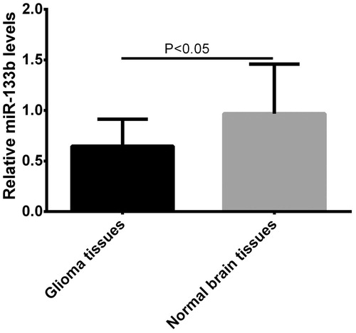
The expression level of miR-133b in 97 pairs of normal brain tissues and glioma tissues was analyzed by real-time quantitative RT-PCR. We found that the expression level of miR-133b was significantly lower in glioma tissues compared with matched non-cancerous brain tissues (p < .05, shown in ). We divided 97 glioma patients into two groups according to the levels of miR-133b expression. The cut-off point was the median expression level of miR-133b (high expression group, n = 49; low expression group, n = 48).
Figure 1. qRT-PCR detection of relative miR-133b expression in glioma tissues and normal brain tissues.
Relationship between miR-133b expression levels and clinicopathological features
The relationship between miR-133b expression level and clinicopathological parameters was evaluated. As shown in , the level of miR-133b in glioma was strongly correlated with Karnofsky Performance Scale (KPS) score (p < .001) and WHO grade (p < .001). However, there was no significant difference between low expression of miR-133b and other clinicopathological characteristics, such as age, gender, smoking status and surgery type.
The correlation of miR-133b expression with survival in glioma
Kaplan–Meier survival and log-rank analysis were performed to evaluate association of miR-133b expression with overall survival of glioma patients. The results indicated that the decreased expression of miR-133b was strongly correlated with shorter overall survival of patients with glioma (log-rank test, p = .03; shown in ). Multivariate analysis for overall survival identified miR-133b levels as an independent prognostic factor in glioma (HR = 3.197, 95%CI: 1.826–8.055, p = .017, shown in ).
Figure 2. Kaplan–Meier curve for overall survival in glioma patients with low versus high miR-133b expression.
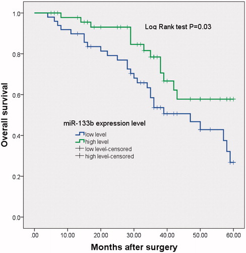
Table 2. Multivariate analysis for overall survival in 97 patients with glioma.
Discussion
Malignant gliomas are the most common primary tumours of the central nervous system with high morbidity and mortality [Citation9]. A combination of surgery, radiotherapy and chemotherapy is widely used to treat gliomas, particularly malignant gliomas. However, the prognosis of the disease remains poor [Citation10]. Therefore, it is very important to identify new biomarkers that can be used in clinicopathological determinants of prognosis and in the choice of better therapeutic strategies.
In recent years, increasing studies have highlighted the importance of microRNAs, a class of small and non-coding RNAs, in the regulation of gene expression [Citation11]. More than 50% of miRNAs are located in cancer-associated genomic break points, and can function as tumour suppressor genes or oncogenes, depending on their targets [Citation12,Citation13]. Recent studies have implicated that the expression signature of these small non-coding RNA molecules can provide insight into the diagnosis and prognosis of human cancers including glioma [Citation14].
Recently, miR-133b dysregulation has been shown to play a key role in several types of human cancer. For example, Chen et al. found that miR-133b was down-regulated in urothelial carcinoma of the bladder (UCB) tissues. Furthermore, down-regulation of miR-133b was associated with aggressive clinicopathological features of UCB and was able to predict unfavorable prognosis in patients with UCB, suggesting that it might serve as feasible biomarker for clinical outcome of UCB patients after surgery and potential therapeutic target in the future [Citation15]. Li et al. found that miR-133b targeted and down-regulated RB1CC1 in prostate cancer cells. The expression of miR-133b and RB1CC1 protein was inversely correlated, and they could be used to predict the risk of biochemical recurrence (BCR) in patients with prostate cancer after radical prostatectomy (RP) [Citation16]. The expression of miR-133b was found to be greatly down-regulated in colorectal cancer, and it was associated with overall survival and metastasis of colorectal cancer [Citation17]. In addition, miR-133b expression inhibited the proliferation of colorectal cancer cells in vitro and in vivo by directly targeting the receptor tyrosine kinase MET [Citation18]. Liu et al. have found that the expression of miR-133b was decreased in non-small-cell lung cancer (NSCLC), and it was associated with tumour stage, the extent of regional lymph node involvement, stage, visceral pleura or vessel invasion and EGFR mRNA expression of NSCLC patients. miR-133b was able to regulate growth, invasion and apoptosis of NSCLC cells by targeting EGFR. Moreover, they also suggest that transfection of miR-133b has therapeutic potential for overcoming resistance to EGFR-TKI in EGFR-addicted NSCLC [Citation19]. Kano et al. found that miR-133b could act as a tumour suppressor in esophageal squamous cell carcinoma by inhibiting FSCN1 expression [Citation20].
The role of miR-133b in glioma has also been investigated in glioma. Previously, Wang et al. showed that miR-133b was markedly down-regulated in clinical glioblastoma specimens, and contributed to arsenic-induced apoptosis in U251 glioma cells by targeting the hERG channel [Citation8]. Furthermore, the study by Li et al. showed that miR-133b was able to suppressed the proliferation and invasion of glioma U87 cells, at least partly by targeting silent information regulator 1 (Sirt1) [Citation7]. However, until now, the clinical significance and prognostic value of miR-133b in glioma have not been investigated. Therefore, we aimed to investigate the clinical significance and prognostic value of miR-133b in glioma.
In this study, the expression level of miR-133b in 97 pairs of normal brain tissues and glioma tissues was analyzed by real-time quantitative RT-PCR. We found that the expression level of miR-133b was significantly lower in glioma tissues compared with matched non-cancerous brain tissues. The relationship between miR-133b expression level and clinicopathological parameters was evaluated. We found that the level of miR-133b in glioma was strongly correlated with KPS score and WHO grade. Kaplan–Meier survival and log-rank analysis indicated that the decreased expression of miR-133b was strongly correlated with shorter overall survival of patients with glioma. Furthermore, multivariate analysis for overall survival identified miR-133b levels as an independent prognostic factor in glioma.
In conclusion, the results of this study confirmed the clinical and prognostic significance of miR-133b in glioma. The down-regulation of miR-133b may play an important role in the progression of glioma and can be used as an independent factor to determine the prognosis of patients with glioma.
Disclosure statement
No potential conflict of interest was reported by the authors.
References
- Rousseau A, Mokhtari K, Duyckaerts C. The 2007 WHO classification of tumors of the central nervous system – what has changed? Curr Opin Neurol. 2008;21:720–727.
- Louis DN, Ohgaki H, Wiestler OD, et al. The 2007 WHO classification of tumours of the central nervous system. Acta Neuropathol. 2007;114:97–109.
- Marumoto T, Saya H. Molecular biology of glioma. Adv Exp Med Biol. 2012;746:2–11.
- Valinezhad Orang A, Safaralizadeh R, Kazemzadeh-Bavili M. Mechanisms of miRNA-mediated gene regulation from common downregulation to mRNA-specific upregulation. Int J Genomics. 2014;2014:970607.
- Croce CM, Calin GA. miRNAs, cancer, and stem cell division. Cell. 2005;122:6–7.
- Treiber T, Treiber N, Meister G. Regulation of microRNA biogenesis and function. Thromb Haemost. 2012;107:605–610.
- Li C, Liu Z, Yang K, et al. miR-133b inhibits glioma cell proliferation and invasion by targeting Sirt1. Oncotarget. 2016;7:36247–36254.
- Wang J, Li Y, Jiang C. MiR-133b contributes to arsenic-induced apoptosis in U251 glioma cells by targeting the hERG channel. J Mol Neurosci. 2015;55:985–994.
- Reardon DA, Rich JN, Friedman HS, et al. Recent advances in the treatment of malignant astrocytoma. JCO. 2006;24:1253–1265.
- Hottinger AF, Stupp R, Homicsko K. Standards of care and novel approaches in the management of glioblastoma multiforme. Chin J Cancer. 2014;33:32–39.
- Kasinski AL, Slack FJ. Epigenetics and genetics. MicroRNAs en route to the clinic: progress in validating and targeting microRNAs for cancer therapy. Nat Rev Cancer. 2011;11:849–864.
- Calin GA, Croce CM. MicroRNA signatures in human cancers. Nat Rev Cancer. 2006;6:857–866.
- Esquela-Kerscher A, Slack FJ. Oncomirs – microRNAs with a role in cancer. Nat Rev Cancer. 2006;6:259–269.
- Guan Y, Mizoguchi M, Yoshimoto K, et al. MiRNA-196 is upregulated in glioblastoma but not in anaplastic astrocytoma and has prognostic significance. Clin Cancer Res. 2010;16:4289–4297.
- Chen X, Wu B, Xu Z, et al. Downregulation of miR-133b predict progression and poor prognosis in patients with urothelial carcinoma of bladder. Cancer Med. 2016;5:1856–1862.
- Li X, Wan X, Chen H, et al. Identification of miR-133b and RB1CC1 as independent predictors for biochemical recurrence and potential therapeutic targets for prostate cancer. Clin Cancer Res. 2014;20:2312–2325.
- Akcakaya P, Ekelund S, Kolosenko I, et al. miR-185 and miR-133b deregulation is associated with overall survival and metastasis in colorectal cancer. Int J Oncol. 2011;39:311–318.
- Hu G, Chen D, Li X, et al. miR-133b regulates the MET proto-oncogene and inhibits the growth of colorectal cancer cells in vitro and in vivo. Cancer Biol Ther. 2010;10:190–197.
- Liu L, Shao X, Gao W, et al. MicroRNA-133b inhibits the growth of non-small-cell lung cancer by targeting the epidermal growth factor receptor. FEBS J. 2012;279:3800–3812.
- Kano M, Seki N, Kikkawa N, et al. miR-145, miR-133a and miR-133b: tumor-suppressive miRNAs target FSCN1 in esophageal squamous cell carcinoma. Int J Cancer. 2010;127:2804–2814.