
Abstract
Glaucoma is one of the leading causes of blind worldwide. Post-operative scar formation of filtering tract was one of the main reasons for failure of glaucoma filtration surgery. In this study, we conducted several experiments to detect the expression of miR-26a in the scar tissue in filtering tract and then detect its potential biological effects as well as its target gene. In our present study, it was found that miR-26a was significantly down-regulated in filtering tract scar. Advanced study on the association between the miR-26a and connective tissue growth factor (CTGF) micro RNA (mRNA) showed that miR-26a was inversely correlated with CTGF mRNA level. Advanced biological studies showed that overexpression of miR-26a could decrease the cell viability and migration ability of human Tenon’s fibroblasts (HTFs) fibrosis in vitro model. It was also found that miR-26a might up-regulate the apoptotic level of HTFs. Through protein expression detection and luciferase reporter assay, it was found that miR-26a could produce functions that directly target the CTGF. In conclusion, the key finding of the current study is that miR-26a can suppress the activation of HTFs by transforming growth factor (TGF)-β by targeting CTGF. This data indicates that miR-26a plays an essential role in the formation of filtering tract scar and function as a potential drug target.
Introduction
Glaucoma is commonly recognized as one of the leading causes of blind worldwide [Citation1]. By now, there were approximately 66.8 million people being afflicted with glaucoma and this number was expected to increase to 80 million by 2020 [Citation2]. At present, filtration surgery is still one of the main clinical methods for the treatment of glaucoma [Citation3]. Post-operative scar formation of filtering tract was one of the main reasons for failure of glaucoma filtration surgery and the failure rate within 2 years was reported to be 15%∼25% [Citation4]. Several different methods, including topical application of mitomycin C and 5-FU, were obtained on enhancing the efficacy of filtration surgery and prevent the incidence of bleb failure [Citation5]. However, the use of these drugs in surgery leads to several potential complications, including filtering bleb leakage, persistent low intraocular pressure, corneal injury and ocular infection [Citation6].
As reported in previous studies, scar formation in the filtering tract of glaucoma surgery is mainly caused by the proliferation of fibroblasts and collagen deposition. The human Tenon’s fibroblasts (HTFs) were active by the surgical stimulation and growth factor exposure and then transformed to myofibroblasts (MFs) which produced secretion and contraction functions [Citation7]. Among all the potential influenced growth factors, the transforming growth factor (TGF-β1) and connective tissue growth factor (CTGF) in the downstream played the key role in the phenotype transition of HTFs and extracellular matrix (ECM) formation [Citation8]. As reported in a previous study, it was found that increased levels of was increased at subconjunctival wounds after filtering surgery [Citation9]. CTGF coordinates the signalling of growth factors and promotes fibrosis [Citation10]. CTGF was the downstream effector of TGF-β1 and it mediated the effect of TGF on the ECM accumulation [Citation11]. In addition to a direct fibrogenic effect, CTGF can exacerbate TGF-β-induced fibrosis by activating TGF-β and SMAD signalling through promoting the association of TGF-β with its receptor. Considering the existence and importance of the TGF/CTGF pathway, it was quite important to detect the potential inhibitor of TGF/CTGF pathway and might produce as potential target of drug development.
MicroRNA (mRNA) is a small non-coding RNA and functions in transcriptional and post-transcriptional regulation of gene expression [Citation12]. In general, mRNAs could combine with their targets’ 3'-untranslated region (3’-UTR) regions and then regulate the target genes by direct degradation of mRNA or by inhibiting protein synthesis, according to the degree of complementarities in combination [Citation13]. Recent studies provide clear evidence that mRNAs are abundant in the ocular [Citation14] and play an important role in the functions and pathological progress in the ocular disorders [Citation15]. Dysregulation of mRNAs is involved in numerous pathological conditions, including fibrosis [Citation16]. As reported in previous studies, miR-26a was reported to be involved in the TGF-β-dependent fibrosis formation of diabetic nephropathy [Citation16] and lung disorders [Citation17]. It was hypothesized that key mRNAs may synergistically target individual critical genes and thus regulate scar formation after glaucoma filtration surgery. In this study, we conducted several experiments to detect the expression of miR-26a in the scar tissue in filtering tract and then detect its potential biological effects as well as its target gene.
Materials and methods
Tissue samples
All the tissue samples were scar samples and subconjunctival Tenon’s capsule tissues. The glaucoma patients included in this study were obtained between 2015.0 1 and ∼2016.07 in Department of Ophthalmology, Affiliated Hospital of Weifang Medical University. The control samples were from the donated samples. A total of 11 glaucoma cases and 11 age-matched controls were included in the analyses. All samples were obtained with informed consent and approved by the institutional review board of Affiliated Hospital of Weifang Medical University. The scar samples were obtained from the patient after the second glaucoma filtering surgery (removal of scar tissue from the filtering bleb local scar tissue and the scar tissues were confirmed by two surgeons together). The subconjunctival Tenon’s capsule tissues of controls were isolated and used for the polymerase chain reaction (PCR) analyses. Once removed, all the tissue samples were stored in dry ice and transported to liquid nitrogen as soon as possible.
RNA isolation and PCR analyses
Total RNA in tissue samples and in vitro models were isolated by using the mRNA Isolation Kit (Ambion, USA) according to the manufacturer’s instructions. The scar and control tissues were obtained after surgical resection and immediately placed in liquid nitrogen. All analysed tissues were homogenized before isolation. The extracted RNA samples were dissolved with 40 μL of nuclease-free water and stored at −80 °C for future use. Concentration and purification of RNA were determined by NanoDrop Spectrophotometer (Thermo Scientific Wilmington, DE, USA) and the value of A260/280 > 2.0, A260/230 > 1.8 were considered relatively high quality. For the PCR analysis, 500 ng of RNA was used to generate cDNA using the miScript reverse transcription kit. 2 μL of total RNA was reverse-transcribed at 42 °C for 30 min using mRNA-specific reverse transcription primers. Reverse transcription quantitative PCR (RT-qPCR) experiments were performed using TaqMan MiRNA Assays (Life Tech, Carlsbad, CA) following manufacturer instructions. During real-time quantitative PCR reaction, 2 μL of reverse transcription products were used as the template. The PCR reaction was performed in triplicate for the mRNA in each sample. The reaction conditions were as follows: 95 °C for one minute, 50 cycles at 95 °C for 15 s and 60 °C for 30 s. Relative expression was determined using the ΔΔCt method and a Ct value over 35 indicated negative amplification.
Cell culture
Tenon’s capsule fibroblasts were isolated from subconjunctival tissue and the tissues were from the elderly cataract patients as previously described [Citation18]. In general, tissue explant method was used in the primary culture. Then, HTFs in 5 ∼ 10 generation were identified by immunohistochemistry by vimentin and keratin using immunofluorescence staining before used in the following experiments. Cells were maintained in Dulbecco’s modified Eagle’s medium containing 10% foetal bovine serum (FBS), 1% L-glutamine (2 mM) and 1% antibiotics at 37 °C in 5% CO2 atmosphere.
mRNA transfection
HTFs were treated with miR-26a mimics (miR-26a precursor) or nonsense control (Invitrogen, CA, USA) using Lipofectamine® 2000 (Invitrogen). The mRNA transfection was conducted according to the manufacturer’s instructions. In general, Lipofectamine 2000 was mixed with the miR-26a vector and then used for advanced analyses. After 24 h of the treatment, the cells in all the independent groups were collected and assayed.
Cell proliferation and viability detection
Cell proliferation was measured using the BrdU proliferation assay. Briefly, 24 h after transfection, cells were cultured in triplicate at 5 × 10³ cells per well the day before BrdU incubation (Ribobio, Guangzhou, China). After BrdU labelling, the cells were treated with 100 ml of 1 × Apollo reaction cocktail, stained with 100 ml of Hoechst 33342 (5 mg/mL) and visualized under a fluorescence microscope (Olympus, Japan). The percentage of EdU positive cells was defined as the proliferation rate. For the cell viability assay, the MTT (3–(4,5-dimethylthiazol-2-yl)-2,5- diphenyltetrazolium bromide) method was obtained for the analyses. All the according to the manufacturer's instruction. After transfection of the HTFs with miR-26a, 20 μL of sterile MTT dye (Sigma-Aldrich, St. Louis, MO, USA) was added and incubated for 4 h at 37 °C. Then, 150 μL of dimethyl sulfoxide was added to each well and the plates were thoroughly mixed for 10 min. Spectrometric absorbance at a wavelength of 492 nm was measured on an enzyme immunoassay analyser (model 680; Bio-Rad Laboratories, Hercules, CA, USA).
Cell migration assays
Migration assay was conducted with Transwell® inserts with 8.0 mm pore size membrane (24-well format, Corning, New York, USA). A total of 1 × 10⁵ cells were resuspended in serum-free medium and seeded to the upper chamber. The lower chambers were filled with complete culture medium containing 10% FBS. After incubation at 37 °C for 24 h, the migrated cells present on the lower side of the membrane were fixed, stained and counted. Each experiment was performed in triplicate.
Evaluation of apoptosis by annexin V/PI
In this study, the apoptotic rates were analysed by Flow Cytometry (Beckman Coulter, CA, USA) using the annexin V-fluorescein isothiocyanate (FITC)/propidium iodide (PI) kit (Roche, Penzberg, Germany). Cells were washed twice in cold 1 PBS and resuspended in Annexin V-binding buffer (BD Pharmingen) at a concentration of 3 × 10⁶ per ml. This suspension (100 μl) was stained with 5 μl of Annexin V-FITC and 5 μl PI. The rate of both early and late apoptosis was calculated and recorded for the analyses. Each test was repeated in triplicate.
Luciferase reporter assay
Luciferase activity assay was performed as previously described [28]. Briefly, HTFs were cultured in the 12-well plate (1 × 10⁵ cells/well), and co-transfected with wild type or mutated 3′-UTRs of CTGF (WT and Mut, respectively) luciferase reporter constructs and miR-26a or control mimic with Lipofectamine 2000. After treatment for 24 h, firefly and Renilla luciferase activities were measured using the dual-luciferase reporter assay system (Promega, Madison, WI, USA).
Western blotting analyses
Proteins were separated on 15% SDS-PAGE gel and then transferred to PVDF membrane. After blocked with 5% non-fat milk, the membrane was incubated with rabbit anti-CTGF polyclonal antibody (1:1000, Proteintech, Chicago, IL, USA) and anti-glyceraldehyde-3-phosphate dehydrogenase (anti-GAPDH) antibody (Abcam, San Francisco, CA, USA, 1:1000 dilution). The secondary antibody was goat anti-rabbit IgG conjugated with horseradish peroxidase (HRP) with a dilution of 1:1000. Protein bands were visualized by enhanced chemiluminescence. Western blotting of GAPDH on the same membrane was used as a loading control. The grey scale of each strap was measured were automatically counted by using Image J software (http://rsbweb.nih.gov/ij/).
Statistical analysis
The data are expressed as the mean ± SD. Differences between the groups were assessed with the non-paired t test. In this study, SPSS 13.0 was used to analyse the cytotoxic (IBM, Chicago, IL). P less than .05 was considered statistically significance.
Results
Down-regulation of miR-26a in filtering tract scar
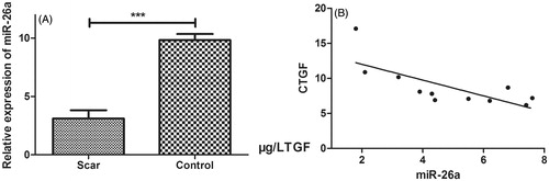
To detect the miR-26a expression pattern in the filtering tract scar, we detect the miR-26a expression levels in 11 scar and subconjunctival Tenon’s capsule tissues of age-matched controls. As showed in , it was found that miR-26a was significantly down-regulated in filtering tract scar tissues (P < .001). In advanced study on the association between miR-26a and CTGF mRNA, the miR-26a and CTGF mRNA in the 11 filtering tract scars were measured. It was found that miR-26a was inversely correlated with CTGF mRNA level (r2 = 0.5413, P = .0099, ).
Figure 1. miR-26a was significantly decreased in filtering tract scar. (A) The miR-26a was significantly down-regulated in filtering tract scar tissues. (B) miR-26a was inversely correlated with CTGF mRNA level. Graph represents the 2-ΔΔCt values ± SD, *P < .05, ***P < .001.
miR-26a inhibits TGF-β-induced HTFs activation
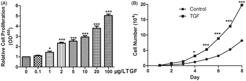
First, the cell viability and proliferation effect of TGF-β on HTFs were detected. As showed in , TGF-β in over 1 μg/L could significantly induce HTFs activation. TGF-β in 1, 2, 5, 10, 20 and 100 μg/L could improve cell viability significantly. It was also found that a dose–response relationship was also detected. Considering that the improvement of cell viability in 10 μg/L was quite significant, the quality concentration of 10 μg/L was obtained in the following experiments. Following study showed that after 10 μg/L TGF-β treatment for over 4 days, the proliferation rate increased significantly (P < 0.05) and the longer after the treatment, the more significantly improvement was detected ().
Figure 2. TGF-β treatment increased the cell viability and proliferation. (A) The cell viability was detected by TGF-β in 0.1, 1, 2, 5, 10, 20, 100 μg/L and TGF-β in 1, 2, 5, 10, 20 and 100 μg/L could improve cell viability significantly. (B) TGF-β treatment for over 4 days, the proliferation rate increased significantly. *P < .05, ***P < .001).
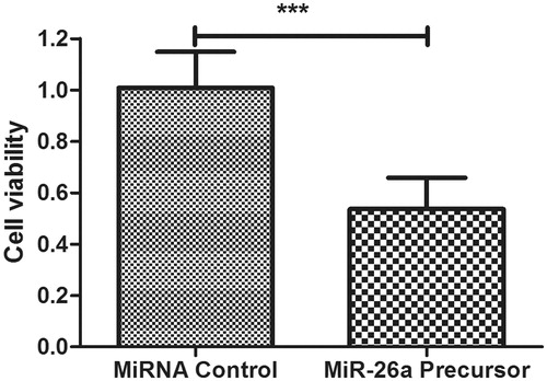
In order to assess the effects of miR-26a on TGF-β-induced HTF fibrosis model, miR-26a precursor was transfected into the in vitro models and cell viability was examined. As showed in , miR-26a precursor was found to be able to down-regulate the proliferation in HTFs treated by TGF-β (P = .026).
Figure 3. Overexpression of miR-26a inhibits HTFs fibrosis in vitro model. The cell viability was determined in HTFs fibrosis in vitro model transfected with miR-2ba precursor or negative control. A450 absorption was assayed after transfection for 24 h. ***P < .001.
miR-26a inhibit HTFs fibrosis cell model migration
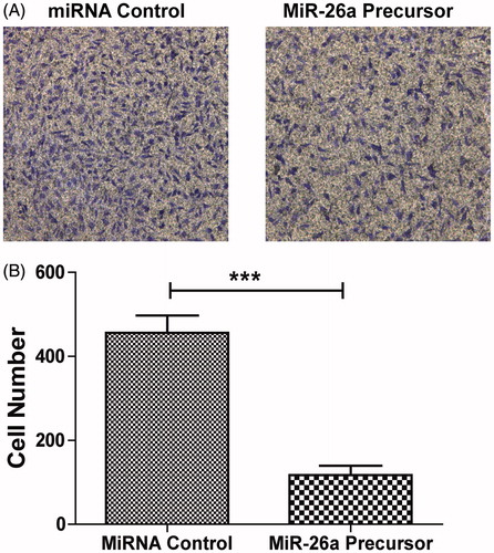
The effect of migration of HTFs was quite important in the formation of HTFs fibrosis, we conducted related experiments. To determine if miR-26a is responsible for HTFs cell model migration, we studied the effects of miR-26a on HTFs fibrosis in vitro model by transwell assay. It was found that miR-26a could strongly reduce HTF cell migration (P < .001, ).
Figure 4. miR-26a inhibited migration of HTFs fibrosis in vitro model. (A) Cell migration ability was analysed by transwell chamber assay 24 h after miR-26a or NC transfection. (B) Quantification of the migratory cells by solubilization of crystal violet. Data represented mean ± SD. ***P < .001.
miR-26a-induced apoptosis of HTFs fibrosis in vitro model
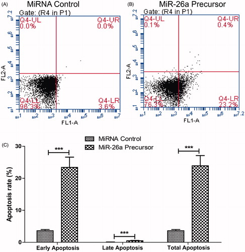
To determine whether the miR-26a could regulate the apoptosis of HTFs fibrosis in vitro model, we performed flow cytometric analysis after transfection of miR-26a precursor mimics. The flow cytometric analysis demonstrated that transfection of HTFs fibrosis in vitro model with miR-26a for 24 h resulted in a significant up-regulated percentage of both early and late apoptosis compared with the control transfected cells (P < .001, ).
Figure 5. miR-26a decreased apoptosis of HTFs fibrosis in vitro model. Cells in each group were collected and stained with Annexin-V/PI. The percentages of early (low right quadrant) and late apoptotic cells (upper right quadrant) were assessed by flow cytometry. (A). flow cytometry plot of control mRNA. (B) flow cytometry plot of miR-26a. (C) percentages of early and late apoptotic cells. Data represented mean ± SD. *** P < .001.
miR-26a directly targets the CTGF in HTFs fibrosis cell model
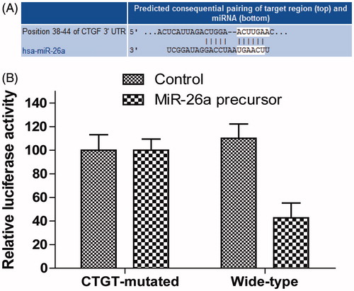
Considering that miR-26a was inversely associated with CTGF mRNA level in the tissue samples, we conducted more studies to find whether miR-26a directly targets the CTGF in HTFs fibrosis cell model. As showed in , it was found that miR-26a treatment could reduce CTGF protein expression significantly compared with control group. To verify the direct target of miR-26a on CTGF, the luciferase reporter assay was conducted to detect whether CTGF was the target gene of miR-26a. As showed in stringent bioinformatics approach, it was identified CTGF was a potential target gene of human miR-26a (). The luciferase reporter assay showed that overexpression of miR-26a led to a reduction of luciferase activity when the reporter construct contained the CTGF 3’-UTR (). In contrast, mutation of the conserved miR-26a-binding motif abrogated the reduced luciferase expression.
Figure 6. miR-26a inhibited CTGF expression. Western blotting analysis of CTGF protein level. GAPDH was used as a loading control.
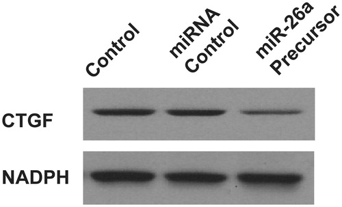
Figure 7. miR-26a directly targets the CTGF in HTFs fibrosis cell model. (A) Computer prediction of miR-26a-binding sites in the 3′-UTRs. (B) wild-type 3′-UTR-reporter or mutant constructs together with miR-26a precursor or controls. Data represented mean ± SD, P < .05.
Discussion
It has been widely demonstrated that mRNAs could regulate diverse biological processes and mRNAs have been studied extensively in fibrosis of different organs [Citation16,Citation19,Citation20]; however, only limited information about mRNAs in the development of HTFs fibrosis and scar formation. As showed in pathophysiological processes of pulmonary fibrosis, down-regulation of miR-26a was detected in the lungs of mice with experimental pulmonary fibrosis [Citation21]. Moreover, a previous study showed that the inhibition of endogenous miR-26a-promoted proliferation and restoration of mature miR-26a inhibited the proliferation of human lung fibroblasts [Citation17]. Such studies indicate that miR-26a plays a protective role in the process of fibrosis. However, the role of miR-26a in the process of scar formation has not been studied in detail.
In our present study, it was found that miR-26a was significantly down-regulated in filtering tract scar. The down-regulation of miR-26a suggests a potential biological effect in the scar formation. Advanced study on the association between the miR-26a and CTGF mRNA showed that miR-26a was inversely correlated with CTGF mRNA level. Considering that miR-26a usually produce effect by down-regulating the target mRNA, CTGF might be regulated by miR-26a. Advanced biological studies showed that overexpression of miR-26a could decrease the cell viability and migration ability of HTFs fibrosis in vitro model. The lower cell viability might be associated with weaker cellular activation, while down-regulated migration ability might affect the migration of cells to damaged sites and the production of ECM. It was also found that miR-26a might increase the apoptotic level of HTFs and thus produce certain anti scarring effects. Through protein expression detection and luciferase reporter assay it was found that miR-26a could produce functions directly targets the CTGF. These findings indicate that miR-26a is a strong antifibrotic mRNA in the scar formation after trabeculectomy and may be considered a potential target for the prevention and treatment of scar formation of filtering tract.
Previous studies have showed that several different functional gene could work as the target gene of miR-26a. In a study based on clinical samples and experimental studies, it was indicated miR-26a performed converse roles in proliferation and metastasis of different gastric cancer cells PTEN was identified as a direct target of miR-26a [Citation22]. In another study, it was reported that ectopic expression of miR-26a enhanced migration and invasion of lung cancer cells. Glycogen synthase kinase-3β was identified as a direct target of miR-26a [Citation23]. It was also detected that decreased miR-26a expression lead to cisplatin resistance and promoted growth and migration in human lung cancer. Enhancer of zeste homolog 2 (EZH2) was identified as the target of miR-26a and miR-26a/EZH2 played significant role in malignant behaviours of lung carcinoma [Citation24]. Importantly, there was a study conducted to identify the detailed mechanism of miR-26a in targeting CTGF on podocytes in diabetic nephropathy [Citation25]. Transfection of miR-26a mimics in cultured human podocytes decreased the CTGF protein level by 50%, and directly inhibited CTGF expression in podocytes, as demonstrated by a reporter assay with the 3'-untranslated region of the CTGF gene. This effect was abolished by a mutant plasmid. miR-26a mimics also inhibited TGF-β1-induced collagen expression, SMAD-binding activity and expression of its host genes CTDSP2 and CTDSPL.
CTGF is one of the most important downstream cytokines of TGF pathway and it exists widely in many kinds of human organs [Citation26]. The biological effects of CTGF were quite widely abundant. In general, CTGF could promote mitosis, improve proliferation, induce chemotaxis, increase cell adhesion, promote ECM synthesis, and function as a key growth factor in the wound healing and tissue repair processes [Citation11,Citation27]. Besides, as a major profibrotic molecule downstream of the TGF-β signalling pathway, CTGF is considered a key factor in differentiation of fibroblasts into MFs and in ECM synthesis [Citation10]. Very low or no expression of CTGF in normal condition, but it was overexpressed in pathological conditions. The overexpression of CTGF is closely related with the occurrence of some proliferative or fibrous disease. There is a certain relationship such as pathological scar, keloid, scleroderma, liver cirrhosis, atherosclerosis, pulmonary fibrosis, renal fibrosis and tumour and migration. Thus, CTGF is considered to be a switch and biological marker for the initiation of fibrosis [Citation28]. In addition, CTGF also has the function of inducing phenotypic transformation in some tissues. All these reveal the important role and key role of CTGF in fibrosis and scar formation. In this study, it was found that activation of miR-26a could suppress expression of CTGF significantly and thus miR-26a produced the potential role in the drug development for filtering tract maintenance.
In conclusion, the key finding of the current study is that miR-26a can suppress the activation of HTFs by TGF-β by targeting CTGF. This data indicates that miR-26a plays an essential role in the formation of filtering tract scar and function as a potential drug target. Understanding the role of miR-26a would provide us importance knowledge in the detection of a novel therapeutic target for the filtering tract scar.
Acknowledgements
None.
Disclosure statement
The authors declare no conflict of interest.
References
- Wallace DM, Clark AF, Lipson KE, et al. Anti-connective tissue growth factor antibody treatment reduces extracellular matrix production in trabecular meshwork and lamina cribrosa cells. Invest Ophthalmol Vis Sci. 2013;54:7836–7848.
- Balendra SI, Shah PA, Jain M, et al. Glaucoma: Hot Topics in Pharmacology. Curr Pharm Des. 2017;23:596–607.
- Riva I, Roberti G, Oddone F, et al. Ahmed glaucoma valve implant: surgical technique and complications. Clin Ophthalmol. 2017;11:357–367.
- Matlach J, Hipp M, Wagner M, et al. A comparative study of a modified filtering trabeculotomy and conventional trabeculectomy. Clin Ophthalmol. 2015;9:483–492.
- Manners T, Salmon JF, Barron A, et al. Trabeculectomy with mitomycin C in the treatment of post-traumatic angle recession glaucoma. Br J Ophthalmol. 2001;85:159–163.
- Yamamoto T, Sawada A, Mayama C, et al. The 5-year incidence of bleb-related infection and its risk factors after filtering surgeries with adjunctive mitomycin C: collaborative bleb-related infection incidence and treatment study 2. Ophthalmology. 2014;121:1001–1006.
- Van de Velde S, Van Bergen T, Vandewalle E, et al. Rho kinase inhibitor AMA0526 improves surgical outcome in a rabbit model of glaucoma filtration surgery. Prog Brain Res. 2015;220:283–297.
- Chen N, Guo D, Guo Y, et al. Paclitaxel inhibits cell proliferation and collagen lattice contraction via TGF-β signaling pathway in human Tenon's fibroblasts in vitro. Eur J Pharmacol. 2016;777:33–40.
- Chung EJ, Sohn YH, Kwon SH, et al. Lithium chloride inhibits TGF-beta1-induced myofibroblast transdifferentiation via PI3K/Akt pathway in cultured fibroblasts from Tenon's capsule of the human eye. Biotechnol Lett. 2014;36:1217–1224.
- Kok HM, Falke LL, Goldschmeding R, et al. Targeting CTGF, EGF and PDGF pathways to prevent progression of kidney disease. Nat Rev Nephrol. 2014;10:700–711.
- Klaassen I, van Geest RJ, Kuiper EJ, et al. The role of CTGF in diabetic retinopathy. Exp Eye Res. 2015;133:37–48.
- Cora D, Re A, Caselle M, et al. MicroRNA-mediated regulatory circuits: outlook and perspectives. Phys Biol. 2017;14:045001.
- Adams BD, Parsons C, Walker L, et al. Targeting noncoding RNAs in disease. J Clin Invest. 2017;127:761–771.
- Liu CH, Wang Z, Sun Y, et al. Retinal expression of small non-coding RNAs in a murine model of proliferative retinopathy. Sci Rep. 2016;6:33947.
- Tkatchenko AV, Luo X, Tkatchenko TV, et al. Large-Scale microRNA expression profiling identifies putative retinal miRNA-mRNA signaling pathways underlying form-deprivation myopia in mice. PLoS ONE. 2016;11:e0162541.
- Yamada Y, Takanashi M, Sudo K, et al. Novel form of miR-29b suppresses bleomycin-induced pulmonary fibrosis. PloS ONE. 2017;12:e0171957.
- Li X, Liu L, Shen Y, et al. MicroRNA-26a modulates transforming growth factor beta-1-induced proliferation in human fetal lung fibroblasts. Biochem Biophys Res Commun. 2014;454:512–517.
- Su Y, Jiang C, Zhang L, et al. Arsenic trioxide inhibits proliferation of rabbit Tenon's capsule fibroblasts after trabeculectomy by downregulating expression of extracellular matrix proteins. Invest Ophthalmol Vis Sci. 2015;56:6663–6670.
- Ooi JY, Bernardo BC, McMullen JR. Therapeutic potential of targeting microRNAs to regulate cardiac fibrosis: miR-433 a new fibrotic player. Ann Transl Med. 2016;4:548.
- Oglesby IK, McKiernan PJ. MiRNA expression in cystic fibrosis bronchial epithelial cells. Methods Mol Biol. 2017;1509:57–69.
- Liang H, Xu C, Pan Z, et al. The antifibrotic effects and mechanisms of microRNA-26a action in idiopathic pulmonary fibrosis. Mol Therap. 2014;22:1122–1133.
- Dey N, Bera A, Das F, et al. High glucose enhances microRNA-26a to activate mTORC1 for mesangial cell hypertrophy and matrix protein expression. Cell Signal. 2015;27:1276–1285.
- Lin G, Liu B, Meng Z, et al. MiR-26a enhances invasive capacity by suppressing GSK3β in human lung cancer cells. Exp Cell Res. 2017;352:364–374.
- Chen J, Xu Y, Tao L, et al. MiRNA-26a contributes to the acquisition of malignant behaviors of doctaxel-resistant lung adenocarcinoma cells through targeting EZH2. Cell Physiol Biochem. 2017;41:583–597.
- Koga K, Yokoi H, Mori K, et al. MicroRNA-26a inhibits TGF-β-induced extracellular matrix protein expression in podocytes by targeting CTGF and is downregulated in diabetic nephropathy. Diabetologia. 2015;58:2169–2180.
- Ubink I, Verhaar ER, Kranenburg O, et al. A potential role for CCN2/CTGF in aggressive colorectal cancer. J Cell Commun Signal. 2016;10:223–227.
- Wang S, Li B, Li C, et al. Potential renoprotective agents through inhibiting CTGF/CCN2 in diabetic nephropathy. J Diabetes Res. 2015;2015:962383.
- Rosenbloom J, Ren S, Macarak E. New frontiers in fibrotic disease therapies: the focus of the Joan and Joel Rosenbloom Center for Fibrotic Diseases at Thomas Jefferson University. Matrix Biol. 2016;51:14–25.