
Abstract
The application of stem cells holds great promises in cell and tissue transplants. This study was designed to compare the hepatogenic differentiation of iPSCs on aligned PES/COL versus random.
Aligned and random PES/COL nanofibrus scaffolds were fabricated by electrospining and their surface modified through plasma treatment and collagen coating. The scaffolds were characterized using scanning electron microscopy (SEM) and ATR-FTIR. Morphology and biochemical activities of the differentiated hepatocyte-like cells (HLCs) were examined after 5 and 20 days of differentiation. Real-Time RT-PCR and ICC showed no significant difference in the mRNA and protein levels of two important definitive endoderm specific markers, including Sox17 and Foxa2 between two scaffolds. However, Real-Time RT-PCR analysis indicated an increase in the expression of Cyp7A1 gene over the period of the differentiation procedure on the aligned nanofibers but there was no difference in other genes such as Albumin and CK19. Moreover, comparison of hepatogenic differentiation evaluated by Albumin production in conditioned media of HLCs differentiated on aligned PES/COL, showed increase expression of these markers after 20 days compared to that of the random nanofibers. Taken together, the results of this study may indicate that aligned PES/COL nanofibrous scaffolds can improve terminal differentiation of HLCs from iPSCs.
Introduction
The application of pluripotent and multipotent stem cells holds great promises in the cell-based therapy and tissue engineering applications. Recently, tissue engineering has introduced new medical cell replacement therapies as an alternative to the traditional transplantation procedures using polymeric biomaterials, which comprises a composite network of nano- and microscale fibers forming highly structured local extracellular matrix (ECM) and microenvironment [Citation1]. Synthetic and natural scaffolds such as nanofibers that trial to mimic the ECM are required to organize the single cells into a higher ordered assembly so as to complete the desired tissue topography and function for tissue engineering applications [Citation2].
Over the past decade, electrospinning is the most generally investigated technique for nanofiber construction with high porosity and high surface area that can be readily accessible controlled during fabrication [Citation3]. Among the various forms of scaffolds available, scaffolds based on polymeric nanofibers which mimic the topography and structure of the ECM offer that can serve as a scaffold for differentiation stem cells into desired lineage [Citation4,Citation5]. Recently, nanofiber structured Polyethersulfone (PES) is reported as a suitable biocompatible biomaterial in artificial liver system and liver tissue engineering [Citation6]. Various sources types of cells such as fibroblasts [Citation7], endothelial cells [Citation8] and mesenchymal stem cells [Citation9] have been reported that can adhere to PES. Human iPSCs derived hepatocyte-like cells (HLCs) have the potential as a new cell source for the creation of a bioartificial and producing an in vitro model for determining the toxicological and metabolic properties of influence drugs in human [Citation10]. Within the last decade, some advancement has been made in developing new protocols to efficiently differentiate pluripotent stem cells to HLCs, by using small molecules and growth factors [Citation11,Citation12]. The many of these studies to date involving HLCs differentiation have been done in two-dimensional (2 D) culture systems. The resulting HLCs indicate typical hepatocyte morphology, perform a wide range of pre-mature hepatocyte functions, and express almost all the pre-mature hepatocyte markers [Citation13]. Despite a many of study about direct differentiation of pluripotent stem cells into HLCs in 2 D, the effective protocols for iPSCs derived HLCs that amplify a high level of biological function on nanofibers scaffolds remain elusive [Citation14]. Therefore, newer strategies need to be considered in hepatocyte differentiation approaches and culture conditions to enhance their biochemical functionality [Citation15]. Considering the lack of optimal in vitro model for the terminal hepatogenic differentiation and to improve hepatocyte cells functionality in vitro, this study was designed to compare the hepatogenic differentiation of iPSCs on aligned PES/COL versus random nanofibers scaffolds. Additionally, by modifying the alignment of the nanofibers, it seems that hepatocyte differentiation and function will improve. To our knowledge, no study has yet addressed this approach.
Materials and methods
Electrospinning
Nanofiberous scaffolds were fabricated by electrospinning method according to the protocol previously reported by Ardeshirylajimi et al. [Citation16]. Briefly, polyethersulfone powder (Ultrason E6020P, MW: 58,000 Da, USA) was dissolved in N, N-dimethylformamide (Merck, USA) at 25%wt. Then, the solution was fed into a 10-ml glass syringe and driven by a syringe pump. A voltage of 20 KV was utilized by a High DC power between the tip of the needle and the collector at a route of 15 cm. For collecting aligned nanofibers, a rotating disk with a linear rate set to 1000 rpm was applied, whereas a rotating disk with a linear rate set to 100 rpm was used for collecting random nanofibers. Various types of collectors were used to generate different types of nanofiberous scaffold assemblies. Random nanofibers were directly rolled up using cover glass slips. A stainless steel frame (with an open void of 2 cm × 5 cm) was utilized as the collector.
Finally, the aligned nanofibers were transferred to cover glass slips by lifting up the fibres from underneath. Random and aligned nanofiber scaffold samples were collected with a rotating mandrel at a rotating speed of 200 rpm for the random sample or 3000 rpm for the aligned sample.
Surface modification
After scaffold fabrication, surface plasma treatment was performed under optimized conditions of 40 kHz low frequency at 30 w with a cylindrical quartz reactor (Diener Electronics, Germany) by introducing pure oxygen into the reaction chamber at 0.4 mbar pressure and then the glow discharge was applied for 5 min. For collagen grafting, plasma-treated sheets were punched and immersed in 1-ethyl-3–(3-dimethylaminopropyl) carbodiimide/N-hydroxysuccinimide (EDC/NHS, country) solution (5 mg/mL) for 12 h. Then, the scaffolds were treated with 1 mg/ml collagen I (Advanced Biomatrix, PureCol) solution at 4 °C overnight. Nanofibrous scaffolds (PES/COL) were soaked overnight in culture medium supplemented with 10% serum, 2X pen/strep (Sigma) and 5X amphotericin B (Sigma) at 37 °C.
Attenuated total reflection Fourier transform infrared spectroscopy
Surface chemical modifications after plasma treatment and collagen grafting were investigated by attenuated total reflection Fourier transform infrared (ATR-FTIR, Bomem MB100, Canada). The spectra were recorded using an Equinox 55 spectrometer (Bruker Optics) equipped with a diamond ATR crystal and a deuterated triglycine sulfate (DTGS) detector.
Characterization of the nanofibers
The polymer constructs were examined using a scanning electron microscopy (SEM; Hitachi S-4500, Japan) with and without stem cells. The cell-polymer constructs were rinsed with phosphate buffered saline (PBS, sigma, USA) before scanning and were fixed in 2.5% glutaraldehyde for 1 h. After dehydration through a graded series of ethanol solutions (30, 50, 70, 80, 90 and 100%) at 37 °C for 10 min per concentration. Finally, cell-polymer constructs were vacuum dried, mounted onto aluminum stubs, and sputter-coated with gold.
iPSCs culture and hepatogenic differentiation
The human iPSCs were kindly donated by Stem Cells Technology Research Center cell bank and were routinely maintained on mitotically inactivated mouse embryonic fibroblast (MEF) as feeder [Citation17,Citation18]. For HLCs induction, monolayers of iPSCs harvested using collagenase were seeded on PES/COL scaffold at a density of 2.5 × 10⁵ cells per well and let them to grow for 24 h in MEF-conditioned media for one week. iPSCs were differentiated into HLCs as previously described [Citation6]. In order to initiate HLCs induction, the culture medium was replaced with 100 ng/ml Activin A (R&D Systems) in RPMI/B27 medium (Invitrogen) for 5 days followed with 20 ng/ml BMP4 (Peprotech) and 10 ng/ml FGF-2 (Invitrogen) in RPMI/B27. Then, the medium was replaced with media composed of RPMI/B27 supplemented with 20 ng/ml HGF (Peprotech) and let them grow under this condition for 5 days. Finally, the cells were treated with 20 ng/ml Oncostatin-M (R&D Systems) in Hepatocyte Culture Media (Lonza) supplemented with SingleQuots (without EGF) for 5 days.
MTT assay
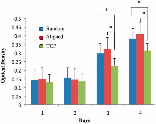
MTT assay was used to evaluate the proliferation and attachment ability of iPSCs on PES/COL nanofibers during 4 days. Briefly, sterilized nanofibers were placed in a 48-well cell culture plate, seeded at a cell density of 4 × 10³ cells per cm2 and incubated at 37 °C, 5% CO2. At 24, 48 and 72 h after cell seeding, 50 μL MTT solution (3-[4,5-dimethyl-thiazolyl-2]-2,5-diphenoltetrazolium bromide) with a final concentration of 5 mg/ml in DMEM was added to each well (n = 3). After 3 h incubation at 37 °C, the supernatant was removed and the formazan crystals were solubilized with dimethylsulfoxide (DMSO). The optical density of the converted dye of each well was read at a wavelength of 570 nm in a micro-plate reader (BioTek Instruments, USA). Data were presented as means ± SD of three repeats.
Immunofluorescence staining
To confirm hepatic differentiation of iPSCs, the differentiated cells were stabilized with 5% paraformaldehyde (PFA; Sigma-Aldrich) for 30 min at 4 °C in definitive endoderm and hepatocyte steps. Then, they got permeable with 0.2% triton X-100 for 10 min, and were blocked in 10% goat serum in PBS for 1 h at 37 °C. For definitive endoderm step, primary antibodies against) human Foxa2 (1:500, Polyclonal rabbit IgG; Millipore, AB4125), Human Sox17 (1:20; Polyclonal Goat IgG, R&D, AF1924 and for HLCs differentiation, albumin (1:200, Santa Cruz, Sc-46293), α-FP (1:200, Santa Cruz, Sc-8108) and antibodies were diluted in 1% BSA in PBS overnight at 4 °C. Secondary antibodies included Alexa fluor 488 donkey anti-goat (1:200; Gibco, A-11058) or Alexa Fluor 594 donkey anti-rabbit (1:200; Gibco, A-21207) for 4 h at 37 °C. The nuclei were counterstained with DAPI (0.1 μg/ml, Sigma-Aldrich, D8417) for 5 min and differentiated cells were analyzed by fluorescence microscope (BX51, Olympus).
RNA extraction and real-time RT-PCR
The total cellular RNA was isolated using Roche High pure RNA isolation kit (Ref: 11828665001). Then, reverse transcription of 1 μg of total RNA to cDNA was carried out with M-MuLV Reverse Transcriptase kit (Fermentas) and a random hexamer primed cDNA synthesis in a total reaction volume of 15 μl. For Real-Time RT-PCR, the reactions were set up in duplicate with SYBR premix ExTaq (Takara) in 96-well optical reaction plates on Step One Plus TM Real-Time PCR machine on 1 μg of cDNA, mixed with 10 μl of SYBR green, 300 nM of forward and reverse primers, and up to 20 μl of ddH2O. The reaction was performed at 95 °C for 2 min, followed by 40 amplification cycles (each 5 s, at 95 °C, 30 s at 60 °C) with fluorescence detection and a final step of melting curve analysis. Primer sequences are illustrated in .
Table 1. Human primers used in real time RT-PCR studies.
Biochemical assays
Samples of conditioned media were collected after 28 days differentiation and stored at 70 °C for albumin secretion was measured through Quantitative enzyme linked immunusorbent assay kit (ELISA) (Bethyl Laboratories, E101, USA) using horseradish peroxidase detection and 3,3′,5,5′-tetramethylbenzidine (TMB) as a substrate as recommended by the manufacturer. The conditioned media of HepG2 and undifferentiated iPSCs (on the day zero) cells were used as positive and negative controls.
Statistical analysis
Statistical analyses were analyzed with ANOVA and student t-test (SPSS 19.0). Student’s t-test was used to determined difference between the two groups. The results were given as mean ± SEM and values of p < .05 were considered to be significant.
Results
Scaffold characterization
SEM micrographs of electrospun nanofibrous scaffolds revealed porous, nanoscaled fibrous structures formed under controlled parameters. Aligned PES nanofibers showed a consistent thickness with fibre diameters of 400–1500 nm ().
Figure 1. Morphology of polyethersulfone nanofibers (×5000). (A) Collagen-coated aligned polyethersulfon, (B) Collagen-coated random polyethersulfon.
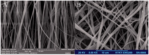
SEM images did not show any changes in the surface morphology of PES nanofiber after plasma treatment in aligned and random nanofibrous scaffolds (). The cells penetrated and adhered well onto the nanofibrous hybrid scaffolds and then expanded on the surface of nanofibers by proliferation during the period of study () and also after four weeks differentiation (), which confirmed biocompatibility of these mats. ATR-FTIR spectroscopy confirmed grafting of collagen onto the surface of PES nanofibers with three peaks at 1649, 2930 and 3325 cm−1 that were related to amide A and amid II bands respectively existing in collagen backbone [Citation6].
Figure 2. (A) SEM analysis of iPSCs culture on aligned PES/COL nanofibers before differentiation. (B) SEM analysis of iPSCs culture on aligned PES/COL nanofibers after 20 days of differentiation.
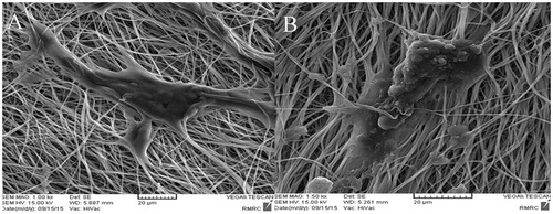
Figure 3. MTT cell proliferation assay of iPSCs on collagen-coated random and aligned polyethersulfone scaffold and tissue culture polystyrene (TCPS) during 1, 2, 3 and 4 days of culture (asterisks show significant difference between the groups at p < .05).
Differentiation of iPSCs to definitive endoderm
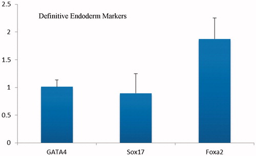
We have used a four-step protocol by modifying a previous method [Citation19], so that iPSCs were routinely cultured without MEF and treated with 100 ng/ml activin-A for 5 days. During this period, we evaluated the endoderm identity of these differentiated cells on nanofibers by ICC and Real Time RT-PCR. Our ICC results showed no difference in the expression of major transcription factors of definitive endoderm such as Foxa2, Sox17, which in differentiated cells on aligned and random nanofibers ( and ). Moreover, endoderm identity of these differentiated cells was further confirmed by Real-Time RT-PCR analysis and the results showed no significant difference in the expression of Sox17, and Foxa2 and GATA4 during differentiation (.
Figure 4. The immunochemical analysis of differentiated iPSc after 5 days for Foxa2 marker on random (A) and aligned (B) nanofibers with nuclear counterstaining (DAPI).
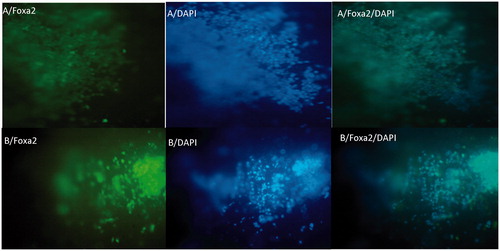
Figure 5. The immunochemical analysis of differentiated iPSc after 5 days for Sox17 markers on random (A) and aligned (B) nanofibers with nuclear counterstaining (DAPI).
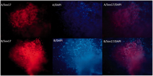
Figure 6. Real time RT-PCR analyses of the expressions level of definitive endoderm genes at day 5 after differentiation of iPSc on aligned PES nanofibers. The expression levels were normalized to control (random nanofibers) group.
Differentiation of iPSCs to hepatocyte cells
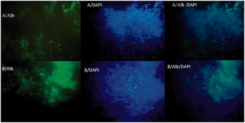
To determine in vitro hepatogenic differentiation of iPSCs on PES/COL nanofibers, the expressions of albumin and αFP was evaluated by ICC analysis after 20 days. As shown in , the differentiated cells were intensely stained for albumin on aligned nanofibers () but not on random nanofibers ().
Figure 7. The immunocytochemical analysis of differentiated iPSCs on day 20 for Albumin marker on random (A) and aligned (B) nanofibers with nuclear counterstaining (DAPI).
Gene expression pattern during hepatogenic differentiation of iPSCs
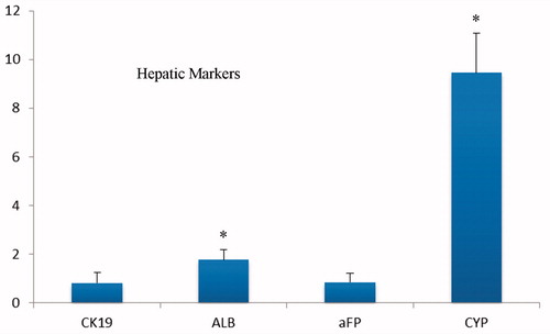
The expression of human liver cell markers, namely, albumin, CK19, AFP and Cyp7A1 was also evaluated by Real Time RT-PCR in the differentiated cells on the surface of nanofibrous scaffolds. Real Time RT-PCR revealed that the expression level of albumin and Cyp7A1 was significantly increased in differentiated cells on align scaffolds comparison to the random PES. However, the expression level of CK19 and AFP genes show no difference (.
Figure 8. Quantitative RT-PCR analyses of the expressions level of hepatic cell genes at day 20 after hepatogenic differentiation of iPSc on collagen-grafted PES nanofibers. The expression levels were normalized to random nanofiber as a control group.
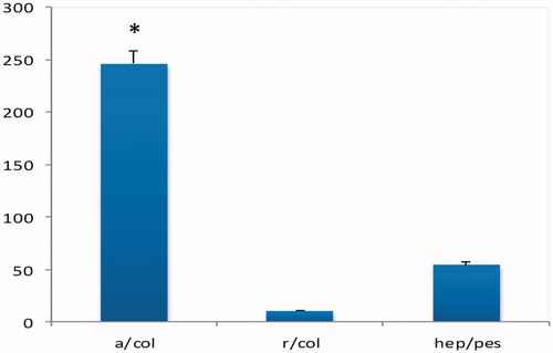
To confirm the staining results, we measured secreted amounts of albumin. The ELISA results verified that culture differentiated cells on aligned scaffold expressed mature hepatic markers including ALB as detected by RT-PCR compare to random scaffold (.
Figure 9. Quantification of albumin produced in hiPSCs after 28 days of hepatic lineage induction compare with undifferentiated hiPSCs and hepG2 as a positivie control.
Discussion
Recent studies report that the iPSCs have therapeutic uses and potential to differentiate into various tissues such as, bone [Citation20], muscular [Citation21], neural [Citation22], pancreas [Citation23], etc. When the iPSCs were generated from fibroblast cells by Yamanaka et al. in 2006 [Citation24], scientists were looking for differentiation strategies for liver failure treatment. The iPSCs are appropriate for cell-based therapy in autologous transplantation because iPSCs can be generated from the patient’s own fibroblasts. Several studies reported that the iPSCs were applied for differentiating to HLCs with various differentiation methods in vitro [Citation19,Citation25]. Cell aggregation in 3 D demonstrate a critical role in maintaining the differentiated function and viability of primary hepatocytes in vitro mimic the ultrastructure and morphology of the native liver lobule [Citation14]. So as an important part of liver tissue engineering, scaffolds provide a place for cell attachment, increased surface area, and support for a large cell mass and specific structures. Hepatocytes are anchorage dependent; therefore, cell-substrate and cell-matrix interactions control aggregation [Citation26]. Cell-substrate interaction and adhesion strength are a basic factor governing the morphology and formation of multicellular aggregates [Citation27]. In order for aggregation to improve, stem cells must initially adhere strongly and sufficiently to the surface for retention and exhibited hepatocyte-like morphology, but also not adhere so compactly that they are unable to later migrate and expand towards one another and then finally disassociate the substrate to create multilayered aggregates.
In the present study, we designed aligned and random PES nanofibers, compared and evaluated its supporting potential and hepatogenic differentiation enhancing. The results indicated that the aligned compared to random nanofiberous performed better in enhancing the hepatogenic maturation and differentiation iPSCs into HLCs. The iPSCs acquired hepatic differentiation by day 28, confirmed by functional assays (), the expression of hepatic markers () and morphological changes (). Differentiation of iPSCs was investigated on electrospun PES nanofiberous with aligned and random topographies. PES nanofiberous showed good biocompatibility with iPSCs. The morphology, proliferation, and differentiation of iPSCs were not dependent on PES topography. Both aligned and random PES nanofibers enhanced the differentiation of iPSCs into hepatocytes but aligned nanofibers directed the cell growth along the maturation. On the other hand, aligned PES nanofibers differentiate the cells and gradually encouraged differentiation into functional hepatocyte. These findings suggest that aligned PES is proper substrates for regulating stem cell differentiation into fully functional hepatic cell lineage for liver tissue regenerative applications. In the current study, the effects of PES nanofibers surface topography have also been studied on iPSCs differentiation. Our results indicated that iPSCs could attach, proliferate, and differentiate on PES nanofiberous. The surface topography of PES nanofibers was also effective on iPSCs morphology, proliferation, and differentiation to hepatocytes. It’s seemed that aligned PES nanofibers reduced the proliferation of iPSCs compared to random PES nanofibers. Aligned PES nanofibers also induced a significant upregulation of hepatocyte markers including Albumin and CYP450 compared to random PES nanofibers, thus suggesting the induction into hepatogenic lineage. Albumin secretion of differentiated hepatocytes was also different on random and aligned PES nanofibers. When the iPSCs were cultured on aligned PES nanofiberous in hepatogenic induction media, the cells remarkably produce more Albumin protein compare with random PES nanofibers. These results showed the importance of electrospun PES nanofiberous nanotopographical cues on the hepatogenic differentiation of iPSCs. The significant effects of aligned nanofibers on enhancing stem cell differentiation have been previously studied [Citation28]. In 2007 Yim et al. indicated that aligned poly (dimethylsiloxan) (PDMS) significantly upregulated differentiated cell markers of human mesenchymal stem cells (hMSCs) compared to non-aligned PDMS. They showed that the changes in differentiated hMSC marker expression were related with the elongation of cytoskeleton and nuclei. They indicated that aligned nanotopography enhances differentiation of stem cells via changing stem cell morphology [Citation28]. Our results indicated that aligned PES nanofiberous apparently changed the morphology of differentiated iPSCs compared to random nanofibers. It seems that the cells cultured on aligned PES nanofiberous elongated along the fibres main axis. This biomechanical reaction may be transmitted to the nucleus by cytoskeletal-linked signalling pathways [Citation29]. This may be a stimulus for enhancing hepatogenic differentiation of iPSCs on aligned PES nanofiberous. Therefore, proliferation of iPSCs cultured on aligned PES nanofiberous was significantly less than that on random PES nanofiberous in hepatogenic induction medium and it can be concluded that although randomly oriented PES nanofibers are suitable for hepatogenic differentiation of iPSCs but aligned PES nanofibers is preferred according to the present results to use in liver tissue engineering applications.
Disclosure statement
There is no conflict of interest for any of the authors.
References
- Goyal R, Guvendiren M, Freeman O, et al. Optimization of polymer-ECM composite scaffolds for tissue engineering: effect of cells and culture conditions on polymeric nanofiber mats. JFB. 2017;8.
- Gholizadeh-Ghaleh Aziz S, Gholizadeh-Ghaleh Aziz S, Akbarzadeh A. The potential of nanofibers in tissue engineering and stem cell therapy. Artif Cells Nanomed Biotechnol. 2016;44:1195–1200.
- Eatemadi A, Daraee H, Zarghami N, et al. Nanofiber: synthesis and biomedical applications. Artif Cells Nanomed Biotechnol. 2016;44:111–121.
- Xie J, Li X, Xia Y. Putting electrospun nanofibers to work for biomedical research. Macromol Rapid Commun. 2008;29:1775–1792.
- Chen F, Li X, Mo X, et al. Electrospun chitosan-P(LLA-CL) nanofibers for biomimetic extracellular matrix. J Biomater Sci Polym Ed. 2008;19:677–691.
- Maymand MM, Soleimanpour-Lichaei HR, Ardeshirylajimi A, et al. Hepatogenic differentiation of human induced pluripotent stem cells on collagen-coated polyethersulfone nanofibers. ASAIO J. 2017;63:316–323.
- Babaeijandaghi F, Shabani I, Seyedjafari E, et al. Accelerated epidermal regeneration and improved dermal reconstruction achieved by polyethersulfone nanofibers. Tissue Eng Part A. 2010;16:3527–3536.
- Unger RE, Huang Q, Peters K, et al. Growth of human cells on polyethersulfone (PES) hollow fiber membranes. Biomaterials. 2005;26:1877–1884.
- Pournaqi F, Ghiaee A, Vakilian S, et al. Improved proliferation and osteogenic differentiation of mesenchymal stem cells on polyaniline composited by polyethersulfone nanofibers. Biologicals. 2017;45:78–84.
- Vasanthan KS, Subramanian A, Krishnan UM, et al. Role of biomaterials, therapeutic molecules and cells for hepatic tissue engineering. Biotechnol Adv. 2012;30:742–752.
- Shan J, Schwartz RE, Ross NT, et al. Identification of small molecules for human hepatocyte expansion and iPS differentiation. Nat Chem Biol. 2013;9:514–520.
- Sullivan GJ, Hay DC, Park I-H, et al. Generation of functional human hepatic endoderm from human induced pluripotent stem cells. Hepatology 2010;51:329–335.
- Han S, Bourdon A, Hamou W, et al. Generation of functional hepatic cells from pluripotent stem cells. J Stem Cell Res Ther. 2012;Suppl 10:1–7.
- Ramasamy TS, Yu JSL, Selden C, et al. Application of three-dimensional culture conditions to human embryonic stem cell-derived definitive endoderm cells enhances hepatocyte differentiation and functionality. Tissue Eng Part A. 2013;19:360–367.
- Chamuleau RA. Artificial liver support in the third millennium. Artif Cells Blood Substit Immobil Biotechnol. 2003;31:117–126.
- Ardeshirylajimi A, Hosseinkhani S, Parivar K, et al. Nanofiber-based polyethersulfone scaffold and efficient differentiation of human induced pluripotent stem cells into osteoblastic lineage. Mol Biol Rep. 2013;40:4287–4294.
- Ardeshirylajimi A, Soleimani M, Hosseinkhani S, et al. A comparative study of osteogenic differentiation human induced pluripotent stem cells and adipose tissue derived mesenchymal stem cells. Cell J. 2014;16:235–244.
- Lahmy R, Soleimani M, Sanati MH, et al. MiRNA-375 promotes beta pancreatic differentiation in human induced pluripotent stem (hiPS) cells. Mol Biol Rep. 2014;41:2055–2066.
- Si-Tayeb K, Noto FK, Nagaoka M, et al. Highly efficient generation of human hepatocyte-like cells from induced pluripotent stem cells. Hepatology. 2010;51:297–305.
- Ardeshirylajimi A, Dinarvand P, Seyedjafari E, et al. Enhanced reconstruction of rat calvarial defects achieved by plasma-treated electrospun scaffolds and induced pluripotent stem cells. Cell Tissue Res. 2013;354:849–860.
- Salani S, Donadoni C, Rizzo F, et al. Generation of skeletal muscle cells from embryonic and induced pluripotent stem cells as an in vitro model and for therapy of muscular dystrophies. J Cell Mol Med. 2012;16:1353–1364.
- Denham M, Dottori M. Neural differentiation of induced pluripotent stem cells. Methods Mol Biol. 2011;793:99–110.
- Enderami SE, Mortazavi Y, Soleimani M, et al. Generation of insulin-producing cells from human-induced pluripotent stem cells using a stepwise differentiation protocol optimized with platelet-rich plasma. J Cell Physiol. 2016.
- Takahashi K, Tanabe K, Ohnuki M, et al. Induction of pluripotent stem cells from adult human fibroblasts by defined factors. Cell. 2007;131:861–872.
- Song Z, Cai J, Liu Y, et al. Efficient generation of hepatocyte-like cells from human induced pluripotent stem cells. Cell Res. 2009;19:1233–1242.
- Berthiaume F, Moghe PV, Toner M, et al. Effect of extracellular matrix topology on cell structure, function, and physiological responsiveness: hepatocytes cultured in a sandwich configuration. FASEB J. 1996;10:1471–1484.
- Chang TT, Hughes-Fulford M. Molecular mechanisms underlying the enhanced functions of three-dimensional hepatocyte aggregates. Biomaterials. 2014;35:2162–2171.
- Yim EK, Pang SW, Leong KW. Synthetic nanostructures inducing differentiation of human mesenchymal stem cells into neuronal lineage. Exp Cell Res. 2007;313:1820–1829.
- Lim SH, Liu XY, Song H, et al. The effect of nanofiber-guided cell alignment on the preferential differentiation of neural stem cells. Biomaterials. 2010;31:9031–9039.