
Abstract
Aim: This study aims to evaluate the antinociceptive effect of combined Achillea millefolium and Origanum extract encapsulated in liposome.
Methods: The effect of Achillea millefolium and Origanum vulgare L. extract, and their liposome-incorporated form was assessed using 3% formalin test in rat. 12 male Wistar rats, 4 in each group, were used in this study, and increasing doses of Achillea millefolium (31.6, 100, 178, and 316 mg/kg) and Origanum vulgare L. extract (5.6, 10, 17.8, and 31.6 mg/kg), and co-administered extract were i.p. administered 10 min before 3% formalin. The mechanisms of action were evaluated for the liposomal encapsulated co-administered extract using N(G)-nitro-l-arginine methyl ester (L-NAME) (3 mg/kg) and naloxone (1 mg/kg).
Results: The interaction index and isobolographic analysis revealed a synergistic effect of the extracts. We observed a lower experimental ED30 as compared to the theoretical ED30. Naloxone also reduced the antinociceptive effect of the liposome encapsulated co-administered extract.
Conclusion: These data suggest that the Achillea millefolium and Origanum vulgare L. extract encapsulated in liposome gave a synergistic effect.
Introduction
Many studies have explored the analgesic effect of higher plants in the past decades [Citation1,Citation2]. In spite of the progress in the advancement of pain therapies, it is still essential to find a new potent analgesia without any side effects that have been noted with the current known analgesic drugs, most importantly for the relief of chronic pain [Citation3].
Achillea millefolium (Asteraceae), popularly known by Anador® or Novalgina® (sodium dipyrone) and Atroveran® (sodium dipyrone, papaverine hydrochloride, and Atropa belladonna) with respect to the Brazilian region [Citation4], is a perennial herb that had been used for centuries in folk medicine in different countries. Teas from the aerial parts of the plant are widely used for treating spasms [Citation5], digestive complaints, pain, and other ailments [Citation6]. Literature shows the presence of phytochemicals such as terpenoids, lactones, sesquiterpene essential oils [Citation7,Citation8], flavonoids di-O-glycosides, di-C-glycosides, flavonoid aglycones, and two caffeic acid derivatives, 3,5-dicaffeoylquinic acid and chlorogenic acid [Citation9,Citation10] in A. millefolium. The alkaloid achilleine was found in this specie.
Oregano, a species of Origanum of the mint family, is a native to Mediterranean region and Europe, Central and Southern Asia [Citation11]. Approximately 40 species of oregano exist, but essential oil extracted from Origanum vulgare L. (ORG) makes this specie the most therapeutically useful. The oil is obtained from the dried flowering herb through steam distillation [Citation12,Citation13]. By tradition, the leaves of ORG are a medicinal plant employed in Iranian folk medicine to suppress pain; however, there are just few documented data about its possible antinociceptive mechanism(s).
Many studies have focused on the application of nanoparticle systems, with particle size between 1 and 100 nm for drug delivery [Citation14]. Nanoparticles have distinctive physicochemical characteristics such as controllable and ultrasmall size, high reactivity, large surface area to volume ratio, and a functionalizable structure [Citation15]. These unique characteristics can be employed to promote the administration of drugs like pain killers, thus bypassing some barriers in traditional antinociceptive therapeutics. Extensive studies have showed that nanoparticles like liposomes, solid lipid nanoparticles, polymeric nanoparticles, and others can be employed in controlled release and antimicrobial drug delivery [Citation16]. Encapsulation offers a way to control solubility, stability, and bioavailability, as well as release of bioactive compounds. Several methods of natural bioactive compounds encapsulation have been demonstrated as an efficient method to increase their in vitro and in vivo absorption [Citation17].
Liposomes are presently the most extensively studied clinically established nanoscale systems for the delivery of drug. They have been widely studied in cosmetic and pharmaceutical industries [Citation16–18]. Their bilayer structure, mimicking cell membrane, permits easy fusion. Their excellent biodegradability, biocompatibility, and possibility to control their surface characteristics and size make them perfect particles in the delivery of drug [Citation18,Citation19]. Liposomal antimicrobial delivery systems have several advantages such as improved solubility, bioavailability and efficacy, reduced toxicity, and increased product stability [Citation18]. Besides their exceptional advantages, liposomes also possess a low encapsulation efficiency, low stability, high manufacturing cost, oxidation or hydrolysis degradation, sedimentation, aggregation, or fusion at the time of storage [Citation19]. Thus, this present study aims at evaluating the antinociceptive synergistic interaction between A. millefolium and ORG encapsulated in liposome in rats and possible involvement of opioid receptors in its antinociceptive mechanism.
Materials and methods
Experimental animals
Twelve male Wistar rats 3–4 months of age (220–280 g) were obtained from Lorestan University of Medical Sciences (LUMS). The animals were kept in an isolated room at a temperature- and light-controlled room (22 ± 2°C; 12-h light/dark cycle) and were given a free access to food and water ad libitum. A day before the commencement of our study, the rats were denied food but not water. Approval from the Ethical Committee of LUMS was acquired for the animals and the guidelines on ethical standards for experimental pain in animal investigations [Citation20].
Preparation of Achillea millefolium and ORG extracts
The leaves of both plants were collected during spring in Yazd City. The plants were authenticated by a botanist at LUMS Biology Department, Iran, as A. millefolium (herbarium no: 158) and ORG (herbarium no: 9428). The leaves were dried under shade and ground into powder. The powder of each leaf obtained was separately refluxed with warm distilled water (below 50 °C) at a ratio of 1/100 ratio for 24 h. The extracts were filtered with Whatman No. 2 filter paper, and its supernatants were concentrated under low pressure at 40 °C using a rotary evaporator. A semisolid mass that yielded 8% W/W was obtained upon evaporation. A stock solution of the extract was prepared by dissolving in sterile saline (protocol from Akindele et al. with slight modification [Citation21]).
Liposome preparation
Liposomes can be fabricated from many species of lipid and by several methods. Phospholipids obtained from soya bean lecithin are often used in liposomal active compound delivery systems because they are readily available at comparatively low cost for upscale production and safe for use making unpurified soya bean phospholipids a better substitute and attractive choice. The major phospholipids extracted from soya bean lecithin are phosphatidylethanolamine, phosphatidylcholine, and phosphatidylinositol [Citation18]. In this study, A. millefolium and ORG were encapsulated into liposome nanoparticles fabricated from soya bean lecithin. Three separate methods for liposome fabrication were tested: thin film evaporation, ethanol injection, and ultrasonication. For liposome formation, a lecithin solution of 8–45 mg/mL with cholesterol addition in the ratio of 9:1 lecithin/cholesterol was used. The dispersion of lipid was sonicated (80 W, 20 kHz) in an ice bath with ultrasonic homogenizer (BANDELIN Electronic GmbH & Co. KG, Berlin, Germany) for few minutes. Lecithin solution of 100 mg/mL was used during ethanol injection. Multilamellar vesicles were prepared with the thin film evaporation. Briefly, the lipid phase (lecithin 13.5 mg/mL with cholesterol addition in the ratio of 8:1 lecithin/cholesterol) was dissolved in chloroform (with or without the encapsulated lipophilic compound), which was then removed in a rotary evaporator under reduced pressure (IKA®-Werke GmbH & Co. KG, Staufen, Germany), therefore obtaining a dry lipid thin film on the flask wall. The film was then hydrated by adding distilled water (with or without the encapsulated hydrophilic compound) under rigorous stirring condition in order to stimulate the vesicle fabrication [Citation18].
Particle size and stability analysis
Particle size and size distribution are the main parameters used for the assessment of nanoparticles physical stability. The dynamic light scattering (DLS) is extensively used to assess the size and size distribution of small particles suspended in liquid medium. The mean particle size, size distribution, and polydispersity index (PDI) are the main parameters of this technique. A PDI value that falls between 0.1 and 0.25 shows a narrow size distribution, while a PDI >0.5 shows a broad size distribution [Citation16]. Although the DLS method provides fast measurement of size distribution and particle size, they are not competence in assessing particle morphology similarly to direct visualization method like microscopy. Laser Doppler electrophoresis is a technique often employed to measure zeta potential. It evaluates electrophoretic mobility of suspended particles in the medium. It is an overall rule of thumb that an absolute value of zeta potential >60 mV gave a good stability, value of 30 mV usually results in good stability, 20 mV is acceptable short-term stability, and <5 mV means rapid particle aggregation [Citation16].
In this study, particle size distribution, average size of particles, and PDI were assessed by colloidal DLS analyzer Zetasizer Nano ZS (Malvern Instruments Ltd., Malvern, UK). The prepared particle morphology was evaluated by light microscope (Labomed Lx 500; Labomed Inc., Los Angeles, CA) and scanning electron microscope (EVO LS 10; Carl Zeiss AG, Oberkochen, Germany). For other applications, the unified particle size (100 nm) was acquired using membrane extruder (LiposoFast, AVESTIN Europe GmbH, Mannheim, Germany).
Encapsulation efficiency
A. millefolium and ORG extracts were encapsulated. Encapsulation efficiency was evaluated spectrophotometrically (UV-Vis spectrophotometer, Thermo Spectronic Helios™ δ; Thermo Fisher UK Ltd., Waltham, MA). The encapsulated components were measured before and after the process of encapsulation. The % of encapsulation efficiency (EE) was then calculated according to the following equation:
Formalin test
This test was used to quantify an eventual anti-inflammatory effect. The rats were placed in transparent plastic chambers and a mirror at a 45° angle was placed to allow a clear view of the paw. The extract and liposome encapsulation of their combination was injected subcutaneously and made into the plantar surface of the rear paws with 30 ml of diluted 3% formalin via a 30-gauge needle. After this procedure, animals were then returned to the previous chambers. The nociceptive character was evaluated as the licking time on the paw injected during the first phase (neurogenic, 0–5 min), and the second phase (inflammatory,15–35 min). At the end of the experiment, mice were sacrificed in a CO2 chamber [Citation22].
Design
Different groups were used for the characterization of the dose–response curve of each individual extract formulation. Increasing doses of A. millefolium (31.6, 100, 178, and 316 mg/kg) or ORG (5.6, 10, 17.8, and 31.6 mg/kg) were given i.p. 10 min before 3% formalin administration. The last group include the co-administered extract. We determined an experimental ED50 value for each extract after obtaining the dose–response curve of each of the drug. The A. millefolium and ORG encapsulated in liposome was analyzed. To evaluate the likely mechanism of the formulation action, the co-administration effect was checked in the presence of naloxone (1 mg/kg), L-NAME (3 mg/kg), or vehicle. These drugs were administered i.p. 10 min before the liposome encapsulated extract combination administration. Nociception was analyzed 10 min later.
Data analysis
Data are represented as mean ± standard error of the mean for four animals per group. The total licking time is equivalent to the second phase, analyzed from 15 to 45 min with regard to formalin administration. Dose–response data are represented as the percentage antinociception of the overall licking time of the second phase of formalin test. The % antinociception was assessed according to the expression [Citation23]: ((vehicle − postcompound)/vehicle) × 100.
The dose–response curves were plotted and experimental points were fitted using least-squares linear regression. The SEM was evaluated as described by Tallarida [Citation24]. The evaluation of the interaction between analgesic was done using isobolographic analysis [Citation23,Citation24]. In this present study, this method was used in evaluating the nature of the interaction between A. millefolium and ORG encapsulated in liposome. The isobolographic analysis assumes that combination of the extract is synthesized from equal potent extract doses of the individual drugs. Thus, from the dose–response curves of each individual extract, the dose causing 50% of the effect (ED50 value) can be assessed. However, considering the maximal effect of 100% as the overall suppression of formalin-induced licking and that A. millefolium and ORG encapsulated in liposome were incompetent to give a 50% reaction, the ED50 value calculation was not feasible. Therefore, the ED30 value was evaluated as ED50 value replacement. As a result, a dose–response curve was acquired by concurrent delivery of A. millefolium and ORG extract encapsulated in liposome in a fixed dose ratio (fixed ratio) with respect to the ED30 values of each individual extract. To assess whether the interaction between A. millefolium and ORG encapsulated in liposome co-administration was antagonistic, additive, or synergistic, the theoretical additive ED30 value (Zadd) was determined from the dose–response curves of each extract that has been individually administered, considering the experimental effect with the co-administration results of the sum of the individual effects of each extract. This theoretical ED30 value was then compared with those of the experimental ED30 value (Zexp) to determine whether there was a statistically significant difference between them [Citation25,Citation26]. The experimental and theoretical ED30 values of the studied combination were compared by calculating the interaction index (γ) as follows:
γ = ED30 value of experimental co-administration/ED30 value of theoretical co-administration.
The γ depicts the ED30 value portion of each extract accounting for the corresponding ED30 value in the extract combination. A γ value of approximately 1 represents a synergistic interaction, a γ value of >1 represents an antagonistic interaction, and a γ value of <1 represents an additive interaction.
Statistical analysis
The Student’s t-test was used in determining the experimentally derived and theoretical additive ED30 values. An experimental ED30 value is statistically significantly lower than theoretical additive ED30 value which represents a synergistic interaction between A. millefolium and ORG extract encapsulated in liposome. A one-way ANOVA was used for analyzing the mechanisms of action, and then Student–Newman–Keuls test was used afterwards. p < .05 was considered as statistically significance.
Results
Encapsulation efficiency of natural extracts
The two plant extracts were encapsulated into liposomes. Encapsulation was successful. Usually, herbal extracts of phenolic components were preferably encapsulated as aqueous extracts. Thus, the types of extraction agent have a crucial influence on the encapsulation efficiency. Another essential factor of encapsulation efficiency is the material employed for particle preparation. Better results of encapsulation efficiency of plant water extracts were achieved more frequently in liposomes than in other particles. In liposome particles prepared by sonication, high encapsulation efficiency was observed.
Stability and size of polysaccharide particles
The size of the prepared liposome encapsulated A. millefolium and ORG was on average 160 nm (125.0–348.3 nm) and a PDI value was not greater than 0.25. Zeta potential of these liposomes in almost all cases appears to be in the range of −30 to −60 mV, which means that particles were stable.
Antinociceptive effects of Achillea millefolium, Origanum extract, and their liposome encapsulated combination
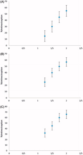
Both A. millefolium and ORG extract encapsulated in liposome decreased the nociceptive behaviour stimulated by the administration of formalin. The maximal A. millefolium effect was 55%, whereas that for ORG extract was approximately 57%. shows the effect of extracts and their combination.
Figure 1. Dose–response curves for the antinociceptive effect of A. millefolium, (A), ORG (B), and A. millefolium–ORG encapsulated in liposome (C) in the second phase of formalin test.
Isobolographic analysis
The optimum effect reached by the highest dose of the extract co-administration encapsulated in liposome was about 66%. The addition of each effects (ED30 value of each drug) suggests that the A. millefolium doses would contribute 30% of its maximum effect (55%), i.e.,16.5%. The maximum effect of ORG extract was 57%; the ED30 value of this drug would give 17.1% of the effect of the co-administration according to the experimental test. So that the total sum of such effects would be approximately 33.6%, a value lower than the maximum effect of the drug combination (66%). Therefore, the combination gave the highest effect. The evaluation of interaction between A. millefolium and ORG extract encapsulated in liposome using the isobologram method demonstrated an antinociceptive synergistic effect in the second phase of the formalin test. The horizontal and vertical bars depict SEM. The slanted line between the y- and x-axes represents the theoretical additive line. A point T at the middle of the line represents the theoretical additive point acquired from ED30 values of each extract as analyzed above. The point represented as E is the observed ED30 value with the co-administration. The experimental ED30 value point in this case is located just below the additive line, which is statistically significantly different from the theoretical ED30 value, depicting a significant synergistic effect (). illustrated the extract combination doses. The theoretical additive ED30 value was 53.2 ± 2.7, while the experimental ED30 value of the A. millefolium and ORG extract encapsulated in liposome co-administration was 11.1 ± 0.1 (p < .05). In addition, the interaction index gave an increase in the efficiency of the extracts for the co-administration (γ= 0.21).
Figure 2. The isobologram antinociceptive synergistic interaction between A. millefolium and ORG extract encapsulated in liposome in the second phase of the formalin test.
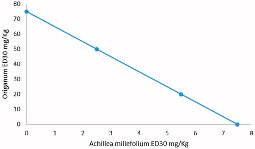
Table 1. Co-administration drug dose used.
Mechanism of action
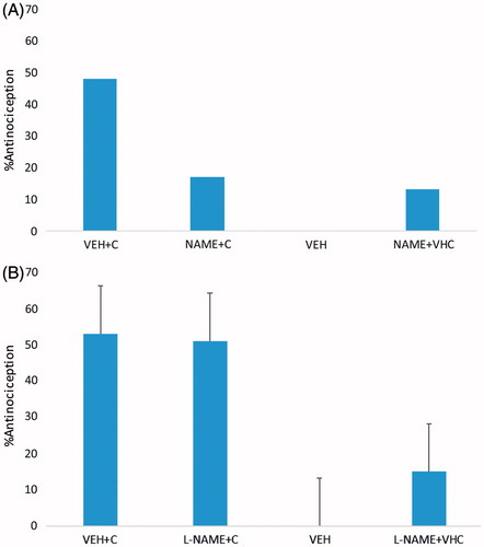
Administration of naloxone, but not L-NAME or vehicle, statistically significantly lowered the co-administrated antinociceptive effect ().
Figure 3. The effect of naloxone (A) and L-NAME (B) on the antinociceptive effect of the A. millefolium and ORG extract encapsulated in liposome combination. Bars are the mean ± SEM for at least six animals. Significantly different (*p < .05) from the co-administration (C), by one-way analysis of variance followed by the Student–Newman–Keuls test.
Discussion
This present study aims at evaluating the antinociceptive synergistic interaction between A. millefolium and ORG extracts encapsulated in liposome in rats and possible involvement of opioid receptors in its antinociceptive mechanism. In the present study, both A. millefolium and ORG extract encapsulated in liposome decreased the nociceptive behaviour stimulated by the administration of formalin. The maximal A. millefolium effect was 55%, whereas that for ORG extract was approximately 57%. shows the effect of extracts and their combination. The optimum effect reached by the highest dose of the extract co-administration encapsulated in liposome was about 66%. The addition of each effects (ED30 value of each drug) suggests that the A. millefolium doses would contribute 30% of its maximum effect (55%), i.e.,16.5%. The maximum effect of ORG extract was 57%; the ED30 value of this drug would give 17.1% of the effect of the co-administration according to the experimental test.
The data from previous studies demonstrated that some species of the genera Achillea and ORG exhibit analgesic and anti-inflammatory effects, such as Achillea ageratum L., Achillea aleppica DC., A. millefolium, and Artemisia vulgaris, are broadly used in popular medicine as analgesics [Citation27], but in Scopus, Google scholar and PubMed search, no researcher has studied the antinociceptive synergistic interaction between A. millefolium and ORG extracts encapsulated in liposome. However, Ahmad et al. [Citation28] did not note an antinociceptive effect for A. millefolium, and Mistieri et al. [Citation29] observed an increase in the tail-flick latency for florid end teas, but not for its leaves. The same investigation failed to show a positive effects for writhing test in both teas. In addition, the formalin test is more suitable for the evaluation of drugs acting on long duration nociceptive stimuli [Citation30]. Thus, the fact that A. millefolium 500 mg/kg had decreased the number of contortions without acting in the formalin test proposes that the extracts have a weak antinociceptive effect with short duration. In their study, the antinociceptive effect of the tested plants was obtained with increased doses of the extracts (500 and 1000 mg/kg). In addition, 500 and 1000 mg/kg doses were well tolerated on initial pharmacological screening, and investigation with lower doses (100 and 250 mg/kg) was unsuccessful in inhibiting the writhing. However, morphine, serving as their positive control, was effective in these models only at high doses (10–20 mg/kg) due to large variability. Phenolic acids, lignans, tannins, triterpenes, and sesquiterpene lactones, are phytochemicals found in A. millefolium [Citation8,Citation4], and they are classes that present analgesic and/or anti-inflammatory-related effects [Citation31–35].
Essential oils might be responsible for the observed antinociceptive effect, as some sesquiterpenes and monoterpenes possess antiinflammatory and/or antinociceptive effects, as is the case of linalool, alpha-amyrin, azulene, and 1,8-cineole, which were present in A. millefolium [Citation36,Citation37]. However, these compounds were not identified in this study, because polar solvent extraction is not suitable for this component extraction. In a study by Pires et al. [Citation38], the results obtained showed that hydroalcohol extracts of A. millefolium and A. vulgaris possessed an adequate antinociceptive peripheral effect, but no anti-inflammatory effect in the formalin test. Because of the high concentration of hydroxybenzoic acid, caffeic acid, and rutin and their identified analgesic effect, it is possible that the results obtained for the hydroalcohol extracts in their study may be traced to these compounds, whose concentration is higher in both extracts. There was possibly synergistic effect among the component in both plants studied.
In a study by Mikaili et al. [Citation39] to evaluate the antinociceptive effect of intracerebroventricular (ICV) microinjection of ORG extract and its probable connection with opioid receptors, it was observed that the co-administration of ORG extract with morphine gave a significant elevation in naloxone and TFL and pretreatment significantly blocks the antinociceptive activity of morphine and ORG. The aqueous extract of ORG in a dose-dependent manner possesses antinociceptive effect in the tail-flick test. In other studies as well, ORG stimulated antinociception may have been facilitated by opioid mechanism. Our findings are similar to Mikaili et al., who showed IP injections of ORG (1, 1.5 and 2 mg/ml) which caused a significant antinociception in acute pain model by tail-flick test [Citation12].
The antinociceptive effect of ORG mechanism(s) of action is yet to be understood; nevertheless, it may be mediated via the chemical composition of the essential oils present in the ORG plant, mainly its carvacrol/thymol components [Citation40]. Others have also demonstrated the antinociceptive effect of carvacrol (CARV) (5-isopropyl-2-methylphenol) in different pain models in mice [Citation41]. Analgesia induced by systemic administration of local anaesthetics has been so far known to rely on a local anaesthesia of the endoreceptors (stabilization of nerve ending nociceptors) [Citation42] and therefore to be of peripheral origin.
The isobolographic analysis of dose–response curves of fixed ratios of drugs gave a rigorous means of evaluating drug–drug interaction [Citation43]. In the present study, we constructed the isobolograms for various time points to determine the nature of the interaction throughout the time-effect course. The mechanism behind the synergism is yet to be elucidated, and the interactions are clearly complex.
In conclusion, the synergistic antinociceptive interaction of A. millefolium and ORG encapsulated in liposome interacts with a higher synergism level. This is possibly mediated through central and peripheral mechanisms relating to inhibition of release and/or actions of vasoactive substances such as kinins, histamine, serotonin, and prostaglandins. This signifies that a combination therapy can be applied in the design of clinical research for pain therapy.
Ethical approval
All applicable international, national, and/or institutional guidelines for the care and use of animals were followed. All procedures performed in studies involving animals were in accordance with the ethical standards of the ethical board of Sari University of Medical Sciences.
Acknowledgements
The authors thank the Department of Medical Biotechnology, School of advance Science in Medicine, TUMS and Department of Anesthesiology, Sari, Iran.
Disclosure statement
The authors declare that they have no conflict of interest.
References
- Almeida RN, Navarro DS, Barbosa-Filho JM. Plants with central analgesic activity. Phytomedicine. 2001;8:310–322.
- Carlini EA. Plants and the central nervous system. Pharmacol Biochem Behav. 2003;75:501–512.
- Calixto JB, Beirith A, Ferreira J, et al. Naturally occurring antinociceptive substances from plants. Phytother Res. 2000;14:401–418.
- Lorenzi H, Matos FJA. Plantas medicinais no Brasil: nativas e exóticas cultivadas. Brazil: Acta Botanica Brasilica; 2002.
- Yaeesh S, Jamal Q, Khan AU, et al. Studies on hepatoprotective, antispasmodic and calcium antagonist activities of the aqueous-methanol extract of Achillea millefolium. Phytother Res. 2006;20:546–551.
- Schulz V, Hänsel R, Blumenthal M, et al. Rational phytotherapy: a reference guide for physicians and pharmacists. Ration Phyther. 2004;1:1–417.
- Agnihotri VK, Lattoo SK, Thappa RK, et al. Chemical variability in the essential oil components of Achillea millefolium Agg. from different Himalayan habitats (India). Planta Med. 2005;71:280–283.
- Heinrich M, Robles M, West JE, et al. Ethnopharmacology of Mexican asteraceae (Compositae). Annu Rev Pharmacol Toxicol. 1998;38:539–565.
- Benedek B, Gjoncaj N, Saukel J, et al. Distribution of phenolic compounds in Middleeuropean taxa of the Achillea millefolium L. aggregate. Chem Biodivers. 2007;4:849–857.
- Innocenti G, Vegeto E, Dall’Acqua S, et al. In vitro estrogenic activity of Achillea millefolium L. Phytomedicine. 2007;14:147–152.
- Mockute D, Bernotiene G, Judzentiene A. The essential oils with dominant germacrene-D of Hypericum perforatum L. growing wild in Lithuania. J Essent Oil Res. 2008;20:128–131.
- Pande C, Mathela CS. Essential oil composition of Origanum vulgare L. ssp. vulgare from the Kumaon Himalayas. J Essent Oil Res. 2000;12:441–442.
- Srihari T, Sengottuvelan M, Nalini N. Dose-dependent effect of oregano (Origanum vulgare L.) on lipid peroxidation and antioxidant status in 1,2-dimethylhydrazine-induced rat colon carcinogenesis. J Pharm Pharmacol. 2008;60:787–794.
- Sharma A, Kumar Arya D, Dua M, et al. Nano-technology for targeted drug delivery to combat antibiotic resistance. Expert Opin Drug Deliv. 2012;9:1325–1332.
- Zhai Y, Zhai G. Advances in lipid-based colloid systems as drug carrier for topic delivery. J Control Release. 2014;193:90–99.
- Wu L, Zhang J, Watanabe W. Physical and chemical stability of drug nanoparticles. Adv Drug Deliv Rev. 2011;63:456–469.
- Basnet P. Nanodelivery systems for improved topical antimicrobial therapy. CPD. 2013;19:7237–7243.
- Akbarzadeh A, Rezaei-Sadabady R, Davaran S, et al. Liposome: classification, preparation, and applications. Nanoscale Res Lett. 2013;8:102.
- Rahimpour Y, Hamishehkar H. Liposomes in cosmeceutics. Expert Opin Drug Deliv. 2012;9:443–455.
- Zimmermann M. Ethical considerations in relation to pain in animal experimentation. Acta Physiol Scand. 1986;554:221–233.
- Akindele AJ, Oladimeji-Salami JA, Usuwah BA. Antinociceptive and anti-inflammatory activities of Telfairia occidentalis Hydroethanolic leaf extract (Cucurbitaceae). J Med Food. 2015;18:1157–1163.
- Leal LKAM, Ferreira AAG, Bezerra GA, et al. Antinociceptive, anti-inflammatory and bronchodilator activities of Brazilian medicinal plants containing coumarin: a comparative study. J Ethnopharmacol. 2000;70:151–159.
- Argüelles C. Peripheral antinociceptive action of morphine and the synergistic interaction with lamotrigine. Anesthesiology. 2002;4:921–925.
- Tallarida RJ. Drug synergism: its detection and applications. J Pharmacol Exp Ther. 2001;298:865–872.
- Tallarida RJ, Stone DJ, McCary JD, et al. Response surface analysis of synergism between morphine and clonidine. J Pharmacol Exp Ther. 1999;289:8–13.
- Tallarida RJ. The interaction index: a measure of drug synergism. Pain. 2002;98:163–168.
- Matos FJ, Machado MI, Alencar JW, et al. Plants used in traditional medicine of China and Brazil. Mem Inst Oswaldo Cruz. 1991;86:13–16.
- Ahmad F, Khan R, Rashid AS. Pharmacological evaluation of medicinal plants for their analgesic activity in mice. Med J Islam Repub Iran. 1996;8:1–7.
- Mistieri MLA, Thomazo NMM, Mataqueiro M, et al. Estudo das propriedades analgsicas da Achillea millefolium L. (Asteraceae). Rev Bras Plantas Med. 2001;3:1–6.
- Alexandre-Moreira MS, Viegas C Jr, Palhares De Miranda AL, et al. Antinociceptive profile of (-)-spectaline: a piperidine alkaloid from Cassia leptophylla. Planta Med. 2003;69:795–799.
- da Silva KL, dos Santos AR, Mattos PE, et al. Chemical composition and analgesic activity of Calophyllum brasiliense leaves. Therapie. 2001;56:431–434.
- Da Silva R, De Souza GHB, Da Silva AA, et al. Synthesis and biological activity evaluation of lignan lactones derived from (-)-cubebin. Bioorganic Med Chem Lett. 2005;15:1033–1037.
- Kuroshima KN, De Campos F, De Souza MM, et al. Phytochemical and pharmacological investigations of Virola oleifera leaves. Z Naturforsch C. 2001; 56:703–706.
- Spessoto MA, Ferreira DS, Crotti AE, et al. Evaluation of the analgesic activity of extracts of Miconia rubiginosa (Melastomataceae). Phytomedicine. 2003;10:606–609.
- Gaertner M, Muller L, Roos JF, et al. Analgesic triterpenes from Sebastiania schottiana roots. Phytomedicine. 1999;6:41–44.
- Peana AT, D’Aquila PS, Chessa ML, et al. Linalool produces antinociception in two experimental models of pain. Eur J Pharmacol. 2003;460:37–41.
- Otuki MF, Vieira-Lima F, Malheiros Â, et al. Topical antiinflammatory effects of the ether extract from Protium kleinii and α-amyrin pentacyclic triterpene. Eur J Pharmacol. 2005;507:253–259.
- Pires JM, Mendes FR, Negri G, et al. Antinociceptive peripheral effect of Achillea millefolium L. and Artemisia vulgaris L.: both plants known popularly by brand names of analgesic drugs. Phyther Phytother Res. 2009;23:212–219.
- Mikaili P, Nezhady MAM. Study of antinociceptive effect of Nepeta meyeri, Raphanus sativus and Origanum vulgare extracts. Int J Acad Res. 2010;2:126–128.
- De Martino L, De Feo V, Formisano C, et al. Chemical composition and antimicrobial activity of the essential oils from three chemotypes of Origanum vulgare L. ssp. hirtum (Link) Ietswaart growing wild in Campania (Southern Italy). Molecules. 2009;14:2735–2746.
- Guimarães AG, Oliveira GF, Melo MS, et al. Bioassay-guided evaluation of antioxidant and antinociceptive activities of carvacrol. Basic Clin Pharmacol Toxicol. 2010;107:949–957.
- Belelli D, Pistis M, Peters JA, et al. General anaesthetic action at transmitter-gated inhibitory amino acid receptors. Trends Pharmacol Sci. 1999;20:496–502.
- Berenbaum MC. What is synergy? Pharmacol Rev. 1989;41:93–141.