
Abstract
The gut microbiota is a vast community of synergistic bacterial species providing health benefits to the host. Imbalances in the gut microbiota (dysbiosis) due to diet, antibiotic use, age and stress contribute to disease development including diabetes, obesity, colon cancer, inflammatory bowel disease, inflammaging and neurodegeneration. Fortunately, a probiotic regime with a diet rich in prebiotics may reverse dysbiosis promoting health and wellness in age. The current study designs, optimizes and tests a novel probiotic and synbiotic formulation consisting of three metabolically active probiotics Lactobacillus plantarum, Lactobacillus fermentum and Bifidobacteria infantis together with a novel polyphenol-rich prebiotic, Triphala. The prebiotic action of Triphala was characterized using in vitro batch cultures, Drosophila melanogaster and a simulated model of the human gastrointestinal tract (SHIME) where in each model, Triphala supported growth of beneficial bacteria while inhibiting pathogenic species. Neither Triphala at 0.5% w/v nor the individual probiotics at 5.0 × 10⁸ to 7.5 × 10⁹ CFU/ml demonstrated toxicity in Drosophila. Interestingly, motility was combinatorially enhanced by the probiotic and synbiotic formulations reflecting the beneficial variations in the gut microbiota. Altogether, the present study shows that probiotics and synbiotics in combination are more effective at modulating the gut microbiota and eliciting biological effects than their components.
Introduction
The gut microbiota is a synergistic community composed of trillions of microorganisms living within the gastrointestinal tract (GIT) important for health through mineral absorption, vitamin synthesis, breakdown of toxins and the extraction of nutrients and energy from foods. Imbalances in the microbiota composition and their metabolites (dysbiosis) aggravate several diseases including diabetes, obesity, inflammatory bowel disease, Crohn’s disease, colon cancer and inflammaging [Citation1–4]. Due to this, many efforts have been made to design probiotic and prebiotic regimes that favourably alter the gut microbiota to support a healthy host; however due to the heterogeneousity of probitotics, an all-exclusive formulation remains to be established.
Probiotics are defined as living bacteria that when taken in adequate amounts elicit health benefits in the host [Citation5]. Although administration of individual probiotics has been shown to be beneficial in many GI associated problems [Citation6], their potential alone is limited as the gut microbiota is an evolved microenvironment that requires many synergistically acting parts. Recently, there has been a trend towards combining several probiotics in addition to specific prebiotics as synbiotics to maximize their benefits. There have been few studies that have directly compared the effectiveness of single probiotic therapies with multi-strain formulations; however, one comprehensive review concluded that multi-strain combinations were better 75% of the time when directly compared to the effectiveness of their individual constituents [Citation7]. Variability in multi-strain formulations comes from the heterogeneousity of disease models, dosing, preparation of probiotics and the formulation themselves making it difficult to directly compare studies from different research groups. In one study, the action of xylooligosaccharides alone or in combination with the probiotic B. lactis in healthy adults was tested and it was shown that although XOS elicited many beneficial effects on GI health, inflammation, plasma lipid profile and induced bifidogenesis, the synbiotic formulation conferred further health benefits due to the discrete effects of the probiotic on the gut microbiota and markers of immune function [Citation8].
In order for probiotics to reach their full potential, their growth needs to be supported by a diet rich in prebiotics: non-digestible fermentable food ingredients that promote the growth of beneficial bacteria in the GIT [Citation9]. Prebiotics have several advantages over probiotics as the latter normally cannot colonize the GIT whereas prebiotics take advantage of beneficial species already present in the gut (autochthonous members) and their fermentation immediately produces beneficial end products. Traditionally, prebiotics were characterized as polysaccharides with a chain of indigestible carbohydrate moieties with unique β- and terminal α-linkages that came from the diet as plant cell-wall polysaccharides and resistant starches [Citation10]. These fibres pass through the upper GI tract unaffected as the host does not have the proper enzymes to initiate hydrolysis and depolymerization of the complex carbohydrates, reach the lower colon intact and stimulate beneficial bacteria’s expansion and metabolic activity [Citation11].
Fermentation of prebiotics results in different groups of metabolites including the short-chain fatty acids (SCFAs). SCFAs are saturated aliphatic organic acids that consist of one to six carbons of which acetate (C2), propionate (C3) and butyrate (C4) are the most abundant [Citation12]. SCFAs can act directly upon the intestinal epithelial cells (IECs) or be transported across the gut epithelial barrier into systemic circulation where they act on more distal organ systems including the pancreas, immune cells and liver [Citation13]. SCFAs have several biological roles, though the most notable is in energy regulation with implications in obesity, insulin resistance and diabetes [Citation14] but also in the control of inflammation through maintenance of gut barrier integrity, especially with butyrate [Citation15].
Recently, the definition of prebiotics has been redefined to “a selectively fermented ingredient that allows specific changes, both in the composition and/or activity in the gastrointestinal microflora that confers benefits upon host well-being and health” [Citation16]. The issue with traditional carbohydrate-derived prebiotics is that they also promote the growth of pathogenic bacteria [Citation17] and have variable bioavailability depending on the composition of the gut microbiota. This expansion includes polyphenolic compounds from various plant sources that have recently been shown to have potent prebiotic activity including green tea, red wine, cranberry, blueberry, grape seed, aloe vera, agave angustifolia, garlic, pennywort, peaches, plums and many more (rev. in [Citation18]). Polyphenols are biotransformed by the gut microbiota, first by deglycosylation followed by the breakdown into simple aromatic carboxylic acids known as phenolic acids [Citation19]. These products have many beneficial effects in the body including the inhibition of pathogenic growth, platelet aggregation and antioxidant protection [Citation20,Citation21] enhancing the natural activity of the probiotics.
Triphala (TFLA) is a polyherbal formulation composed of equal quantities of Emblica offcinalis (amalaki), Terminalia chebula (haritaki) and Terminalia belerica (bibhitaki) known in the traditional medical practice in India (Ayurveda) for its positive effects on digestive distress [Citation22]. Recently, the ethnomedicinal claims of TFLA have be confirmed by researchers to include anti-oxidant, anti-inflammatory, maintenance of serum cholesterol levels, analgesic, wound healing, hypoglycaemic and chemopreventative effects [Citation23].
The present report focuses on the characterization of the prebiotic activity of TFLA plant extract and its effectiveness when combined with three metabolically active probiotics that together create a potent synbiotic formulation. The formulations’ activity will be tested in in vitro batch cultures, in a simulated model of the human GIT (SHIME) and in an in vivo model system. Drosophila melanogaster was chosen for the model to explore both toxicity and gut microbiota dynamics. Despite being a small insect, Drosophila carries remarkable complexity in terms of its molecular, cellular and biological signalling processes, which closely resembles the human system, albeit to a simpler extent [Citation24]. Drosophila also host a diverse microbiota dominated by the families Lactobacillaceae and Enterococcaceae in the phylum Firmicutes and the alpha and gamma classes of Proteobacteria represented by the Acetobacteraceae and Enterobacteriaceae families [Citation25]. The core species that are found throughout both wild and lab-reared flies include Lactobacillus plantarum and Acetobacter pomorum while L. brevis and E. faecalis were frequently associated with all groups of flies [Citation26].
Methods
Triphala extracts
The dried components of Triphala (TFLA; Emblica officinalis, Terminalia bellirica and Terminalia chebula) were obtained from the Ayurvedic Pharmacy at Banaras Hindu University in Varanasi, India. All experiments were conducted with same raw ingredients and the same preparation to minimize batch to batch inconsistencies. Each component was individually weighed and combined in equal parts (by weight) before being manually crushed and ground with a mortar and pestle. Extractions were done by gently agitating 5 g of the TFLA powder in 100 ml of either water, water–HCl (85:15 v/v), methanol, methanol–HCl (85:15 v/v), ethanol or ethanol–HCl (85:15 v/v) for 72 h at room temperature. Following extraction and filtration, the solvent phase was filtered and evaporated with a roto-vaporator (IKA, RV-10, Wilmington, NC) under vacuum pressure at 40 °C to complete dryness. The dry mass was recorded, reconstituted to make a 10% w/v solution in double-distilled water and stored at 4 °C until further processing.
Characterization of Triphala extracts’ constituents
Total phenolic content in each of the extracts was determined using the Folin–Ciocalteu method as previously described compared to gallic acid standards [Citation27]. Total flavonoid content was measured using sodium nitrite and aluminium chloride as previously described [Citation28]. The level of condensed tannins was determined using the butanol–HCl method [Citation29] while hydrolysable tannins were quantified by converting the hydrolysable tannins to gallic acid esters and measuring the gallic acid equivalents with potassium iodide [Citation30].
Probiotic cultivation
The three probiotic strains, Lactobacillus plantarum NCIMB 8826 (Lp8826), Lactobacillus fermentum NCIMB 5221 (Lf5221) and Bifidobacteria longum spp. infantis NCIMB 702255 (Bi702255) were obtained from NCIMB culture collection (Aberdeen, Scotland, UK). Cells were cultured in Man–Rogosa–Sharpe (MRS) media obtained from Sigma Aldrich (Oakville, Canada) at 37 °C on MRS-agar plates or in liquid media. As constant culturing was required to carry out all experiments, bacterial stocks were renewed from the frozen stock in 20% w/v glycerol bi-weekly in order to maintain culture purity. The probiotic formulation contained equal parts of Lp8826, Lf5221 and Bi702255 each at 1.0 × 10⁹ CFU/ml totalling 3.0 × 10⁹ CFU/ml. The synbiotic formulation contained, in addition to the three probiotic strains, 0.5% w/v TFLA supplementation prepared as a water extract.
Drosophila melanogaster husbandry
Drosophila melanogaster (Oregon R) were procured from the Bloomington Drosophila Stock Center (Indiana University, Bloomington, IN). Flies were reared on a standard cornmeal–sucrose–yeast media without active yeast culture in controlled conditions with a 12 h:12 h light–dark cycle at 20 °C. To prepare the inoculated Drosophila bottles, overnight probiotic culture were quantified, reduced, resuspended in physiological saline and incorporated into cooled, yet liquid, media to a final concentration of 1.0 × 10⁸ to 1.0 × 10¹⁰ CFU/ml media. This is a verified method of oral-inoculation in flies and it was verified that bacterial cells remained viable in the Drosophila media for up to 2 weeks before a detectable loss of concentration by daily CFU counting. Nevertheless, flies were transferred to freshly inoculated bottles every 3–4 days during the course of an experiment to limit fungal growth and contamination.
Assessment of in vitro prebiotic activity
The activity of TFLA water extract was assessed in comparison to three positive controls: glucose, inulin and short-chain fructooligosaccharides (scFOS). Each supplement was used as the sole energy source in a minimal Man–Rogosa–Sharpe (mMRS) media that contained no carbohydrate other than the testing supplement [Citation31]. Both aerobic and anaerobic bacterial strains were inoculated at 1% in the respective media and placed at 37 °C in either an aerobic (Lactobacillus and pathogenic strains) or an anaerobic (Bifidobacteria strains) conditions. Every hour after inoculation, bacterial content was quantified using spectrophotometry and the media’s pH was tested.
Determining toxicity of Triphala and probiotics in Drosophila
Immediately after eclosion, Drosophila were placed on media with the respective probiotic, prebiotic or combination. The weight of flies was determined after 10 days of exposure to the testing media and measured by weighing 10 flies after light anesthetization with ether. Longevity was determined by isolating five groups of 10 freshly eclosed flies in a vial containing the respective supplementation. The remaining living flies were scored daily until no flies were remaining. Motility was assessed using the negative geotaxis test where 10 newly eclosed flies were kept on their respective testing media for 10 days before being lightly anesthetized with ether and transferred to an empty vial. Flies were allowed 1 h to acclimatize to the vial after which they were gently tapped to the bottom and the time it took for five flies to reach a 10 cm mark was recorded.
Drosophila melanogaster immunological response
To determine the immunological response of Drosophila to the exposure of probiotic bacteria, newly eclosed Drosophila were transferred to normal media for 5 days after which Drosophila were transferred to an agar-only vial for 4 h to synchronize feeding. After 4 h, Drosophila were transferred to bottles inoculated with the respective amount of individual probiotic and samples of 15 flies were taken at 1, 3, 6 and 12 h after inoculation. Drosophila were rinsed once in a 10% bleach solution, thrice in distilled water and stored at –80 °C until use. RNA extraction was completed using the Trizol reagent (ThermoFisher, Waltham, MA) according to the manufacturer’s instructions, followed by cDNA synthesis with the High-Capacity cDNA synthesis kit (ThermoFisher, Waltham, MA). Quantification of the immunological genes was conducted with SybrGreen (EvaGreen qPCR Mastermix, Diamed, Mississauga, Canada) real-time quantitative PCR (Eco Real-Time PCR System, Illumina, San Diego, CA) and normalized to the amount of ribosomal protein (Rp49) using the 2–ddCT method. Primer sequences are summarized in .
Table 1. Primer sequences for immunological genes in Drosophila.
Quantification of Drosophila gut microbiota populations
To test for variations in the gut microbiota, newly eclosed Drosophila were placed on their respective supplemented media for 10 days before being tested for gut microbial populations. Flies were anaesthetized with ether, placed in a 10% bleach solution for 10 s to remove surface bacteria and rinsed in distilled water three times. Twenty-five flies were homogenized in an ice-cold Tris–EDTA–Triton X-100 extraction buffer and treated with 20 mg/ml lysozyme for 4 h at 42 °C. Total microbial DNA was extracted from the lysozyme-treated solution using the Biobasic All-4-One kit (Markham, Canada) following manufacturer’s instructions. The 16S region of the microbial genome was amplified (10 cycles) using conventional Taq-PCR (BioBasic, Markham, Canada) with 16S-specific amplification primers (27F and 1429R) (see ). Quantification of a battery of gut microbial genuses and species was conducted using SybrGreen (EvaGreen qPCR Mastermix, Diamed, Mississauga, Canada) real-time quantitative PCR (Eco Real-Time PCR System, Illumina, San Diego, CA) and normalized to the total amount of microbial DNA (Universal Primer). Primers, their sequences and annealing temperatures are listed in .
Table 2. Primers for quantification of Drosophila microbiota.
Simulated model of the human gastrointestinal tract (SHIME)
The SHIME was used to characterize the effects of individual probiotics, prebiotic and their combination on the human microbiota. The SHIME is an automated model of the human GIT consisting of five bioreactors connected in series representing the stomach, small intestine and the ascending, transverse and descending colon, respectively. Movement of reactor content between the vessels was automated with National Instruments software and hardware and emulated three feedings per 24 h period. Each bioreactor was kept at a designated pH and temperature using real-time feedback loops. Food containing carbohydrates, proteins and mucin was released into the stomach thrice daily as was pancreatic solution into the small intestine containing bile acids (oxgall; BD, Mississauga, Canada) and pancreatin. The SHIME was inoculated with a 10% faecal slurry from two healthy male volunteers without antibiotic or probiotic usage in the past 6 months. One volunteer followed a vegetarian diet while the other volunteer followed a meat-based diet. After allowing 3 weeks of stabilization of the gut microbiota, either the probiotics, 0.5% (w/v) TFLA, the probiotic or synbiotic formulation was added to the food source. Samples from the ascending, transverse and descending colon were taken twice a week for analysis.
Microbial quantification of SHIME samples
A bacterial pellet from 2 ml of sample from the SHIME was used for DNA extraction and the protocol as described in the in vivo Drosophila model was followed. Real-time PCR was used for the microbial quantification and the primers used are outlined in .
Table 3. Primers for quantification of human microbiota.
Quantification of short-chain fatty acids
Levels of butyric, acetic and propionic acids in each of the compartments of the SHIME were determined by high-performance liquid chromatography (HPLC) using a Varian 335 model (Agilent, Fort Worth, TX). The analysis was performed on a HPLC ion-exclusion column: Rezek ROA-Organic Acid H + (8%), 25 × 0.46 cm in series with SecurityGuard Cartridges (Phenomenex, Torrance, CA). The HPLC system consisted of a ProStar 335 diode array detector set at 210 nm and a ProStar 410 autosampler monitored using the Varian Star 6 Chromatography Workstation. Degassed 5 mM H2SO4 was used as the mobile phone at a flow rate of 0.5 ml/min. The injection volume was 10 μl and the analysis was carried out at room temperature. Before analysis, the samples were thawed, mixed at a ratio of 4:5 (v/v) with an internal standard of 50 mM 2-ethylbutyric acid. Quantification was against standard curves for each metabolite and identification was made by comparison to the standards’ retention time.
Statistical analysis
All statistical analyses were carried out using [R] computational program. For between-group comparisons (TFLA constituents, Drosophila longevity and gut microbiota components), the Student t-test was applied. For the remaining data sets, either 1- or 2-way ANOVAs were conducted using Tukey’s post hoc analysis to control for multiple comparisons for between-sample comparisons. Significance was determined as either p < .05 or p < .01.
Results
Constituents of various Triphala extracts
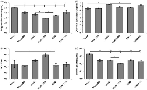
The method of extraction can greatly affect the activity of an herbal product. TFLA was extracted in six different solvents (water, water–HCl 85:15 v/v, methanol, methanol–HCl 85:15 v/v, ethanol or ethanol–HCl 85:15 v/v) to determine which gave the highest level of different phytochemicals. Using the Folin–Ciocalteu test for total polyphenols, the simple water extract contained 20% more polyphenols than the next highest groups, water–HCl and ethanol–HCl (). Total flavonoid content did not vary greatly; however, the methanol extract contained the highest level of flavonoids followed by ethanol–HCl while the rest of extracts remained the same (). Condensed tannins were found with the highest concentration in the methanol–HCl extract, followed by the water and methanol extracts with similar extraction frequencies (). Finally, hydrolysable tannins were most highly extracted in water with all the other solvents to a similar extent (). Since there is more evidence supporting the role of polyphenol-rich plants with prebiotic potential over the other components tested, the water extract was used for the remainder of the analysis.
Figure 1. Total phenolic, flavonoid and tannin constitution in Triphala extracts. Triphala extracts were made using water, water:HCl, methanol (MeOH), MeOH:HCl, ethanol (EtOH) or EtOH:HCl solutions as extract media. Total (a) phenolic, (b) flavonoid, (c) condensed tannin and (d) hydrolysable tannins were assessed using various colourimetric assays. Each group contained n = 3 independent trials while significant differences are indicated by *p < .05 and **p < .01 between groups.
Characterization of the prebiotic activity of Triphala: in vitro assessment
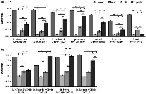
To determine whether TFLA could be used as an energy source to promote the growth of beneficial gut bacteria, mMRS media were made without any carbohydrate source so that the effect of TFLA or the growth controls glucose, inulin or scFOS could be tested. In isolation, five aerobic Lactobacillus strains, two aerobic pathogenic strains (S. aureus and E. coli) and four anaerobic Bifidobacteria strains were grown in the mMRS broth with their respective supplement. As expected, glucose supported the highest level of growth in each of the Lactobacillus, pathogenic and Bifidobacteria strains (). mMRS supplemented with TFLA supported better growth than either inulin or scFOS (). L. plantarum had the highest growth confluency of the Lactobacillus species followed by L. delbrunckii, L. reuteri, L. casei and L. fermentum. Similarly, with the anaerobic Bifidobacteria group, glucose elicited the greatest growth response in most cases however TFLA supported growth that was either the same (B. bifidum) as the scFOS group or greater (B. infantis, B. breve and B. longum). TFLA also supported the growth of two of the strains (B. infantis and B. breve) to the same extent as the glucose control (). Unlike many prebiotics, TFLA did not support elevated growth of the two tested pathogenic species (E. coli and S. aureus) making TFLA a superior prebiotic of choice, especially considering the enrichment of Bifidobacteria species.
Figure 2. Characterization of the prebiotic activity of Triphala compared to known controls in both aerobic and anaerobic species. The prebiotic activity of Triphala was assessed in a variety of (a) aerobic and (b) anaerobic species using in vitro isolated cultures. Each bacterial strain was incubated in mMRS supplemented with either glucose (dark grey), inulin (light grey), scFOS (medium grey) or Triphala (black) as the only fermentable source of energy. Each group contained n = 5 independent samples and significance is indicated as *p < .05 and **p < .01 between groups.
Evaluation of Triphala’s toxicity in Drosophila melanogaster
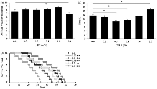
To go forward in the assessment of TFLA as a prebiotic agent in a Drosophila in vivo model, TFLA’s toxicity in the fly was first assessed. To the authors knowledge, TFLA has never been tested in Drosophila before; therefore, in order to establish the best tolerable dose of TFLA in Drosophila to maximize biological impact, toxicity testing was deemed necessary. TFLA, in doses from 0.0 to 2.0% (w/v), was incorporated into the Drosophila media and basic physiological factors were assessed including weight, motility and longevity (). There was little variation in the total Drosophila weight except in the 2.0% TFLA group where there was a significant reduction (). Motility was also affected, both positively and negatively. The greatest improvement in motility occurred in the 0.5% group with a 25% increase in motility whereas in the 2.0% group, there was a significant 26% increase in time to complete the task, indicating impaired motility (). Finally, paralleling the motility results, Drosophila longevity was increased dramatically by almost 50% following 0.5% TFLA supplementation (). Longevity progressively lessened as the concentration of TFLA increased from 0.5% to 2.0%, where at 2.0%, longevity was reduced to 33 days compared to 49 in the untreated controls. Overall, TFLA at a dosage of 0.5% not only showed zero toxicity, but demonstrated significant physiological benefits while at 2.0% TFLA dosage, a toxicity was observed.
Figure 3. Toxicity of Triphala in Drosophila melanogaster. After eclosion, Drosophila were placed on media supplemented with different concentrations (0–2% w/v) of Triphala water extracts. A variety of physiological parameters were assessed including (a) average body weight, (b) motility through the negative geotaxis test and (c) longevity. All groups contained n = 5 independent samples and significance is indicated as *p < .05 and **p < .01.
Triphala has prebiotic activity in Drosophila melanogaster
The variations in the gut microbiota of Drosophila were assessed when flies were placed on media supplemented with TFLA with a dosage range of 0–2.0%. Many reports have indicated that Drosophila raised in a laboratory environment have a reduced microbiota dominated by the genera Acetobacter and Lactobacillus. A. malorum, a potent methanol producer, was slightly elevated with 0.5% TFLA supplementation and repressed in the 1% and 2% groups (). A. pomorum, a bacterium important for anti-viral defence, NFκB induction and insulin signalling showed little variation until 1% group when there was a significant reduction in expression. There was also little change in the A. cerevisiae group, a producer of organic acids, except a slight elevation following 0.5% supplementation. A. aceti, the prominent producer of acetic acid, was slightly reduced in all TFLA groups from 0.2 to 0.75%; however, there was a more prominent loss at 1.0% and 2.0%. A. pasteurianus, also important for acetic acid production, was relatively unchanged spare a small decrease in the 1.0% and 2.0% groups. Considering the Lactobacillus genus, the bacterium key to nutrient allocation L. brevis, was elevated at 0.5% and reduced at 1% supplementation. Also, L. plantarum, which maintains gut epithelial homeostasis, is critical for amino acid synthesis and promotes longevity in flies [Citation32], was significantly elevated by all concentrations of TFLA above 0.5%; however, the 0.5% supplementation elicited a greater increase in L. plantarum than the 2.0% group. Finally, the immunosuppressant Enterococcus spp. were reduced by each concentration of TFLA except 2.0% while the Gluconobacter genus, a direct resource competition to the acetic acid bacteria, was reduced at 0.2 and 0.5% and elevated at 1.0 and 2.0%.
Figure 4. Triphala induces variations in Drosophila gut microbiota populations. After 14 days exposure to the various concentrations (0–2% w/v) of Triphala-treated media, quantification of the Drosophila gutx microbiota was assessed using real-time PCR. Each group contains n = 5 independent samples and significance is marked as *p < .05 relative to the 0% Triphala control.

Probiotics have no physiological toxicity in Drosophila melanogaster
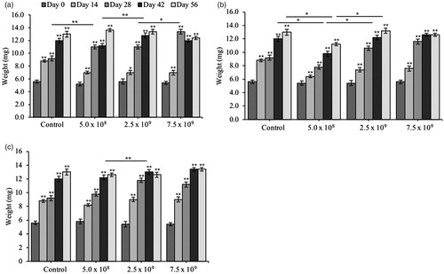
The probiotic bacteria used in the current study are not autochthonous to Drosophila; therefore, to establish that the Drosophila are able to tolerate the probiotics and not recognize them as foreign pathogenic invaders, a series of basic toxicity screens were conducted. Probiotic toxicity in Drosophila was tested by inoculating fly media with several doses (1.0 × 10⁸ CFU/ml to 1.0 × 10¹⁰ CFU/ml) of each probiotic and variations in weight, motility and longevity over time were recorded. As expected, the Drosophila’s weight consistently increased until Day 42 where in most treatment groups, it plateaued (). In the Lp8826 treatment group, there was an increase in weight at Day 28 in all dosage groups, including a slight elevation in the 2.5 × 10⁹ CFU/ml group at Day 42 (). Lf5221 inoculation also showed only minor variations in weight between groups with slight reductions with the 5.0 × 10⁸ CFU/ml dose at Days 42 and 56 compared to all other dosage groups (). Finally, Bi702255 only had an increase in weight at Day 42 in the 2.5 × 10⁹ CFU/ml group compared to the untreated controls (). Dosages up to 1.0 × 10¹⁰ CFU/ml were also tested however there were few differences between the 7.5 × 10⁹ and 1.0 × 10¹⁰ CFU/ml groups so results were not reported.
Figure 5. Triphala is not toxic to variations in weight in Drosophila melanogaster. After eclosion, Drosophila were exposed to media containing a dose-curve of the probiotic bacteria (a) L. plantarum NCIMB 8826 (Lp8826), (b) L. fermentum NCIMB 5221 (Lf5221) and (c) B. longum subsp. infantis NCIMB 702255 (Bi702255) and variations in the Drosophila’s total weight was recorded weekly from Day 0 to Day 56. Each group contained n = 5 independent samples and significance is marked as *p < .05 and **p < .01. Stars indicate significance relative to the normal-media control where bars indicate variations between groups.
Motility represents the flies’ overall fitness and can be a good measure of substance toxicity. Following Lp8826 inoculation, there was a significant reduction in the time it took Drosophila to reach the 10 cm mark in the negative geotaxis test in the 2.5 × 10⁹ CFU/ml group beginning at Day 21 through to Day 49, with the latter being the greatest improvement out of all the doses (). There was no variation in the 5.0 × 10⁸ CFU/ml group and the 7.5 × 10⁹ CFU/ml group demonstrated an improvement of motility beginning at Day 35. Lf5221 supplementation elicited a significant decrease in time in all treatment groups with the 2.5 × 10⁹ and 7.5 × 10⁹ CFU/ml groups having a similar effect on climbing ability with an increase in motility beginning at Day 21. Finally, all doses of Bi702255 improved the climbing ability of Drosophila, with the 2.5 × 10⁹ CFU/ml group having the greatest beneficial effect initiating a significant improvement beginning at Day 28. Dosages up to 1.0 × 10¹⁰ CFU/ml were also tested however there were few differences between the 7.5 × 10⁹ and 1.0 × 10¹⁰ CFU/ml groups so results were not reported.
Table 4. Motility of Drosophila over time after exposure to various probiotic bacteria.
Longevity was also assessed as a measure of toxicity and large variations were recorded depending on the probiotic type and dosage (). Longevity was recorded as the time required for death of 50% and 100% of the sample fly population. Increases in longevity were recorded in all probiotic groups beginning at 2.5 × 10⁹ CFU/ml while Lf5221 first showed an improvement at 1.0 × 10⁹ CFU/ml in the 100% mortality group. Lf5221 at 7.5 × 10⁹ and 1.0 × 10¹⁰ CFU/ml had the greatest beneficial effect on longevity with an increase by almost 20 days over untreated controls; an increase that was significantly greater than both Lp8826 and Bi702255. Similar to the weight and motility groups, the probiotic bacteria did not elicit any toxic effect on Drosophila longevity, and there was rather a significant improvement in longevity supporting the beneficial physiological effects elicited by the probiotics.
Table 5. Longevity of Drosophila fed various probiotic bacteria.
Considering that only L. plantarum is found among the commensal bacteria in Drosophila, Lf5221 was tested for immune activation to determine if the flies were recognizing the foreign probiotics as commensal or a pathogenic invader. The Drosophila intestinal immune system primarily consists of an innate immune response. Immune deficiency (IMD) is activated in response to gram-negative and some gram-positive bacterial insults resulting in the downstream activation of Relish and consequently a transcriptional program that kills invading microbes [Citation33]. After being raised on sterile media for 5-days, the young Drosophila were transferred to media inoculated with 5.0 × 10⁸ to 7.5 × 10⁹ CFU/ml of Lf5221. After 1 h, IMD expression in each of the dosage groups was elevated compared to the control media (). By 6 h after transfer, the level of IMD expression in each of the dosage groups had returned to the level of control. PIMS (PGRP-LC-interacting inhibitor of IMD signalling) is a negative regulator of IMD and is normally upregulated in the presence of commensal bacteria in order to suppress the immune response [Citation34]. PIMS expression following Lf5221 inoculation remained elevated from 1 to 6 h after media transfer in all dosage groups () indicating that the Drosophila were recognizing the foreign bacteria as commensal and likely acting to repress the IMD expression as reported in the 3 and 6 h groups.
Figure 6. Probiotics are not recognized as invading pathogens in Drosophila melanogaster. To assess the immunoreactivity of Drosophila after immediate exposure to the probiotic bacteria, 5-day old Drosophila were transferred to media inoculated with various concentrations of Lf5221. After 1, 3, 6 and 12 h, samples of Drosophila were taken and the expression of the immunological genes (a) immune deficiency (IMD), (b) PGRP-LC-interacting inhibitor of IMD signalling (PIMS), (c) dual oxidase (Duox) and (d) defensin were assessed using real-time PCR. Each group contained n = 5 independent samples and significance is marked as *p < .05 and **p < .01. Stars indicate significance relative to the normal-media control where bars indicate variations between groups.
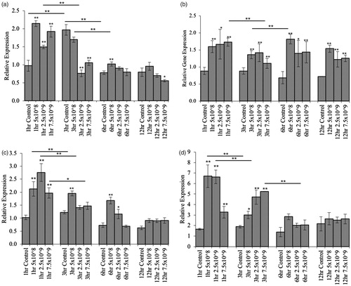
Another level of humeral indiscriminate line of defence in Drosophila is dual oxidase (Duox) which is produced in response to elevated uracil levels, a metabolite released by several pathogenic species [Citation35]. After 1 h, Duox expression was elevated by all dosages of Lf5221 though quickly reduced by 3 h (). By 12 h post transfer, all of the dosage groups were reduced to the level of controls. Defensin is an anti-microbial peptide (AMP) sensitive to the invasion of gram-positive bacteria which is activated downstream of the Toll-like pathway [Citation36]. As expected, there was a significant elevation in defensin expression after 1 h of exposure to the gram-positive Lf5221, however this elevation was quickly reduced after 3 h in both the 5.0 × 10⁸ and 2.5 × 10⁹ CFU/ml groups and by 6 h in the 7.5 × 10⁹ CFU/ml group (). By 12 h, the elevation in Defensin was completely normalized to the level of control indicating that no sustained immune response.
The formulation of probiotic and prebiotics has a combinatorial effect on Drosophila physiological health
TFLA and each of the individual probiotics in the Drosophila model organism elicited beneficial effects on Drosophila’s basic physiological traits including weight and motility. In order to maximize these benefits, a probiotic formulation was conceived that contained equal amounts of each probiotic Lp8826, Lf5221 and Bi702255 totalling a dosage of 3.0 × 10⁹ CFU/ml (i.e. 1.0 × 10⁹ of each of the individual probiotics) as well as a synbiotic formulation with the addition of 0.5% (w/v) TFLA. Each dosage was determined based on the most beneficial concentration in each of the physiological tests. Motility was improved by the combination of probiotics and prebiotics. An improvement in motility was recorded in each of the treatment groups at Day 21 (). By Day 35, both the probiotic and synbiotic formulations had a marked improvement compared to the Lf5221 and TFLA-alone treatments, which was propagated until the end of the study at Day 49.
Table 6. Motility of Drosophila subjected to probiotics and/or prebiotic combinations.
Table 7. Variations in the human gut microbiota following prebiotic and/or probiotic treatment.
The combination of probiotics and prebiotics beneficially alters the composition of the Drosophila gut microbiota
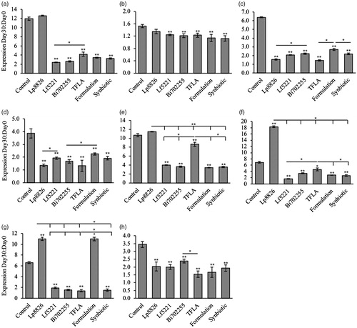
In aging Drosophila, the quantity and diversity of bacteria in the GIT are known to increase as the immunological systems that are in place to control the level of GIT species weaken and the intestinal barrier begins to break down [Citation37]. This phenomenon was clearly present in the control groups of aging Drosophila () as each of the gut microbial populations examined demonstrated a strong upregulation after 30 days. The almost 12-fold elevation in A. malorum in controls was reduced to 2–3 fold by each of the treatment groups except Lp8826 which had no effect (). A. pasteurianus was elevated by 1.6-fold in controls and was unaffected by Lp8826, but slightly reduced by all other treatment groups (). A. aceti was elevated by over 6-fold controls, which was significantly reduced to 1.5–3 fold by each of the treatment groups (). A. tropocalis, elevated almost 4-fold in controls, was reduced by each of the treatment groups to between 1- and 2-fold (). A. cerevisiae, elevated over 10-fold in controls, was reduced to between 3- and 4-fold by all groups except Lp8826 which had no effect and TFLA which was higher than the other treatment groups at a 8.6-fold elevation (). L. brevis was elevated by almost 7-fold in controls by Day 30, which was further increased by Lp8826, likely due to the external supplementation, but reduced by all other treatment groups (). L. plantarum was similarly elevated by almost 7-fold in controls, which was reduced by all treatments except the Lp8826 and probiotic formulation, both of which contained supplemental Lp8826 (). Finally, regarding Gluconobacter spp., there was a keen upregulation by 3.5-fold in controls, which was reduced by all treatment groups (). Overall, the probiotic, prebiotic and the probiotic and synbiotic formulations controlled the elevation in Drosophila microbiota in aging flies indicating that the immune system of Drosophila was greatly benefitted by the treatment. However, there was no drastic difference between the individual probiotic and prebiotic supplementations compared to the probiotic and synbiotic formulations.
Figure 7. The probiotic and/or prebiotics alone or in combination alter the composition of the Drosophila microbiota. Variations in the aging Drosophila gut microbiota were assessed in flies inoculated with each of the individual probiotics (Lf5221, Lp8826 or Bi702255), TFLA or the probiotic or synbiotic formulations. Variations in the gut microbiota are represented by the ratio of relative gene expression from Day 0 to Day 30. Bacterial populations examined include (a) A. malorum, (b) A. pasteurianus, (c) A. aceti, (d) A. tropocalis, (e) A. cerevisiae, (f) L. breve, (g) L. plantarum and (h) Gluconobacter spp. Each group contained n = 5 independent samples and significance is marked as *p < .05 and **p < .01. Stars indicate significance relative to the normal-media control where bars indicate variations between groups.
The human microbiota is benefitted by supplementation of probiotics, prebiotics and their combination
Using a humanized in vitro model of the GIT (SHIME), the impact of TLFA, Lf5221 the probiotic and synbiotic formulation on the composition of the human gut microbiota was compared over 3 weeks of treatment. The ratio of the two major phyla Bacteriodetes and Firmicutes is a marker of metabolic health with an increasing ratio indicating a healthier metabolic constitution [Citation38]. Both TFLA and Lf5221 treatment in the SHIME modestly elevated the Bacteriodetes to Firmicutes ratio between 1.5- and 3.6-fold, with the TFLA treatment being slightly more effective than Lf5221 alone (). The probiotic formulation further elevated this ratio above 3-fold while the synbiotic formulation significantly elevated the Bacteriodetes to Firmicutes ratio above the probiotic formulation in the transverse and descending colon. Actinobacteria, a beneficial phylum including the Bifidobacteria genus, was moderately elevated in the ascending and descending colon following TFLA treatment with a slight increase in the ascending colon following Lf5221 treatment. The probiotic formulation elicited the greatest increase in the ascending and descending colon while the synbiotic formulation elevated Actinobacteria the greatest in the transverse colon. Examining Bifidobacteria spp., a beneficial bacterial genus responsible for a high level of fibre fermentation and SCFA production, was positively affected by TFLA treatment though minimally by Lf5221. Both the probiotic and synbiotic formulations positively affected Bifidobacteria levels in all colonic compartments while the synbiotic formulation in the descending colon had the greatest impact. The beneficial Lactobacillus group was moderately elevated by TFLA and Lf5221 supplementation; however, the probiotic and to a greater extent the synbiotic formulation elevated Lactobacillus spp. levels; the most highly affected group by any of the treatment regimes. The mucin-producing Prevotella genus in the Bacteriodetes phyla was moderately upregulated by TFLA and the synbiotic formulation in all compartments, though reduced by Lf5221 and only elevated in the descending colon following the probiotic formulation. Similarly, for the butyrate-producing Ruminococcus genus, there were significant elevations by TFLA and to a greater extent the synbiotic formulation in all compartments while Lf5221 and the probiotic formulation had a less dramatic impact, and only in the descending colon. The Clostridium cluster XIVa with potent anti-inflammatory and butyrate-producing activity was slightly elevated in the transverse colon following TFLA treatment, though only consistently upregulated following supplementation with the synbiotic formulation. The Enterococcus genus containing many pathogenic pro-inflammatory species was significantly reduced by TFLA, the probiotic and synbiotic formulations though was elevated in the transverse and descending colon following Lf5221 treatment. The Staphylococcus genus contains pathogenic species whose proportions were significantly reduced by TFLA, the probiotic and synbiotic formulations with the synbiotic formulation having the most consistent effect. Finally, E. coli, another pathogenic species, was reduced most effectively by Lf5221, the probiotic and synbiotic formulations and the descending colon following TFLA treatment. Overall, the synbiotic formulation was the most consistently effective at benefitting the composition of the human gut microbiota confirming its combinatorial action.
The probiotic and synbiotic formulation benefits the production of SCFAs in the human gut microbiota
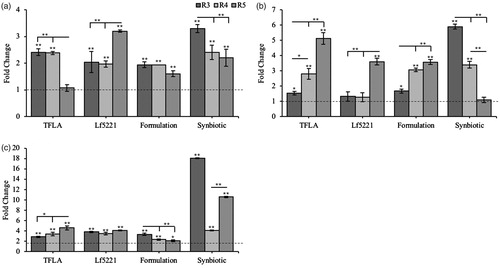
Supplementation with both probiotics and prebiotics not only changes the composition of the relative bacterial populations in the GIT, but also influences their metabolic activity, which is reflected by the proportion of SCFA production. Overall, acetate production was elevated most significantly by the synbiotic formulation, particularly in the ascending colon (). TFLA also elicited a strong upregulation of acetate in the ascending colon, slightly higher than the Lf5221 and probiotic formulation while Lf5221 had a strong effect on acetate production in the descending colon. Similarly, the synbiotic formulation has a very strong impact on propionate production in the ascending colon compared to the other treatment groups whereas TFLA alone had the greatest impact in the descending colon (). Butyrate production was very highly upregulated by the synbiotic formulation in the ascending (18-fold) and descending colons (10-fold), and to a lesser extent in the transverse colon (4-fold) albeit greater than any of the other treatment groups (). TFLA and Lf5221 had similar impacts on butyrate production with TFLA invoking 3- and 5-fold elevations in the ascending and descending legs of the colon, respectively and 3- and 2-fold in the ascending and descending colons of the Lf5221 group. The probiotic formulation similarly elevated the butyrate levels to 3-, 2- and 2-fold in the ascending, transverse and descending colons, respectively. Mirroring the effects on the composition of the gut microbiota, the synbiotic formulation was the most effective at elevating SCFA production, in particular butyrate which is associated with a healthy gut microbial composition.
Figure 8. The probiotic and synbiotic formulations alter SCFA production in a humanized model of the GIT. Supernatant from the humanized in vitro model of the GIT was isolated and the levels of key SCFAs (a) acetate, (b) propionate and (c) butyrate were assessed using isocratic HPLC analysis in the ascending, transverse and descending colon compartments. Quantification is represented as the change in SCFA production from Day 0 to Day 21. Each group contains n = 5 independent samples and significance is marked as *p < .05 and **p < .01. Stars indicate significance relative to the normal-media control where bars indicate variations between groups.
Discussion
There are many lifestyle factors that can push the gut microbiota into a state of dysbiosis, which promotes and aggravates disease. This creates a strong need for the development effective probiotic and synbiotic formulations that stabilize the gut microbiota to promote a healthy constitution. The present study designs an optimized probiotic formulation containing three bioactive probiotics as well as a synbiotic formulation with the addition of a newly characterized polyphenol-rich prebiotic, TFLA. The individual probiotic and TFLA components all demonstrated some beneficial effects on the composition of the gut microbiota and the broad physiological traits of Drosophila; however, together, the probiotic and synbiotic formulations demonstrated a greater, combinatorial effect on all parameters tested.
Recently, the definition of prebiotics was expanded to include health benefiting polyphenols [Citation39]: phytochemicals produced by plants in response to stress. Some polyphenols promote the expansion of beneficial microbes, namely Lactobacillus spp. and Bifidobacteria spp. and suppress the growth of harmful species including Clostridiales and Enterobacteriales [Citation18] earning them the title as a prebiotic. Polyphenols can be metabolized by different groups of microbiota and often require a host of beneficial species to be converted into bioactive phenolic acids. For example, Bacteriodetes have a greater number of glycan-degrading enzymes than the Firmicutes and Clostridium and Eubacterium are the principle microbiota involved in the metabolism of isoflavones, flavonols (quercetin), flavones and flavan-3-ols (catechin and epicatechin) [Citation40].
The major constituents identified in TFLA include gallic acid, ellagic acid and chebulinic acid; however, it also contains other bioactive compounds including flavonoids (quercetin and luteolin), saponins, anthraquinones, fatty acids and various carbohydrates [Citation41]. It has been suggested that gallic acid, quercetin and its breakdown products could promote the growth of Bifidobacteria and Lactobacillus while inhibiting the growth of E. coli [Citation42]. Indeed, aqueous extracts of TFLA, more than ethanolic extracts, were shown to have antimicrobial action against antibiotic-resistant bacteria isolated from human subjects [Citation43]; however, it has never been shown before that crude TFLA extracts can act as a prebiotic in complex microbial mixtures.
In the present study, we demonstrated that TFLA has potent prebiotic activity in vitro, in vivo and in a humanized model of the GIT. TFLA did not support the growth of either gram-positive (S. aureus) or gram-negative (E. coli) pathogenic bacteria, indicating that TFLA creates an intestinal milieu that is non-conducive to pathogenic growth. One reason could be the elevated production of SCFAs that lower luminal pH and inherently increases the presence of toxic compounds like amines, ammonia and phenolic acids due to the reduction of peptide degradation that consequently inhibits pathogenic bacterial growth [Citation44]. In addition, TFLA could directly or indirectly affect the epithelial immune response by prompting lymphocytes and Paneth cells to increase the production of antimicrobial peptides, microbicidal factors or oxidants aimed at destroying pathogenic species or alter the gut microbiota such that it is outcompeting the pathogenic species for space and resources. TFLA water extract at 0.5% w/v had no measured toxicity in the broad physiological traits of Drosophila, and even benefitted the fly’s motility measures. This increase in fitness in aging flies could be reflected by the prebiotic action of TFLA on the gut microbiota which was shown at 0.5% to increase several Acetobacter spp. (A. malorum, A. cerevisiae), L. breve and L. plantarum while reducing the prevalence of the acting pathobionts Enterococcus spp. and Gluconobacter spp. Indeed, the composition of the gut microbiota in Drosophila has been indicated to influence fitness in age, especially in the first week after eclosion [Citation45,Citation46]. Although controversial, it has also been observed that removal of the gut microbiota late during adult life also increases longevity [Citation45] which confides in the fact that aged flies have a reduced ability to fight infection [Citation47]. Nevertheless, the presence of gut microbiota impacts the utilization of nutrition, metabolic homeostasis and insulin signalling, three key factors in aging [Citation48]. This phenomenon could explain why treatment with both the probiotic, TFLA prebiotic and their combinations increased lifespan while simultaneously reducing the specific bacterial load in the aging Drosophila, as outlined in the present study. Further, it was shown that L. plantarum supplementation in nutrient deficient media was sufficient to rescue the increase in developmental time in Drosophila via a dTOR-dependent mechanism [Citation49] while in a normal Drosophila diet, Lf5221 induced a dose-dependent decrease in developmental time via a ferulic-acid and dTOR-dependent mechanism [Citation50] demonstrating the importance of the Drosophila gut microbiota in the management of energy homeostasis.
In the present study, the synbiotic formulation erected the most consistently beneficial effects on the composition of gut microbiota in the SHIME. Confirming the in vitro and in vivo results, TFLA alone was shown to have a modest impact on increasing populations of beneficial bacteria while decreasing the pathogenic Enterococcus spp., Staphylococcus spp. and E. coli. The largest impact of TFLA was observed on the elevation of Bifidobacteria spp., with only slight effects on the butyrate-producing Ruminococcus and Clostridium cluster XIVa groups. Interestingly, the impact of TFLA on the human gut microbiota was more drastic that Lf5221, which had little impact on Prevotella, Ruminococcus, Clostridium cluster XIVa and Staphylococcus species. This supports the idea that prebiotic supplementation has a higher efficacy at changing the composition of the human microbiota by encouraging cross-feeding of beneficial species that are already present in the gut microbiota. In addition, the synbiotic formulation induced a very significant increase in the butyrate-producing species as well as strong downregulation of pathogenic species, which was confirmed with the SCFA assessment. Butyrate production is particularly important for host health as it was previously shown to have potent histone deacetylation (HDAC), anti-inflammatory, antioxidant and energy regulation activities and an important consideration for several chronic diseases including neurodegeneration, diabetes, obesity and cardiovascular disease [Citation51].
Probiotic treatments can be offered as individual strains, as a formulation of several probiotics or in combination with prebiotics making a synbiotic formulation. Although individual probiotic strains have potent medicinal effects, their combination has the potential to simultaneously target several aspects of the heterogeneous disease pathogenesis while also acting combinatorially to increase their effectiveness. The present study is among the first studies to directly compare the effectiveness of probiotics and a prebiotic in isolation to a formulation containing the probiotics or together as a synbiotic. As suggested by the several studies regarding probiotic and synbiotic formulations, the sum of its components is greater than their constituents [Citation52]. We have confirmed this hypothesis as the synbiotic formulation was the most effective at inducing positive variations in the gut microbiota and these changes were reflected in the broad physiological traits of Drosophila including elevated fitness and motility in age.
Conclusions
Although probiotics and prebiotics in isolation offer significant health benefits, multi-strain formulations boost their effectiveness combinatorially as the gut microbiota is a large dynamic system that requires intricate cross-communication to have significant clinical benefit. The current study designed an optimized probiotic formulation containing three complimentary metabolically active probiotics, Lf5221, Lp8826 and Bi702255, in addition to a novel polyphenol-rich prebiotic, Triphala. Together, this formulation increased the motility of Drosophila by inducing significant beneficial effects on the flies’ gut microbiota. Further, in a humanized model of the GIT, the present probiotic and synbiotic formulation induced the expansion of several health-benefitting species while suppressing pathogenic ones, with the synbiotic formulation having a higher impact on the gut microbiota than its components. Maintaining the health of the gut microbiota is critical to manage the development of several chronic diseases including diabetes, obesity, metabolic syndrome, inflammatory bowel diseases and neurodegeneration where pharmacological therapies have shown limited success as they only target the disease symptoms. Usage of optimized synbiotic formulations as presented in this study offers a new generation of treatments for energy-regulating and chronic diseases as they simultaneously target multiple aspects of the disease pathology offering a sustainable, affordable and risk-free solution to disease management.
Disclosure statement
The work presented in this article has been filed as a provisional patent (62/629832) with the company Proviva Pharma of which SW and SP are cofounders. The authors received no financial contribution from the company.
Additional information
Funding
Related Research Data
References
- Lee W-J, Hase K. Gut microbiota-generated metabolites in animal health and disease. Nat Chem Biol. 2014;10:416–424.
- Gu Q-Y, Zhang J, Feng Y-C. Role of NLRP3 inflammasome in Bifidobacterium longum-regulated visceral hypersensitivity of postinfectious irritable bowel syndrome. Artif Cells Nanomed Biotechnol. 2016;44:1933–1937.
- Chow KM, Liu ZC, Prakash S, et al. Free and microencapsulated Lactobacillus and effects of metabolic induction on urea removal. Artif Cells Blood Substit Immobil Biotechnol. 2003;31:425–434.
- Urbanska AM, Bhathena J, Cherif S, et al. Orally delivered microencapsulated probiotic formulation favorably impacts polyp formation in APC (Min/+) model of intestinal carcinogenesis. Artif Cells Nanomed Biotechnol. 2016;44:1–11.
- Bravo JA, Julio-Pieper M, Forsythe P, et al. Communication between gastrointestinal bacteria and the nervous system. Curr Opin Pharmacol. 2012;12:667–672.
- Wilkins T, Sequoia J. Probiotics for gastrointestinal conditions: a summary of the evidence. Am Fam Phys. 2017;96:170–178.
- Chapman CMC, Gibson GR, Rowland I. Health benefits of probiotics: are mixtures more effective than single strains? Eur J Nutr. 2011;50:1–17.
- Belenguer A, Duncan SH, Calder AG, et al. Two routes of metabolic cross-feeding between Bifidobacterium adolescentis and butyrate-producing anaerobes from the human gut. Appl Environ Microbiol. 2006;72:3593–3599.
- Gibson GR, Roberfroid MB. Dietary modulation of the human colonic microbiota: introducing the concept of prebiotics. J Nutr. 1995;125:1401–1412.
- Roberfroid M, Gibson GR, Hoyles L, et al. Prebiotic effects: metabolic and health benefits. Br J Nutr. 2010;104(Suppl.2):S1–S63.
- Whelan K. Mechanisms and effectiveness of prebiotics in modifying the gastrointestinal microbiota for the management of digestive disorders. Proc Nutr Soc. 2013;72:288–298.
- Hijova E, Chmelarova A. Short chain fatty acids and colonic health. Bratisl Lek Listy. 2007;108:354–358.
- Kimura I, Inoue D, Hirano K, et al. The SCFA receptor GPR43 and energy metabolism. Front Endocrinol. 2014;5:85.
- Besten den G, van Eunen K, Groen AK, et al. The role of short-chain fatty acids in the interplay between diet, gut microbiota, and host energy metabolism. J Lipid Res. 2013;54:2325–2340.
- Cani PD, Possemiers S, Van de Wiele T, et al. Changes in gut microbiota control inflammation in obese mice through a mechanism involving GLP-2-driven improvement of gut permeability. Gut. 2009;58:1091–1103.
- Roberfroid M. Prebiotics: the concept revisited. J Nutr. 2007;137: 830S–837S.
- Goh YJ, Klaenhammer TR. Genetic mechanisms of prebiotic oligosaccharide metabolism in probiotic microbes. Annu Rev Food Sci Technol. 2015;6:137–156.
- Duda-Chodak A, Tarko T, Satora P, et al. Interaction of dietary compounds, especially polyphenols, with the intestinal microbiota: a review. Eur J Nutr. 2015;54:325–341.
- Aura A-M. Microbial metabolism of dietary phenolic compounds in the colon. Phytochem Rev. 2008;7:407–429.
- Kim D-H, Jung EA, Sohng IS, et al. Intestinal bacterial metabolism of flavonoids and its relation to some biological activities. Arch Pharm Res. 1998;21:17–23.
- Lee HC, Jenner AM, Low CS, et al. Effect of tea phenolics and their aromatic fecal bacterial metabolites on intestinal microbiota. Res Microbiol. 2006;157:876–884.
- Peterson CT, Denniston K, Chopra D. Therapeutic uses of Triphala in Ayurvedic medicine. J Altern Complement Med. 2017;23: 607–614.
- Baliga MS, Meera S, Mathai B, et al. Scientific validation of the ethnomedicinal properties of the Ayurvedic drug Triphala: a review. Chin J Integr Med. 2012;18:946–954.
- Wong AC-N, Vanhove AS, Watnick PI. The interplay between intestinal bacteria and host metabolism in health and disease: lessons from Drosophila melanogaster. Dis Model Mech. 2016;9:271–281.
- Erkosar B, Storelli G, Defaye A, et al. Host-intestinal microbiota mutualism: “Learning on the Fly”. Cell Host Microbe. 2013;13:8–14.
- Chandler JA, Lang JM, Bhatnagar S, et al. Bacterial communities of diverse Drosophila species: ecological context of a host-microbe model system. PLoS Genet. 2011;7:e1002272.
- Soong Y-Y, Barlow PJ. Antioxidant activity and phenolic content of selected fruit seeds. Food Chem. 2004;88:411–417.
- Zhishen J, Mengcheng T, Jianming W. The determination of flavonoid contents in mulberry and their scavenging effects on superoxide radicals. Food Chem. 1999;64:555–559.
- Porter L, Hrstich L, Chan B. The conversion of procyanidins and prodelphinidins to cyanidin and delphinidin. Phytochemistry. 1985;25:223–230.
- Hartzfeld PW, Forkner R, Hunter MD, et al. Determination of hydrolyzable tannins (gallotannins and ellagitannins) after reaction with potassium iodate. J Agric Food Chem. 2002;50:1785–1790.
- Watson D, O'Connell Motherway M, Schoterman MHC, et al. Selective carbohydrate utilization by lactobacilli and bifidobacteria. J Appl Microbiol. 2013;114:1132–1146.
- Liu X, Hodgson JJ, Buchon N. Drosophila as a model for homeostatic, antibacterial, and antiviral mechanisms in the gut. PLoS Pathog. 2017;13:e1006277.
- Ferrandon D, Imler J-L, Hetru C, et al. The Drosophila systemic immune response: sensing and signalling during bacterial and fungal infections. Nat Rev Immunol. 2007;7:862–874.
- Lhocine N, Ribeiro PS, Buchon N, et al. PIMS modulates immune tolerance by negatively regulating Drosophila innate immune signaling. Cell Host Microbe. 2008;4:147–158.
- Lee K-A, Kim S-H, Kim E-K, et al. Bacterial-derived uracil as a modulator of mucosal immunity and gut-microbe homeostasis in Drosophila. Cell. 2013;153:797–811.
- Imler J-L, Bulet P. Antimicrobial peptides in Drosophila: structures, activities and gene regulation. Chem Immunol Allergy. 2005;86: 1–21.
- Clark RI, Salazar A, Yamada R, et al. Distinct shifts in microbiota composition during Drosophila aging impair intestinal function and drive mortality. Cell Rep. 2015;12:1656–1667.
- Ley RE, Turnbaugh PJ, Klein S, et al. Microbial ecology: human gut microbes associated with obesity. Nature. 2006;444:1022–1023.
- Anhe FF, Roy D, Pilon G, et al. A polyphenol-rich cranberry extract protects from diet-induced obesity, insulin resistance and intestinal inflammation in association with increased Akkermansia spp. population in the gut microbiota of mice. Gut. 2015;64:872–883.
- Selma MV, Espin JC, Tomas-Barberan FA. Interaction between phenolics and gut microbiota: role in human health. J Agric Food Chem. 2009;57:6485–6501.
- Belapurkar P, Goyal P, Tiwari-Barua P. Immunomodulatory effects of Triphala and its individual constituents: a review. Indian J Pharm Sci. 2014;76:467–475.
- Boto-Ordonez M, Urpi-Sarda M, Queipo-Ortuno MI, et al. High levels of Bifidobacteria are associated with increased levels of anthocyanin microbial metabolites: a randomized clinical trial. Food Funct. 2014;5:1932–1938.
- Biradar YS, Jagatap S, Khandelwal KR, et al. Exploring of antimicrobial activity of Triphala Mashi—an Ayurvedic formulation. Evid Based Complement Alternat Med. 2008;5:107–113.
- Slavin J. Fiber and prebiotics: mechanisms and health benefits. Nutrients. 2013;5:1417–1435.
- Brummel T, Ching A, Seroude L, et al. Drosophila lifespan enhancement by exogenous bacteria. Proc Natl Acad Sci USA. 2004;101:12974–12979.
- Ren C, Webster P, Finkel SE, et al. Increased internal and external bacterial load during Drosophila aging without life-span trade-off. Cell Metabol. 2007;6:144–152.
- Ramsden S, Cheung YY, Seroude L. Functional analysis of the Drosophila immune response during aging. Aging Cell. 2008; 7:225–236.
- Taguchi A, White MF. Insulin-like signaling, nutrient homeostasis, and life span. Annu Rev Physiol. 2008;70:191–212.
- Storelli G, Defaye A, Erkosar B, et al. Lactobacillus plantarum promotes drosophila systemic growth by modulating hormonal signals through TOR-dependent nutrient sensing. Cell Metabol. 2011;14:403. 14:
- Westfall S, Lomis N, Kahouli I, et al. Ferulic acid produced by Lactobacillus fermentum influences developmental growth through a dTOR mediated mechanism. Appl Microbiol Biotechnol. 2016;8:272–284.
- Bourassa MW, Alim I, Bultman SJ, et al. Butyrate, neuroepigenetics and the gut microbiome: can a high fiber diet improve brain health? Neurosci Lett. 2016;625:56–63.
- MacPherson CW, Shastri P, Mathieu O, et al. Genome-wide immune modulation of TLR3-mediated inflammation in intestinal epithelial cells differs between single and multi-strain probiotic combination. PLoS One. 2017;12:e0169847.