
Abstract
Growing evidence have probed a stimulatory influence of Notoginsenoside R1 (NGR1) with osteoblastic probability. miR-23a plays a crucial role in osteoblast differentiation. Whereas whether there exists a miRs-related mechanism by which NGR1 promotes preosteoblast differentiation remains unexplored. We pre-treated MC3T3-E1 with NGR1 to anatomize Runx-2 and Osx expression as well as ALP activity. Phosphorylation of regulators was evaluated by Western blot. SB203580 and Ruxolitinib were used to reduce the phosphorylation of regulators. The effects of NGR1 on miR-23a were verified by qRT-PCR. We analyzed the expression of Runx-2 and Osx, ALP activity as well as phosphorylation of regulators in MC3T3-E1 stimulated with NGR1 and transfected with miR-23a inhibitor. We found that NGR1 enhanced Runx-2 and Osx expression as well as ALP activity in a concentration-dependent manner. NGR1 might exhibit an efficacious promotion on Runx-2, Osx and ALP activity by increased phosphorylation of MAPK, JAK1, and STAT3. NGR1 resulted in miR-23a overexpression which positively modulated Runx-2 and Osx expression as well as ALP activity. Our results showed that miR-23a inhibitor reduced the phosphorylation of MAPK, JAK1 and STAT3 in MC3T3-E1 pre-treated with NGR1. In conclusion, NGR1 exhibited an efficacious promotion on preosteoblast differentiation by up-regulating miR-23a through MAPK and JAK1/STAT3 pathways.
NGR1 induces MC3T3-E1 differentiation;
miR-23a is positively regulated by NGR1;
NGR1 regulates MAPK/JAK1/STAT3 through miR-23a.
Highlights:
Introduction
Bone fracture healing is a complex biological process that sequentially involves inflammatory response, primary cartilaginous callus formation, revascularization, calcification, finally bone repair and remodelling [Citation1]. Even if fracture patients are in the state of clinical treatment, there are still considerable cases of non-union and delayed union. To date, a variety of treatment therapies have emerged, for instance, surgical intervention, mechanical forces, pharmacotherapy, cell therapy and molecular biology therapy [Citation2–5], as well as a combination of several methods. Bioactive agents in combination with targeted therapeutics have drawn researchers’ attention, especially through modulating microRNAs (miRs) and signalling pathways for accelerating osteoblastogenesis [Citation6,Citation7]. Given that preosteoblasts are bone-forming cells, it provides us a new insight into accelerating fracture healing by stimulating cells differentiation for bone formation.
Notoginsenoside R1 (NGR1) is a natural triterpene saponin compound detected in the traditional Chinese medicine Panax notoginseng [Citation8]. Its molecular structure is shown in . NGR1 has been reported to exert multiple pharmacological effects on the cell biological activities, for instance, cardioprotective effects against stress injuries [Citation9], pro-angiogenic activity [Citation10], and neuroprotection [Citation11]. Currently, a growing number of works have probed a stimulatory influence of NGR1 on cells with osteoblastic probability, for instance, preosteoblasts [Citation12] and osteoblasts [Citation13]. Accordingly, it has been revealed that Panax notoginseng saponin stimulates osteogenesis process including osteoblastic proliferation, differentiation and mineralization indicated by enhanced alkaline phosphatase (ALP) activity, mineralization and osteoblast-related molecules [Citation14]. The mechanisms by which NGR1 promotes bone generation are centred on several signalling pathways [Citation15], whereas it is still unexplored that whether there exists a miRs-related mechanism by which NGR1 promotes preosteoblast differentiation.
Figure 1. Molecular structure of NGR1.
miR-23a are generally involved in angiogenesis [Citation16], haematopoiesis [Citation17], and cellular senescence [Citation18]. It also plays critical roles in modulating the balance between osteoblast and adipocyte differentiation in bone marrow mesenchymal stem cells [Citation19]. Zeng et al. revealed that miR-23a cluster directly represses a negative regulator of transforming growth factor-β Prdm16 with the alternative of sclerostin expression by RNA-sequencing analysis [Citation20]. Intriguingly, miR-23a has previously been revealed to directly target and regulate Run-related transcription factor 2 (Runx-2) for suppressing NGR1-induced angiogenesis which is positively correlated with cancer development [Citation21]. Nevertheless, it is still undefined that whether NGR1 modulates miR-23a in preosteoblast cells. Hence, we focused attention on the regulatory effects of NGR1 on miR-23a.
NGR1 coordination of the signalling pathways and miRs, in preosteoblasts, might be conducive in bone formation for fracture therapy. As a consequence, we hypothesized that NGR1 was able to promote differentiation of preosteoblasts through triggering signalling pathways by regulating miRs expression. To verify our assumption, we primarily assessed the acceleration of NGR1 on the differentiation of MC3T3-E1 cells. Subsequently, we anatomized the alteration of Runx-2 and Osterix (Osx) expression, ALP activity, and phosphorylated expression of regulatory factors in signalling pathways.
Materials and methods
MC3T3-E1 cells culture and treatment
The mouse preosteoblast cells MC3T3-E1 were obtained from the American Type Culture Collection (ATCC, Manassas, VA). MC3T3-E1 cells were maintained in α-minimum essential medium (α-MEM; Gibco, NY) replenished with foetal bovine serum (FBS) (10%; Gibco), penicillin (100 units/mL; Invitrogen, Carlsbad, CA), and streptomycin (100 mg/mL; Invitrogen) in a humidified atmosphere containing 5% CO2 at 37 °C. MC3T3-E1 cells were stimulated with NGR1 diluted with dimethyl sulphoxide (DMSO; Sigma, St. Louis, MO) in 0–50 μmol/L for 48 h. For repressing mitogen-activated protein kinase (MAPK) signalling pathway, MC3T3-E1 cells were treated with SB203580 (SB) (10 μmol/L; Sigma). As for Janus kinase 1/signal transducer and activator of transcription 3 (JAK1/STAT3) pathway, we stimulated MC3T3-E1 cells with Ruxolitinib (RU) (300 nmol/L; Sigma) to block this signalling pathway.
Transfection
miR-23a inhibitor and its corresponding negative control (NC) were synthesized by GenePharma (Shanghai, China). MC3T3-E1 cells were transfected with miR-23a inhibitor or NC with Lipofectamine 3000 (Invitrogen) with reference to its description.
Alkaline phosphatase activity assay
MC3T3-E1 cells were lysed with M-PER mammalian protein extraction reagent (0.1 mol/l; Pierce, Appleton, WI) for 30 min after rinsed with pre-cold phosphate-buffered saline (PBS; Sigma) twice. After centrifugation (104 × g, 15 min), the supernatant was determined with p-nitrophenyl phosphate (pNPP; Sigma) at 405 nm. Particularly, the sample (50 μL) was blended with 50 μL pNPP (1 mg/mL) dissolved with 1 mol/l diethanolamine buffer containing 0.5 mmol/L MgCl2 (pH 9.8) (Sigma), and then incubated at 37 °C for 15 min on bench shaker. For termination reaction, 200 μL NaOH (2 mol/l) was added into the reaction mixture (1:1, v/v). BCA™ Protein Assay Kit (Pierce) was applied to quantify total protein content. Calf intestinal alkaline phosphatase (Sigma) was applied for quantification as a standard.
Western blotting assay
For Western blotting assay, proteins were initially abstracted from MC3T3-E1 cells with RIPA lysis buffer (Beyotime, Shanghai, China). Protease inhibitors (Roche, Guangzhou, China) were applied in the extraction process. Protein concentrations were examined with BCATM Protein Assay Kit (Pierce). The proteins were separated by sodium dodecyl sulphate-polyacrylamide gel electrophoresis (SDS-PAGE) afterwards. Isolated proteins were transferred to polyvinylidene difluoride (PVDF) membranes (Millipore, Billerica, MA). The membranes were blocked with bovine serum albumin (BSA; Millipore) for 2 h and incubated with the following antibodies: anti-Runx-2 (ab23981, 1 µg/mL), anti-Osx (ab209484, 1:1000) (Abcam, Cambridge, UK), anti-MAPK (9102, 1:1000), anti-p-MAPK (91021, 1:1000), anti-JAK1 (3344, 1:1000), anti-p-JAK1 (74129,1:1000), anti-STAT3 (12640, 1:1000), anti-p-STAT3 (94994, 1:1000) and β-actin (4967, 1:1000) (Cell Signaling Technology, Danvers, MA) at 4 °C overnight. The membranes were probed with secondary antibodies (7074, 1:5000) (Cell Signaling Technology) for 1 h after washing three times. Membranes were transferred into Bio-Rad ChemiDocTM XRS system (Bio-Rad, Shanghai, China) and covered with 200 μL Immobilon Western Chemiluminescent HRP Substrate (Millipore). The protein signalling was visualized and quantified with Image LabTM software (Bio-Rad). β-actin was applied for relative quantification as an internal control.
qRT-PCR analysis
The expression of miR-23a at RNA level was analyzed by quantitative reverse transcription PCR (qRT-PCR). Total RNA was isolated from MC3T3-E1 cells with Trizol reagent (Life Technologies Corporation, Carlsbad, CA). Besides, Taqman MicroRNA Reverse Transcription Kit and Taqman Universal Master Mix II (Applied Biosystems, Foster City, CA) were used for quantifying miR-23a. U6 was used as a housekeeping gene.
Statistical analyses
Each experiment was performed for three times. The results were expressed as mean ± standard deviation (SD). Statistical analyses were conducted with GraphPad Prism Software (Graph Pad Software, La Jolla, CA). We performed unpaired or paired two-tailed Student’s t-test for comparisons between two groups. One-way analysis of variance (ANOVA) was employed for multiple comparisons. We recognized the statistically significant results when p values were < .05.
Results
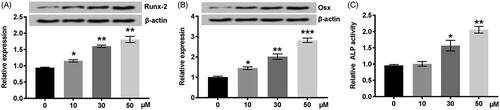
Runx-2 and Osx expression, as well as ALP activity, were enhanced by NGR1 in vitro
For ascertaining the significance of increased Runx-2 and Osx expression as well as ALP activity in the preosteoblast cells pre-treated with NGR1, we stimulated MC3T3-E1 cells with NGR1 at different concentrations (10, 30, and 50 μmol/L). We detected that Runx-2 and Osx protein levels were markedly enhanced in MC3T3-E1 cells (p < .05, .01 or .001) (). Similarly, ALP activity was significantly enhanced paralleled with that of the control group (p < .05 or p < .01) (). Specifically, the simulated concentration of NGR1 was positively related with Runx-2 and Osx expression and ALP activity. These results suggested that NGR1 promoted preosteoblast differentiation verified by increased Runx-2 and Osx expression as well as ALP activity.
Figure 2. NGR1 promoted MC3T3-E1 cells differentiation in a dose-dependent manner. (A) The expression of Runx-2 was elevated by NGR1 at protein levels. (B) The expression of Osx was increased in MC3T3-E1 cells pre-treated with NGR1. (C) ALP activity was enhanced by NGR1. MC3T3-E1 cells were pre-treated with NGR1 (0–50 μmol/L) for 48 h. NGR1: Notoginsenoside R1; Osx: Osterix; ALP: alkaline phosphatase. *p < .05, **p < .01, or ***p < .001 compared to control.
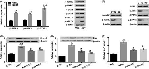
NGR1 promoted Runx-2 and Osx expression and ALP activity by activating MAPK and JAK1/STAT3 signalling pathways
In order to address that NGR1 stimulated preosteoblast cells differentiation through modulating MAPK and JAK1/STAT3 signalling pathways, we initially examined the phosphorylated expression of regulatory factors of MAPK and JAK1/STAT3 signalling pathways at protein levels. Our results validated that NGR1 directly activated MAPK and JAK1/STAT3 signalling pathways (p < .01 or p < .001) (). We next blocked MAPK and JAK1/STAT3 pathways by stimulating MC3T3-E1 cells with SB and RU, separately. The results that the phosphorylated expression of MAPK, JAK1, and STAT3 was reduced revealed that MAPK and JAK1/STAT3 signalling pathways were inactivated (). As we observed in , the expression of Runx-2 and Osx was accordingly suppressed by SB and RU in MC3T3-E1 cells pre-treated with NGR1. Notably, SB and RU similarly reversed the modulatory effects of NGR1 on ALP activity in MC3T3-E1 cells (p < .05) (). As a consequence, NGR1 promoted Runx-2 and Osx expression and ALP activity by activating MAPK and JAK1/STAT3 signalling pathways.
Figure 3. Runx-2, Osx, and ALP activity were enhanced by NGR1 by improving phosphorylation of MAPK, JAK, and STAT3.(A) The phosphorylated expression of MAPK, JAK1, and STAT3 was increased in preosteoblast MC3T3-E1 cells treated with NGR1. (B) SB and RU repressed the phosphorylation of MAPK, JAK1 and STAT3, respectively. (C) SB and RU decreased the protein expression of Runx-2 induced by NGR1. (D) SB and RU inhibited the protein expression of Osx induced by NGR1. (E) SB and RU inactivated ALP activity elevated by NGR1. MC3T3-E1 cells were treated with NGR1 (50 μmol/L) for 48 h in the NGR1 group; MC3T3-E1 cells were not treated with NGR1 in the CTRL group; MC3T3-E1 cells were treated with NGR1 (50 μmol/L) and SB (10 μmol/L) or RU (300 nmol/L) in the NGR1 + SB or NGR1 + RU group, respectively. NGR1: Notoginsenoside R1; Osx: Osterix; ALP: alkaline phosphatase; SB: SB203580; RU: Ruxolitinib; CTRL: control; p-: phosphorylated-; t-: total-; MAPK: mitogen-activated protein kinase; JAK1: janus kinase 1; STAT3: signal transducer and activator of transcription 3. *p < .05, **p < .01, or ***p < .001 compared to CTRL; #p < .05 compared to NGR1.
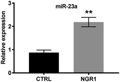
miR-23a elevated the expression of Runx-2 and Osx and ALP activity by up-regulating miR-23a
Given that miR-23a accelerates differentiation of osteoblasts [Citation20,Citation22] and NGR1 could modulate biological processes by regulating the expression of miRs [Citation23], we considered that there might be a modulatory relationship between NGR1 and miR-23a. On the base of our findings, that NGR1 promoted Runx-2 and Osx expression and enhanced ALP activity, we further confirmed that the expression of miR-23a was markedly enhanced by NGR1 (p < .01; ).
Figure 4. NGR1 up-regulated the expression of miR-23a. MC3T3-E1 cells were treated with NGR1 (50 μmol/L) for 48 h in the NGR1 group; MC3T3-E1 cells were not treated with NGR1 in the CTRL group. NGR1: Notoginsenoside R1; CTRL: control; miR-23a: microRNA-23a. **p < .01 compared to CTRL.
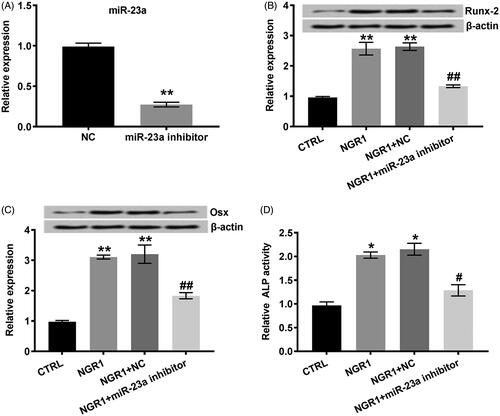
Since miR-23a was increased in MC3T3-E1 cells pre-treated with NGR1, we hypothesized that NGR1 might promote the expression of Runx-2 and Osx as well as ALP activity through up-regulating the expression of miR-23a. For substantiating our assumption, we silenced the expression of miR-23a by transfection of miR-23a inhibitor and stimulated transfected MC3T3-E1 cells with NGR1. Our results showed that miR-23a inhibitor significantly reduced the expression of miR-23a in MC3T3-E1 cells (p < .01; ). Moreover, down-regulated miR-23a obviously restrained Runx-2 and Osx expression (p < .01) (). Similarly, ALP activity increased by NGR1 was reduced by miR-23a inhibitor (p < .05) (). Summarily, NGR1 enhanced Runx-2 and Osx expression and ALP activity by elevating miR-23a expression.
Figure 5. NGR1 accelerated the differentiation of MC3T3-E1 cells by increasing the expression of miR-23a. (A) The expression of miR-23a was down-regulated by miR-23a inhibitor. (B) miR-23a inhibitor reduced the protein expression of Runx-2 induced by NGR1. (C) miR-23a inhibitor decreased the protein expression of Osx induced by NGR1. (D) miR-23a inhibitor inactivated the activity of ALP stimulated by NGR1. MC3T3-E1 cells were treated with NGR1 (50 μmol/L) for 48 h in the NGR1 group; MC3T3-E1 cells were not treated with NGR1 in the CTRL group; MC3T3-E1 cells were treated with NGR1 and transfected with miR-23a inhibitor in the NGR1 + miR-23a inhibitor group; MC3T3-E1 cells were treated with NGR1 and the corresponding negative control of miR-23a in the NGR1 + NC group. miR-23a: microRNA-23a; NGR1: Notoginsenoside R1; CTRL: control; NC: negative control; Osx: Osterix; ALP: alkaline phosphatase. *p < .05 or **p < .01 compared to CTRL; #p < .05 or ##p < .01 compared to NGR1 + NC.
Down-regulated miR-23a expression blocked MAPK and JAK1/STAT3 signalling pathways triggered by NGR1
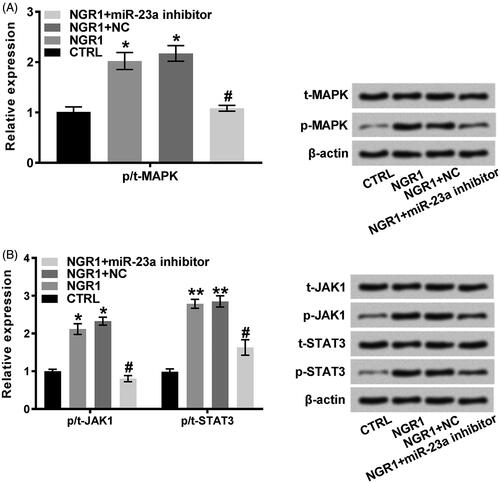
We have elucidated that NGR1 elevated the phosphorylation of MAPK, JAK1, and STAT3. The observation that Runx-2 and Osx expression and ALP activity straightly enhanced by NGR1 by triggering MAPK and JAK1/STAT3 signalling pathways and promoting miR-23a expression raised our hypothesis that miR-23a might participate in activating MAPK and JAK1/STAT3 signalling pathways, which were triggered by NGR1. Consequently, we examined the phosphorylated expression of MAPK, JAK1, and STAT3 in MC3T3-E1 cells after suppressing the expression of miR-23a by transfecting MC3T3-E1 cells with miR-23a inhibitor. Our results revealed that reduced miR-23a expression significantly inhibited phosphorylation of MAPK, JAK1 and STAT3 in MC3T3-E1 cells treated with NGR1 (p < .05; )). Analyzing the above evidence, we might distinctly conclude that NGR1 triggered MAPK and JAK1/STAT3 signalling pathways by elevating miR-23a expression.
Figure 6. NGR1 enhanced the phosphorylated expression of MAPK, JAK1 and STAT3 by promoting the expression of miR-23a. (A) miR-23a inhibitor repressed the phosphorylation of MAPK promoted by NGR1. (B) miR-23a inhibitor decreased the phosphorylated expression of JAK1 and STAT3 in the MC3T3-E1 cells pre-treated with NGR1. MC3T3-E1 cells were treated with NGR1 (50 μmol/L) for 48 h in the NGR1 group; MC3T3-E1 cells were not treated with NGR1 in the CTRL group; MC3T3-E1 cells were treated with NGR1 and transfected with miR-23a inhibitor in the NGR1 + miR-23a inhibitor group; MC3T3-E1 cells were treated with NGR1 and the corresponding negative control of miR-23a in the NGR1 + NC group. NGR1: Notoginsenoside R1; NC: negative control; p-: phosphorylated-; t-: total-; MAPK: mitogen-activated protein kinase; JAK1: janus kinase 1; STAT3: signal transducer and activator of transcription 3. *p < .05 or **p < .01 compared to CTRL; #p < .05 or ##p < .01 compared to NGR1 + NC.
Discussion
In our present work, we have elucidated that NGR1 promoted preosteoblast differentiation. On the basis of its positive effects, our research provided a compelling conclusion for a potential mechanism by which NGR1 accelerated differentiation by activating MAPK and JAK1/STAT3 pathways. Furthermore, our observation revealed that miR-23a exerted a crucial function in differentiation. It is appreciated that down-regulated miR-23a blocked MAPK and JAK1/STAT3 pathways which were triggered by NGR1.
Although the pro-osteogenic function of NGR1 has been evaluated in osteoblast in vitro in a previous study [Citation13], its role in preosteoblast is a compelling question. Since ALP is a late differentiation marker of osteoblasts from preosteoblasts, playing pivotal roles in bone mineralization and suggesting osteoblast activity [Citation24,Citation25], we dissected ALT activity in MC3T3-E1 cells treated with NGR1. The increased ALP activity suggested that NGR1 promoted preosteoblast differentiation which was also proved by enhancement of Runx-2 and Osx expression. Runx-2 has been reported to induce osteoblast differentiation [Citation26]. Osx is identified as an essential multifunctional regulator in bone growth [Citation27]. Hence, NGR1 possessed a pro-osteogenic function. In addition, NGR1 promoted preosteoblasts differentiation in a dose-dependent manner. MAPK and JAK1/STAT3 signalling pathways are involved in multiple type cells differentiation [Citation28–30]. Our observation, that reduced phosphorylated expression of MAPK, JAK1 and STAT3 by pharmacological treatment repressed Runx-2 and Osx expression as well as ALP activity, confirming our hypothesis that NGR1 might accelerate differentiation by triggering MAPK and JAK1/STAT3 signalling pathways. That is, pre-treatment with NGR1 could enhance differentiation of preosteoblast which plays crucial roles in bone formation and fractures healing, indicating a potential therapeutic cure for bone injury.
Noticeably, we detected the higher expression of miR-23a in preosteoblast cells pre-treated with NGR1. Consistent with our findings, previous studies suggested that aberrant expression of miR-23a cluster provokes dysfunction of osteocyte differentiation [Citation20]. Although NGR1 has been proved to exert a pro-osteogenic function in osteoblast differentiation, the mechanism is undefined, especially whether there is a correlation between miR-23a and NGR1. In order to explore whether NGR1 regulated miR-23a expression for influencing the production of Runx-2 and Osx and ALP activity, we suppressed miR-23a expression by transfecting preosteoblasts with miR-23a inhibitor. Our results further elucidated that up-regulating miR-23a expression, NGR1 promoted preosteoblast differentiation. Recent findings demonstrated that miR-23a cluster is necessary for sustaining stage-specific HoxA factor expression which could physically interact with Runx-2 during preosteoblast differentiation [Citation31]. Hasasan et al. reported that Runx-2 negatively modulates miR-23a cluster expression and they provided direct evidence for a potential mechanism that miR-23a cluster targets SATB2 which has been proved to synergize with Runx-2 to facilitate bone formation [Citation22]. Similarly, our findings demonstrated there might exhibit a negative feedback effect of miR-23a on the expression of Runx-2. As for Osx, there are scarce reports about direct regulation of miR-23a on Osx. Therefore, we supposed that NGR1 might up-regulate miR-23a expression which circuitously promotes Osx expression.
The regulatory effects of miRs on signalling pathways, especially studies have validated that miR-23a targets MAPK and JAK/STAT signalling pathways in prostate cancer [Citation32], provoked our attention. Considering MAPK and JAK1/STAT3 signalling pathways are involved in osteoblast cells differentiation process [Citation30,Citation33,Citation34], we focused on the alternation of phosphorylated expression of MAPK, JAK1, and STAT3 which were modulated by a concomitant synergism between NGR1 and miR-23a inhibitor, subsequently verified in our studies. The results showed that miR-23a inhibitor reduced the phosphorylation of MAPK, JAK1, and STAT3. Mechanistically, we confirmed that NGR1 may potentiate MAPK and JAK1/STAT3 signalling pathways by up-regulating miR-23a expression.
Taken together, we have validated that NGR1 promoted preosteoblast cells differentiation. Based on its effective acceleration on preosteoblast cells differentiation, we proposed a potential mechanism that NGR1 exerted up-regulatory effects on Runx-2, Osx and ALP activity by increasing miR-23a expression. Further, NGR1 activated MAPK and JAK1/STAT3 signalling pathways by up-regulating the expression of miR-23a.
Disclosure statement
No potential conflict of interest was reported by the authors.
References
- Bahney CS, Zondervan RL, Allison P, et al. Cellular biology of fracture healing. J Orthop Res. 2018. Available from: https://doi.org/10.1002/jor.24170
- Furuta T, Miyaki S, Ishitobi H, et al. Mesenchymal stem cell-derived exosomes promote fracture healing in a mouse model. Stem Cells Transl Med. 2016;5:1620–1630.
- Taiani JT, Buie HR, Campbell GM, et al. Embryonic stem cell therapy improves bone quality in a model of impaired fracture healing in the mouse; tracked temporally using in vivo micro-CT. Bone. 2014;64:263–272.
- Haffner N, Antonic V, Smolen D, et al. Extracorporeal shockwave therapy (ESWT) ameliorates healing of tibial fracture non-union unresponsive to conventional therapy. Injury. 2016;47:1506–1513.
- Fischer V, Haffner-Luntzer M, Amling M, et al. Calcium and vitamin D in bone fracture healing and post-traumatic bone turnover. Ecm. 2018;35:365–385.
- Shan Z, Cheng N, Huang R, et al. Puerarin promotes the proliferation and differentiation of MC3T3-E1 cells via microRNA-106b by targeting receptor activator of nuclear factor-kappaB ligand. Exp Ther Med. 2018;15:55–60.
- Huang Z, Cheng C, Wang J, et al. Icariin regulates the osteoblast differentiation and cell proliferation of MC3T3-E1 cells through microRNA-153 by targeting Runt-related transcription factor 2. Exp Thera Med. 2018;15:5159–5166.
- Guan J, Lai CM, Li SP. A rapid method for the simultaneous determination of 11 saponins in Panax notoginseng using ultra performance liquid chromatography. J Pharm Biomed Anal. 2007;44:996–1000.
- Yu Y, Sun G, Luo Y, et al. Cardioprotective effects of Notoginsenoside R1 against ischemia/reperfusion injuries by regulating oxidative stress-and endoplasmic reticulum stress- related signaling pathways. Sci Rep. 2016;6:21730.
- Yang BR, Hong SJ, Lee SM, et al. Pro-angiogenic activity of notoginsenoside R1 in human umbilical vein endothelial cells in vitro and in a chemical-induced blood vessel loss model of zebrafish in vivo. Chin J Integr Med. 2016;22:420–429.
- Meng X, Sun G, Ye J, et al. Notoginsenoside R1-mediated neuroprotection involves estrogen receptor-dependent crosstalk between Akt and ERK1/2 pathways: a novel mechanism of Nrf2/ARE signaling activation. Free Radic Res. 2014;48:445–460.
- Liu Y, Lin Z, Guo J, et al. Notoginsenoside R1 significantly promotes in vitro osteoblastogenesis. Int J Mol Med. 2016;38:537–544.
- Wang T, Wan D, Shao L, et al. Notoginsenoside R1 stimulates osteogenic function in primary osteoblasts via estrogen receptor signaling. Biochem Biophys Res Commun. 2015;466:232–239.
- Ji Z, Cheng Y, Yuan P, et al. Panax notoginseng stimulates alkaline phosphatase activity, collagen synthesis, and mineralization in osteoblastic MC3T3-E1 cells. In Vitro Celldevbiol-Animal. 2015;51:950–957.
- Zhao S, Yan L, Li X, et al. Notoginsenoside R1 suppresses wear particle-induced osteolysis and RANKL mediated osteoclastogenesis in vivo and in vitro. Int Immunopharmacol. 2017;47:118–125.
- Hsu YL, Hung JY, Chang WA, et al. Hypoxic lung cancer-secreted exosomal miR-23a increased angiogenesis and vascular permeability by targeting prolyl hydroxylase and tight junction protein ZO-1. Oncogene. 2017;36:4929–4942.
- Kurkewich JL, Hansen J, Klopfenstein N, et al. The miR-23a∼27a∼24-2 microRNA cluster buffers transcription and signaling pathways during hematopoiesis. PLoS Genet. 2017;13:e1006887.
- Rock K, Tigges J, Sass S, et al. miR-23a-3p causes cellular senescence by targeting hyaluronan synthase 2: possible implication for skin aging. J Investig Dermatol. 2015;135:369–377.
- Guo Q, Chen Y, Guo L, et al. miR-23a/b regulates the balance between osteoblast and adipocyte differentiation in bone marrow mesenchymal stem cells. Bone Res. 2016;4:16022.
- Zeng HC, Bae Y, Dawson BC, et al. MicroRNA miR-23a cluster promotes osteocyte differentiation by regulating TGF-beta signalling in osteoblasts. Nat Comms. 2017;8:15000.
- Wu XD, Guo T, Liu L, et al. MiR-23a targets RUNX2 and suppresses ginsenoside Rg1-induced angiogenesis in endothelial cells. Oncotarget. 2017;8:58072–58085.
- Hassan MQ, Gordon JA, Beloti MM, et al. A network connecting Runx2, SATB2, and the miR-23a∼27a∼24-2 cluster regulates the osteoblast differentiation program. Proc Natl Acad Sci USA. 2010;107:19879–19884.
- Jia C, Xiong M, Wang P, et al. Notoginsenoside R1 attenuates atherosclerotic lesions in ApoE deficient mouse model. PLoS One. 2014;9:e99849.
- Millan JL. Alkaline Phosphatases: Structure, substrate specificity and functional relatedness to other members of a large superfamily of enzymes. Purinergic Signal. 2006;2:335–341.
- Balcerzak M, Hamade E, Zhang L, et al. The roles of annexins and alkaline phosphatase in mineralization process. Acta Biochimica Polonica. 2003;50:1019–1038.
- Komori T. Regulation of bone development and extracellular matrix protein genes by RUNX2. Cell Tissue Res. 2010;339:189–195.
- Zhou X, Zhang Z, Feng JQ, et al. Multiple functions of Osterix are required for bone growth and homeostasis in postnatal mice. Proc Natl Acad Sci USA. 2010;107:12919–12924.
- Wang K, Wang C, Xiao F, et al. JAK2/STAT2/STAT3 are required for myogenic differentiation. J Biol Chem. 2008;283:34029–34036.
- Hirano T, Ishihara K, Hibi M. Roles of STAT3 in mediating the cell growth, differentiation and survival signals relayed through the IL-6 family of cytokine receptors. Oncogene. 2000;19:2548–2556.
- Dalagiorgou G, Piperi C, Adamopoulos C, et al. Mechanosensor polycystin-1 potentiates differentiation of human osteoblastic cells by upregulating Runx2 expression via induction of JAK2/STAT3 signaling axis. Cell Mol Life Sci. 2017;74:921–936.
- Godfrey TC, Wildman BJ, Beloti MM, et al. The microRNA-23a cluster regulates the developmental HoxA cluster function during osteoblast differentiation. J Biol Chem. 2018;293:17646–17660.
- Aghaee-Bakhtiari SH, Arefian E, Naderi M, et al. MAPK and JAK/STAT pathways targeted by miR-23a and miR-23b in prostate cancer: computational and in vitro approaches. Tumor Biol. 2015;36:4203–4212.
- Rodriguez-Carballo E, Gamez B, Ventura F. p38 MAPK signaling in osteoblast differentiation. Front Cell Dev Biol. 2016;4:40.
- Murakami K, Kobayashi Y, Uehara S, et al. A Jak1/2 inhibitor, baricitinib, inhibits osteoclastogenesis by suppressing RANKL expression in osteoblasts in vitro. PLoS One. 2017;12:e0181126.