
Abstract
Notoginsenoside R1 (NGR1) is a major bioactive ingredient of Radix notoginseng that has promise in treating several diabetes-related diseases, like diabetic nephropathy and diabetic cardiomyopathy. This work is designed to explore if NGR1 has potential in treating diabetic peripheral neuropathy (DPN). A cell model of DPN was made by stimulating RSC96 cells with high glucose. The effects of NGR1 preconditioning were examined by conducting CCK-8 assay, flow cytometry, ROS assay and Western blot. The downstream effector and signalling of NGR1 were then explored by qRT-PCR and Western blot. High glucose incubation led to cell viability loss, apoptosis facilitation, caspase-3 and PARP cleavage, and ROS generation of RSC96 cells. Meanwhile, expression of NGF and BDNF was declined by high glucose. Preconditioning NGR1 significantly protected RSC96 cells against high glucose-evoked damage. And NGR1 prevented high glucose-induced expression of miR-503. The beneficial functions of NGR1 towards high glucose-injured RSC96 cells were impeded by miR-503 overexpression. Further, NGR1 activated PI3K/AKT and β-catenin signalling through a miR-503-dependent way. This paper illustrated the neuroprotective and neurotrophic function of NGR1 in RSC96 cells. The beneficial function may due to its regulation on miR-503 expression and the downstream signalling such as PI3K/AKT and β-catenin.
Introduction
Diabetic peripheral neuropathy (DPN) is one of the most frequent and troublesome complications of diabetes. It accounts for substantial morbidity and mortality and results in huge health care costs and poor quality of life [Citation1]. There are varied clinical presentations of DPN with the involvement of proximal or distal peripheral sensory and motor nerves as well as autonomic nerves [Citation2]. A population survey in Qena Governorate found that, the prevalence of DPN in patients with type I diabetes was ∼8.7% and in type II was 0.3% [Citation3]. The management of DPN varies with different types of diabetes that patients harboured. Specifically, glycaemic control is a main management for DPN patients with type I diabetes, and lifestyle modification is an option therapeutic approach for type II [Citation4]. However, many therapeutic managements for DPN have failed which calls for more effective therapies for this disease in clinic.
Radix notoginseng is a traditional Chinese medicine used from ancient China to treat cardiovascular diseases. Notoginsenoside R1 (NGR1) is one of the major bioactive ingredients of R. notoginseng. Increasing amount of literatures have shown its pharmacological effects, including anti-inflammatory, antioxidant, anti-apoptotic, and neuro-protective properties [Citation5–8]. Because of that, NGR1 has been considered to have promise in treating various diseases, like chronic atrophic gastritis [Citation9], cerebral hypoxia [Citation10], Alzheimer’s disease [Citation11], and so forth. Of note, NGR1 is beneficial for several diabetes-related diseases, including diabetic nephropathy [Citation12], diabetic cardiomyopathy [Citation13] and diabetic retinopathy [Citation14]. However, no studies have reported the function of NGR1 in treating DPN.
MicroRNAs (miRNAs) are endogenously expressed, small noncoding RNAs that blocking protein translation or inducing mRNA degradation via binding to the 3'UTR of specific target mRNAs. miRNAs have been recognized as critical players in modulating physiological and pathological processes, like cell growth, differentiation, proliferation, inflammation and death. Recent papers have linked miRNA expression with the pathogenesis of DPN. For instance, miR-146a overexpression was able to inhibit DPN progression through increasing neurological function and reducing inflammatory response [Citation15]. Likewise, miR-25 acted as a protective gene in DPN to reduce the generation of ROS [Citation16]. MiR-503 is a hyperglycaemia-sensitive miRNA that can be highly expressed in diabetic patients [Citation17]. Besides that, miR-503 was considered as a factor in regulating neuro-damage [Citation18], which suggested it acted as a potential therapeutic target in DPN.
The present paper designed to study the function of NGR1 in treating DPN. To this end, a cell model of DPN was made by stimulating neuronal Schwann cell line RSC96 with hyperglycaemic condition. Further, this study worked on investigating the regulatory effect of NGR1 on miR-503 expression, to provide a possible explanation of NGR1’s function.
Materials and methods
Cell culture
RSC96 cells (ATCC, Manassas, VA) were cultivated in DMEM medium (ATCC) at 37 °C in a dampen atmosphere with 5% CO2. The complete culture medium was made by adding 10% foetal bovine serum (Gibco, Grand Island, NY).
Establishment of cell injury model and treatment
To make hyperglycaemic condition, cells were cultured in complete culture medium supplemented with 100 mM glucose (Sigma-Aldrich, St. Louis, MO) [Citation19]. The cells were maintained at hyperglycaemic condition for 48 h, and the cells were classed as high glucose (HG) group. The cells cultured in complete culture medium which containing 25 mM glucose were classed as the normal glucose (NG) group.
Notoginsenoside R1 (NGR1) ≥98% (HPLC) was brought from Sigma-Aldrich (St. Louis, MO). Powder NGR1 was dissolved in DMSO and diluted by culture medium before use until the concentration of DMSO was less than 0.1%. Cells were treated by 10–100 μM NGR1 for 48 h before HG treatment.
Transfection
miR-503 mimic (sense: 5'-UAGCAGCGGGAACAGUACUGCAG-3', antisense: 5'-GCAGUACUGUUCCCGCUGCUAUU-3') and its respective control (NC mimic) (5'-UCACAACCUCCUAGAAAGAGUAGA-3') were from GenePharma (Shanghai, China). Transfection was done by lipofectamine 3000 (Invitrogen, Carlsbad, CA) and lasted for 48 h.
qRT-PCR
About 5 × 10⁶ cells per sample were harvested following treatment or transfection. Total RNAs were extracted by miRNeasy Mini Kit (Qiagen, Shenzhen, China). The expression of miR-503 was examined by using Mir-X™ miRNA First Strand Synthesis Kit and Mir-X™ miRNA qRT-PCR TB Green™ Kit (both from Takara, Dalian, China). U6 was applied as reference control.
Cell viability
RSC96 cells in 96-well plates (5000 cells/well) were treated by HG, NGR1 or both, after which cell viability was examined by adding with 10 μL CCK-8 solution (Dojindo Molecular Technologies, Kyushu, Japan). The sample was incubated at 37 °C for 2 h, and then the absorbance was tested at 450 nm by Microplate Reader (Bio-Rad, Hercules, CA).
Apoptosis assay
RSC96 cells in 6-well plates (5 × 10⁵ cells/well) were treated by HG, NGR1 or both. Then, cells were harvested in an eppendorf tube and apoptotic cells were stained by Annexin V-FITC/PI Apoptosis Detection Kit (Sangon Biotech, Shanghai, China). Flow cytometry analysis was done in FACS Calibur (Becton Dickinson, San Jose, CA).
ROS assay
RSC96 cells in 6-well plates (5 × 10⁵ cells/well) were treated by HG, NGR1 or both. Then, 10 μM DCFH-DA (Sigma-Aldrich, St. Louis, MO) was added into each well and incubated for 1 h away from light. The fluorescent intensity was examined by the flow cytometer at 488 and 521 nm to evaluate ROS generation.
Western blot
RSC96 cells in 6-well plates (5 × 10⁵ cells/well) were treated by HG, NGR1 or both. Then, cells were melting in RIPA lysis buffer (Sangon Biotech, Shanghai, China) and proteins in lysate were extracted through repeated centrifugation at 4 °C, 1200 g for 5 min. Purity of protein in extracts was examined by BCA method and the proteins were resolved over SDS-PAGE and transferred onto PVDF membranes (Sangon Biotech, Shanghai, China). Following primary antibodies against pro-caspase-3 (orb35276; dilution 1:1000), cleaved-caspase-3 (orb106556; dilution 1:1000), pro-PARP (orb88930; dilution 1:1000), cleaved-PARP (orb323122; dilution 1:2000), NGF (orb379540; dilution 1:1000), BDNF (orb519283; dilution 1:500), PI3K (orb395443; dilution 1:1000), p-PI3K (orb338965; dilution 1:500), AKT (orb29949; dilution 1:500), p-AKT (orb344403; dilution 1:1000), β-catenin (orb500798; dilution 1:1000) and β-actin (orb181785; dilution 1:500, Biorbyt, San Francisco, CA) were utilized in immunoblotting. Positive bands were developed by BeyoECL Moon (Beyotime, Shanghai, China).
Statistics
Data were presented as mean ± SD. Statistics were done in SPSS 19.0 software (Chicago, IL) with ANOVA or Student’s t-test. Statistical differences were set at p < .05 and indicated as asterisks in figures.
Results
RSC96 cells were injured by HG
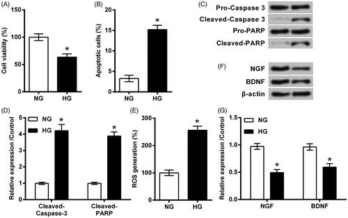
RSC96 cells were cultivated in HG condition for 48 h. As a result, cell viability was repressed and apoptosis was induced by HG as relative to NG (both p < .05, ). In the meantime, caspase-3 and PARP were clearly cleaved by HG as relative to NG (both p < .05, ). Besides that, ROS generation was increased (p < .05, ) while expression of NGF and BNDF was repressed (p < .05, ) by HG when compared to NG. All these data implied the damage evoked by HG in RSC96 cells.
Figure 1. RSC96 cells were injured by high glucose (HG). RSC96 cells were cultivated in HG condition for 48 h. Cells in normal glucose (NG) were considered as control. (A) Cell viability, (B) apoptosis rate, (C,D) cleavage of caspase-3 and PARP, (E) ROS generation, and (F,G) expression of neurotrophic proteins were examined by CCK-8 assay, flow cytometry, Western blot and DCFH-DA probe. Data were presented as mean ± SD (n = 3). *p < .05 (Student’s t-test).
NGR1 pre-conditioning ameliorated HG-evoked injury in RSC96 cells
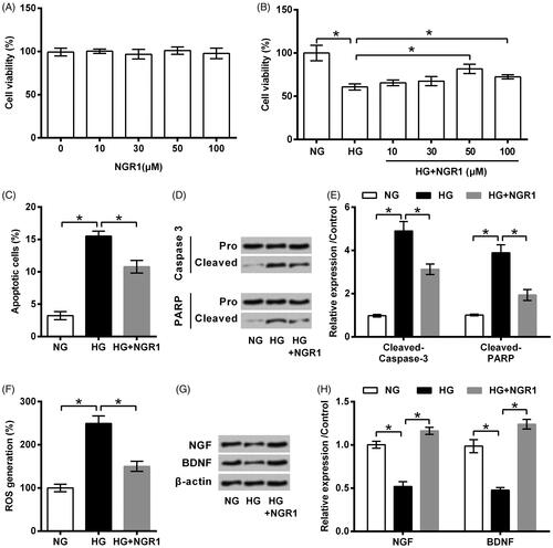
NGR1 with various dosages ranged from 10 to 100 μM was utilized to treat RSC96 cells. Cell viability was examined to evaluate the cytotoxicity of NGR1. As seen in , viability of RSC96 cells was unchanged by NGR1 treatment (p > .05). However, NGR1 could ameliorate HG-induced viability loss to some extent (p < .05, ). Considering 50 μM resulted in a highest viability among these dosages in HG-treated cell, 50 μM was applied as optimum dosage for use in following experiments. displays that HG-evoked apoptosis was also attenuated by NGR1 pre-conditioning, as apoptosis rate was declined and cleavage of caspase-3 and RARP was repressed significantly (all p < .05). The same trend was observed in , HG-induced ROS generation and neurotrophic factor suppression were attenuated when pre-treating with NGR1 (all p < .05).
Figure 2. Notoginsenoside R1 (NGR1) pre-conditioning ameliorated high glucose (HG)-evoked injury in RSC96 cells. (A) NGR1 with various dosages ranged from 10 to 100 μM was utilized to treat RSC96 cells. (B) RSC96 cells were pre-treated with NGR1 and then stimulated by HG. Cell viability was examined by CCK-8 kit. 50 μM was applied as optimum dosage for use in following experiments. (C) Apoptosis rate, (D,E) cleavage of caspase-3 and PARP, (F) ROS generation and (G,H) expression of neurotrophic proteins were examined by flow cytometry, Western blot and DCFH-DA probe. Data were presented as mean ± SD (n = 3). *p < .05 (ANOVA).
NGR1 prevented HG-induced miR-503 expression
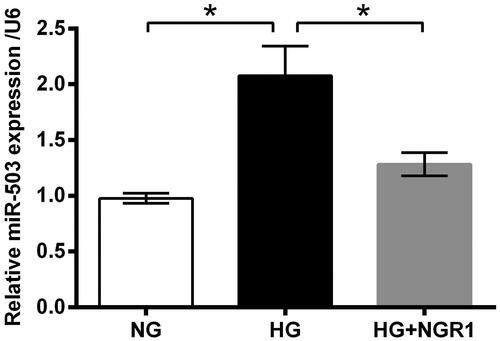
Next, the regulatory effect of NGR1 on miR-503 expression was explored. qRT-PCR data in displayed that miR-503 expression was elevated by HG treatment (p < .05). Nonetheless, NGR1 largely repressed miR-503 expression even under HG-treated condition (p < .05).
Figure 3. Notoginsenoside R1 (NGR1) prevented high glucose (HG)-induced miR-503 expression. RSC96 cells were pre-treated with NGR1 and then stimulated by HG. Expression of miR-503 was examined by qRT-PCR. Data were presented as mean ± SD (n = 3). *p < .05 (ANOVA).
NGR1 pre-conditioning ameliorated HG-evoked injury in RSC96 cells through miR-503
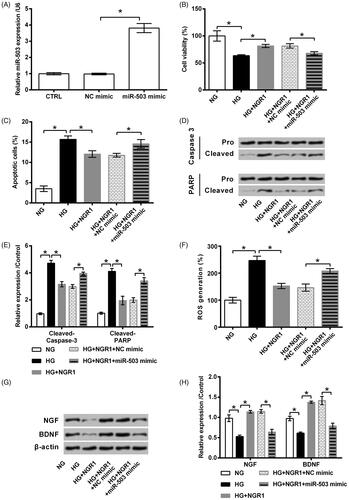
Next, the importance of miR-503 in NGR1’s protective function was explored. To this end, miR-503 expression in RSC96 cells was elevated by miR-503 mimic transfection (p < .05). Following data displayed that, the protective functions of NGR1 against HG-evoked damage in RSC96 cells were partially flatted by miR-503 overexpression. As compared to transfection with NC mimic, transfection of cells with miR-503 mimic before NGR1 plus HG treatment could significantly decline cell viability (p < .05, ), promote apoptosis (p < .05, ), accelerate ROS generation (p < .05, ) and repress neurotrophic factor expression (p < .05, ).
Figure 4. Notoginsenoside R1 (NGR1) pre-conditioning ameliorated high glucose (HG)-evoked injury in RSC96 cells through miR-503. (A) miR-503 mimic and its respective control (NC mimic) were transfected into RSC96 cells. Transfection efficiency was examined by qRT-PCR. The transfected or non-transfected cells were then treated by NGR1, HG or both. (B) Cell viability, (C) apoptosis rate, (D,E) cleavage of caspase-3 and PARP, (F) ROS generation, and (G,H) expression of neurotrophic proteins were examined by CCK-8 assay, flow cytometry, Western blot and DCFH-DA probe. Data were presented as mean ± SD (n = 3). *p < .05 (ANOVA).
NGR1 activated PI3K/AKT and β-catenin signalling through miR-503
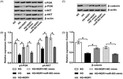
To decode the deep reason for NGR1’s protective function, we focused on investigating two signalling pathways (PI3K/AKT and β-catenin) which are closely associated with pathogenesis of DPN [Citation20,Citation21]. As seen in , HG repressed PI3K and AKT phosphorylation and down-regulated β-catenin expression as related to NG (all p < .05). NGR1 pre-conditioning ameliorated HG-induced alterations of above mentioned proteins (p < .05). Besides, NGR1 did not prevent HG-induced these alterations when miR-503 was overexpressed by miR-503 mimic transfection (p < .05).
Figure 5. Notoginsenoside R1 (NGR1) activated PI3K/AKT and β-catenin signalling through miR-503. miR-503 mimic and its respective control (NC mimic) were transfected into RSC96 cells. The transfected or non-transfected cells were then treated by NGR1, high glucose (HG) or both. Expression of (A, B) PI3K, AKT and (C,D) β-catenin was examined by Western blot. Data was presented as mean ± SD (n = 3). *p < .05 (ANOVA).
Discussion
A large amount of data evidenced that, the whole neuron from perikaryon to the terminal is the target of diabetes [Citation4]. Once exposed to hyperglycaemic condition, peripheral axons and Schwann cells are damaged first. And, the dysfunctional Schwann cells will lead to nerve injury-related DPN [Citation22]. So, recent studies have utilized Schwann cells as the experiment materials to better understand the pathogenesis of DPN [Citation23,Citation24]. The present study stimulated RSC96 cells with high glucose to mimic a cell model of DPN. As seen in the result section, hyperglycaemic condition led to cell viability loss, apoptosis and ROS generation, indicating RSC96 cells were clearly damaged. Additionally, the function of RSC96 cell was disturbed as evidenced by the declined expression of neurotrophic factors (NGF and BDNF). Of note, NGR1 preconditioning in this experimental system significantly protected RSC96 cells against high glucose-evoked damage. And, the neuroprotective properties of NGR1 may due to the down-regulated expression of miR-503 and the activated PI3K/AKT and β-catenin signalling pathways.
Apoptosis is a common feature occurred in diabetes and in its complications. During the progression of diabetes, duration of hyperglycaemic exposure induces neuronal apoptotic death which leads to nerve dysfunction and ultimately neuropathy. Besides that, hyperglycaemic condition increases ROS level which subsequently induces DNA damage of Schwann cells [Citation25]. It has been reported that, NGR1 could protect neurons against apoptotic death evoked by several stimulations, including oxygen-glucose deprivation/reoxygenation [Citation10], amyloid-β [Citation26], and high-glucose [Citation27]. The present work was consistence with these findings, indicating the neuroprotective function of NGR1 in a cell model of DPN. As revealed from in vitro experiments, NGR1 protected RSC96 cells against high glucose-induced viability loss, apoptosis and ROS generation.
The main function of Schwann cells is producing neurotrophic factors to maintain the health of axons. Dysfunction of axons reduces theirs communication between nervous system and leads to peripheral nerve degeneration [Citation28]. A recently published paper has demonstrated that, NGR1 treatment increased BDNF expression, suggesting the neurotrophic function of NGR1. The data in this study illustrated that NGR1 was not only able to increase BDNF expression, but also increase NGF expression in RSC96 cells.
Recently, several promising therapies are in ongoing experimental trials, including oestrogen treatment [Citation29]. Under pathological conditions, oestrogen processes neuroprotective and neurotrophic effects [Citation30]. NGR1 is a phytoestrogen that is isolated from R. notoginseng. Recent studies have mentioned the potential of NGR1 in treating several complications of diabetes, like diabetic nephropathy [Citation12], diabetic cardiomyopathy [Citation13], and diabetic retinopathy [Citation14]. However, the present paper for the first time evidenced NGR1 as a promising approach for DPN. As mentioned above, both neuroprotective and neurotrophic functions of NGR1 were revealed. These findings were in line with a previous study, in which the neuroprotective function of NGR1 was reported in an oxygen-glucose deprivation/reoxygenation injury model [Citation31].
Since NGR1 is one of the widely studied natural compounds of traditional herbal Chinese medicine, the underlying mechanisms of its function has been preliminary studied. A small fraction of studies believed that NGR1 exerted its function via regulating miRNA expression pattern [Citation32]. For the selected examples, miR-26a [Citation33], miR-23a [Citation34], and miR-132 [Citation35] are found as the targets of NGR1. Herein, we observed that miR-503 was another target of NGR1, as its expression was negative regulated by NGR1. And the beneficial function of NGR1 in high glucose-injured RSC96 cells was impeded by miR-503 overexpression. But, further studies are required to discover other miRNAs of which NGR1 works as an anti-DPN agent.
PI3K/AKT is a critical signal transduction pathway that is significantly involved in the pathogenesis of DPN. In animal model of DPN, PI3K/AKT pathways were repressed and then the expression of neurogenesis-related factors was down-regulated [Citation36]. Besides, activation of PI3K/AKT pathway has promise in the management of DPN, as it is capable of controlling chronic pain [Citation37]. Other than PI3K/AKT pathway, β-catenin pathway has been mentioned to be critical in DPN. High glucose decreases β-catenin expression [Citation38]. And the silenced β-catenin expression blocks the cytoprotection of erythropoietin in endothelial cells during experimental diabetes [Citation39]. Here, studies found that NGR1 was able to activate PI3K/AKT and β-catenin signalling under high glucose condition through down-regulating miR-503. This was in line with a previous finding, in which PI3K was reported as a target of miR-503 [Citation40]. It seems that miR-503 modulated PI3K/AKT signalling through directly targeting PI3K. Also, the inhibitory role of miR-503 in β-catenin signalling has been confirmed previously [Citation41,Citation42]. Altogether, the finding hinted us that NGR1 conferred its anti-DPN property possibly through down-regulating miR-503 and then modulating these two signalling.
To conclude, the present paper illustrated the neuroprotective and neurotrophic function of NGR1 in RSC96 cells. The beneficial function may due to its regulation on miR-503 expression and the downstream signalling such as PI3K/AKT and β-catenin. Those findings may be valuable for scientists in the related field to develop novel treatment strategies for DPN.
Disclosure statement
The authors declare that they have no conflict of interest.
Data availability
The datasets used and/or analyzed during the current study are available from the corresponding author on reasonable request.
References
- daCosta DiBonaventura M, Cappelleri JC, Joshi AV. A longitudinal assessment of painful diabetic peripheral neuropathy on health status, productivity, and health care utilization and cost. Pain Med. 2011;12(1):118–126.
- Liu Y, Sebastian B, Liu B, et al. Sensory and autonomic function and structure in footpads of a diabetic mouse model. Sci Rep. 2017;7(1):41401.
- Khedr EM, Fawi G, Allah Abbas MA, et al. Prevalence of diabetes and diabetic neuropathy in *Qena Governorate: population-based survey. Neuroepidemiology. 2016;46(3):173–181.
- Feldman EL, Callaghan BC, Pop-Busui R, et al. Diabetic neuropathy. Nat Rev Dis Primers. 2019;5(1):41.
- Zhang J, Ding L, Wang B, et al. Notoginsenoside R1 attenuates experimental inflammatory bowel disease via pregnane X receptor activation. J Pharmacol Exp Ther. 2015;352(2):315–324.
- Liu WJ, Tang HT, Jia YT, et al. Notoginsenoside R1 attenuates renal ischemia–reperfusion injury in rats. Shock (Augusta, Ga). 2010;34(3):314–320.
- Meng X, Sun G, Ye J, et al. Notoginsenoside R1-mediated neuroprotection involves estrogen receptor-dependent crosstalk between Akt and ERK1/2 pathways: a novel mechanism of Nrf2/ARE signaling activation. Free Radic Res. 2014;48(4):445–460.
- Huang G, Zou B, Lv J, et al. Notoginsenoside R1 attenuates glucose-induced podocyte injury via the inhibition of apoptosis and the activation of autophagy through the PI3K/Akt/mTOR signaling pathway. Int J Mol Med. 2017;39(3):559–568.
- Luo C, Sun Z, Li Z, et al. Notoginsenoside R1 (NGR1) attenuates chronic atrophic gastritis in rats. Med Sci Monit. 2019;25:1177–1186.
- Tu L, Wang Y, Chen D, et al. Protective effects of notoginsenoside R1 via regulation of the PI3K-Akt-mTOR/JNK pathway in neonatal cerebral hypoxic-ischemic brain injury. Neurochem Res. 2018;43(6):1210–1226.
- Li Z, Li H, Zhao C, et al. Protective effect of notoginsenoside R1 on an APP/PS1 mouse model of Alzheimer’s disease by up-regulating insulin degrading enzyme and inhibiting Abeta accumulation. CNSNDDT. 2015;14(3):360–369.
- Zhang B, Zhang X, Zhang C, et al. Notoginsenoside R1 protects db/db mice against diabetic nephropathy via upregulation of Nrf2-mediated HO-1 expression. Molecules (Basel, Switzerland). 2019;24(2):10.
- Zhang B, Zhang J, Zhang C, et al. Notoginsenoside R1 protects against diabetic cardiomyopathy through activating estrogen receptor alpha and its downstream signaling. Front Pharmacol. 2018;9:1227.
- Fan C, Qiao Y, Tang M. Notoginsenoside R1 attenuates high glucose-induced endothelial damage in rat retinal capillary endothelial cells by modulating the intracellular redox state. Drug Des Dev Ther. 2017;11:3343–3354.
- Liu XS, Fan B, Szalad A, et al. MicroRNA-146a mimics reduce the peripheral neuropathy in type 2 diabetic mice. Diabetes. 2017;66(12):3111–3121.
- Zhang Y, Song C, Liu J, et al. Inhibition of miR-25 aggravates diabetic peripheral neuropathy. Neuroreport. 2018;29(11):945–953.
- Caporali A, Meloni M, Vollenkle C, et al. Deregulation of microRNA-503 contributes to diabetes mellitus-induced impairment of endothelial function and reparative angiogenesis after limb ischemia. Circulation. 2011;123(3):282–291.
- Sheikhbahaei S, Manizheh D, Mohammad S, et al. Can MiR-503 be used as a marker in diabetic patients with ischemic stroke? BMC Endocr Disord. 2019;19(1):42.
- Luo Q, Feng Y, Xie Y, et al. Nanoparticle-microRNA-146a-5p polyplexes ameliorate diabetic peripheral neuropathy by modulating inflammation and apoptosis. Nanomed: Nanotechnol Biol Med. 2019;17:188–197.
- Li R, Wu Y, Zou S, et al. NGF attenuates high glucose-induced ER stress, preventing Schwann cell apoptosis by activating the PI3K/Akt/GSK3β and ERK1/2 pathways. Neurochem Res. 2017;42(11):3005–3018.
- Resham K, Sharma SS. Pharmacologic inhibition of porcupine, disheveled, and beta-catenin in Wnt signaling pathway ameliorates diabetic peripheral neuropathy in rats. J Pain: Off J Am Pain Soc. 2019. [Epub ahead of print]. DOI: 10.1016/j.jpain.2019.04.010
- Gumy LF, Bampton ET, Tolkovsky AM. Hyperglycaemia inhibits Schwann cell proliferation and migration and restricts regeneration of axons and Schwann cells from adult murine DRG. Mol Cell Neurosci. 2008;37(2):298–311.
- Du W, Wang N, Li F, et al. STAT3 phosphorylation mediates high glucose-impaired cell autophagy in an HDAC1-dependent and -independent manner in Schwann cells of diabetic peripheral neuropathy. FASEB J: Off Publ Federation Am Soc Exp Biol. 2019;33(7):8008-8021.
- Xu DD, Li WT, Jiang D, et al. 3-N-butylphthalide mitigates high glucose-induced injury to Schwann cells: association with nitrosation and apoptosis. Neural Regen Res. 2019;14(3):513–518.
- Park C, Choi EO, Kim GY, et al. Protective effect of Baicalein on oxidative stress-induced DNA damage and apoptosis in RT4-D6P2T Schwann cells. Int J Med Sci. 2019;16(1):8–16.
- Ma B, Meng X, Wang J, et al. Notoginsenoside R1 attenuates amyloid-beta-induced damage in neurons by inhibiting reactive oxygen species and modulating MAPK activation. Int Immunopharmacol. 2014;22(1):151–159.
- Zhai Y, Meng X, Luo Y, et al. Notoginsenoside R1 ameliorates diabetic encephalopathy by activating the Nrf2 pathway and inhibiting NLRP3 inflammasome activation. Oncotarget. 2018;9(10):9344–9363.
- Cashman CR, Hoke A. Mechanisms of distal axonal degeneration in peripheral neuropathies. Neurosci Lett. 2015;596:33–50.
- Saravia FE, Beauquis J, Revsin Y, et al. Hippocampal neuropathology of diabetes mellitus is relieved by estrogen treatment. Cell Mol Neurobiol. 2006;26(4–6):943–957.
- Chakraborty TR, Cohen J, Yohanan D, et al. Estrogen is neuroprotective against hypoglycemic injury in murine N38 hypothalamic cells. Mol Med Rep. 2016;14(6):5677–5684.
- Wang Y, Tu L, Li Y, et al. Notoginsenoside R1 alleviates oxygen-glucose deprivation/reoxygenation injury by suppressing endoplasmic reticulum calcium release via PLC. Sci Rep. 2017;7(1):16226.
- Jia C, Xiong M, Wang P, et al. Notoginsenoside R1 attenuates atherosclerotic lesions in ApoE deficient mouse model. PLoS One. 2014;9(6):e99849.
- Liu J, Hou C, Chen X, et al. Notoginsenoside R1 protects human renal proximal tubular epithelial cells from lipopolysaccharide-stimulated inflammatory damage by up-regulation of miR-26a. Chem Biol Interact. 2019;308:364–371.
- Wang C, Sun H, Zhong Y. Notoginsenoside R1 promotes MC3T3-E1 differentiation by up-regulating miR-23a via MAPK and JAK1/STAT3 pathways. Artif Cells Nanomed Biotechnol. 2019;47(1):603–609.
- Fu C, Yin D, Nie H, et al. Notoginsenoside R1 protects HUVEC against oxidized low density lipoprotein (Ox-LDL)-induced atherogenic response via down-regulating miR-132. Cell Physiol Biochem. 2018;51(4):1739–1750.
- Chen L, Gong HY, Xu L. PVT1 protects diabetic peripheral neuropathy via PI3K/AKT pathway. Eur Rev Med Pharmacol Sci. 2018;22(20):6905–6911.
- Chen SP, Zhou YQ, Liu DQ, et al. PI3K/Akt pathway: a potential therapeutic target for chronic pain. CPD. 2017;23(12):1860–1868.
- Wang R, Gao D, Zhou Y, et al. High glucose impaired estrogen receptor alpha signaling via β-catenin in osteoblastic MC3T3-E1. J Steroid Biochem Mol Biol. 2017;174:276–283.
- Chong ZZ, Hou J, Shang YC, et al. EPO relies upon novel signaling of Wnt1 that requires Akt1, FoxO3a, GSK-3beta, and beta-catenin to foster vascular integrity during experimental diabetes. CNR. 2011;8(2):103–120.
- Yan W, Wu Q, Yao W, et al. MiR-503 modulates epithelial–mesenchymal transition in silica-induced pulmonary fibrosis by targeting PI3K p85 and is sponged by lncRNA MALAT1. Sci Rep. 2017;7(1):11313.
- Li W, Li J, Mu H, et al. MiR-503 suppresses cell proliferation and invasion of gastric cancer by targeting HMGA2 and inactivating WNT signaling pathway. Cancer cell Int. 2019;19:164.
- Li Q, Li C, Xi S, et al. The effects of photobiomodulation therapy on mouse pre-osteoblast cell line MC3T3-E1 proliferation and apoptosis via miR-503/Wnt3a pathway. Lasers Med Sci. 2019;34(3):607–614.