
Abstract
Introduction
The diagnosis of a perilymphatic fistula (PLF) has long been a challenge, as its presentation is variable and can mimic other vestibular disorders. Currently, it is suggested that resolution of symptoms after treatment should be used as a gold standard in diagnosing PLF. Recently, improvement in Magnetic Resonance Imaging (MRI) have caused PLF to be diagnosed more consistently with a higher sensitivity and specificity, preceding surgery.
Case report
We report a 43-year-old patient, who was seen with progressive vertigo since 15 years, triggered by bending forward, and lasting from 20 min to 2 h. During these spells, she would also have pulse-synchronous tinnitus and mild left-sided mixed hearing loss. Physical examination was normal and vestibular function tests did not reveal evidence of peripheral or central audiovestibular dysfunction. Delayed acquisition MRI, with 3-dimensional fluid attenuated inversion recovery (3D FLAIR) of the inner ear displayed a left ‘round window sign’ (RWS): focal enhancement in the round window niche. There were no signs of vestibular or cochlear hydrops to suggest Meniere’s syndrome. Her symptoms resolved after surgery.
Conclusion
RWS on 3D FLAIR MRI sequence seems to be a sensitive and specific pre-operative diagnostic phenomenon for the detection of PLF.
Introduction
A perilymphatic fistula (PLF) is a rare condition in which the fluid from the inner ear leaks into the middle ear, often through the round or oval window [Citation1–5]. Diagnosis of PLF remains difficult and is often missed or misdiagnosed [Citation1,Citation4,Citation5]. Patients commonly experience a combination of vertigo, hearing loss, aural fullness and tinnitus, precipitated or provoked by increased pressure [Citation1–3]. A positive fistula sign, meaning nystagmus induced after applying positive pressure through the ear canal, could aid diagnosis, although its sensitivity is said to vary widely from 0–77% [Citation1,Citation4,Citation5]. Pure-tone audiometry (PTA) can show conductive, sensorineural and fluctuating hearing loss in varying degrees [Citation1]. High resolution computed tomography (HRCT) may show specific signs of pneumolabyrinth, or non-specific signs such as round window opacification, but shows no abnormalities in most cases [Citation1]. Exploratory tympanotomy is often considered to be the most accurate way to confirm the diagnosis, even though positive identification rates vary markedly (24–100%) [Citation1,Citation3]. Surgical repair with fascia or fat graft is usually effective. Recently, improvements in Magnetic Resonance Imaging (MRI) have caused PLF to be diagnosed more consistently with a higher sensitivity and specificity, preceding surgery [Citation2,Citation4]. We report the imaging of a case in which PLF was diagnosed accurately, before being confirmed by resolution of symptoms after surgical treatment.
Case report
A 43-year-old female was referred to the multidisciplinary balance clinic by her neurologist. She had been suffering from episodic vertigo, lasting several hours, triggered by bending forward for the last 15 years. Simultaneously, she would often experience mild hearing loss and aural pressure in her left ear. She denied vomiting, hearing loss, headache, migraine features or posterior circulation symptoms at the time. Over the past year, she described fluctuant hearing loss, aural fullness and tinnitus lasting between 20 min and 2 h, specifically whilst driving. Furthermore, she reported to hear a whooshing, pulse-synchronous tinnitus, when she turned from lying on her right side to her left side, which would last several minutes. Otoscopy was normal, and full neuro-otological examination showed no abnormalities.
Pure-tone audiometry (PTA) showed a lowered bone threshold of 30 and 20 dB at 250 and 500 Hz, respectively. On the middle to high frequencies her bone threshold was normal, but a mild air-bone gap was seen of 10–20 dB. She had normal hearing in her right ear. Tympanometry was normal. Extensive vestibular function tests showed no dysfunction. Delayed acquisition MRI with 3-dimensional fluid attenuated inversion recovery (3D FLAIR) of the inner ear displayed a left ‘round window sign’ (RWS), characterised as a high signal covering the round window ( and ). Pre-operative Computed Tomography (CT) scan was normal, besides round window opacification.
Figure 1. (a) Axial 3D REAL IR image through the basal turn inferior segment (arrow) and round window. Note the enhancement of the perilymphatic space in the basal turn with focal enhancement also seen in the round window niche - the ‘round window sign’ (arrow head). (b) Axial 3D SPACE image demonstrates T2 hyperintense signal within the round window niche (arrow) at the site of the presumed perilymphatic round window fistula. (c) Cone beam CT image demonstrates soft tissue opacification within the round window niche (arrow) at the site of the presumed perilymphatic round window fistula.
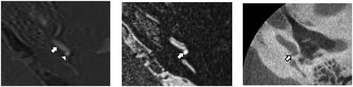
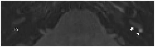
Figure 2. Axial 3D FLAIR image through the basal turn inferior segment (filled arrow) and round window. Note the enhancement of the perilymphatic space in the basal turn with focal enhancement also seen in the round window niche (arrow head). Compare the contralateral round window niche where there is no corresponding enhancement (open arrow).
During surgery, a piece of tragal cartilage was harvested and used to obliterate the round window niche, despite perilymph leakage not being clearly evident. Post-operative recovery was uneventful. More than two months after her surgery, the patient was seen back at our Ear, Nose and Throat (ENT) clinic where she denied having had further vertigo or imbalance after surgery. PTA revealed that her mixed hearing loss had worsened slightly, to approximately 35 dB over all frequencies. At follow-up one year post-operatively, she remained symptom free with no recurrence of vertigo or disequilibrium. Her hearing had remained stable and she was getting good benefit from a hearing aid.
Discussion
Diagnosis of PLF has long been challenging, as its presentation is variable and can mimic other vestibular disorders [Citation1,Citation3]. Association of vertigo with pressure-increasing events may point towards a third window syndrome and CT can exclude superior canal dehiscence [Citation2–4]. Fluctuating hearing loss is suggested to have an important predictive value, but might only be present in around 10% of patients [Citation3]. Previous authors were able to find predisposing factors for PLF, excluding stapedectomy, in 68% of patients, with most people having had barotrauma (23.9%), head trauma (19.7%) or chronic ear disease (16.9%), before developing symptoms [Citation1,Citation3–5].
Our patient had been seen by local ENT and neurology services before being referred to a tertiary vestibular centre. Common vestibular disorders causing episodic vertigo/disequilibrium such as vestibular migraine and Meniere’s syndrome were excluded in this patient as she did not have the key features required by the diagnostic criteria for these conditions [Citation6]. Vestibular function tests, such as videonystagmography (VNG), video head-impulse test (vHIT), cervical vestibular evoked myogenic potentials and caloric testing, are not diagnostic of PLF [Citation3,Citation4]. The Fraser test, PTA performed after the patient has lain down for a half hour, might not be sensitive, but is easily executed and can help secure PLF diagnosis [Citation3]. On CT, disorientation of the footplate and a pneumolabyrinth are the most common signs in oval window fistula patients [Citation5]. Exploratory tympanotomy can be negative for PLF, as in this case, in up to 25–78% of patients [Citation1,Citation3]. Obliteration of both windows is reported to lead to an improvement of auditory and vestibular symptoms in 40% and 87–95% of patients, respectively [Citation3,Citation4]. Although, as in the presented patient, conductive hearing loss after obliteration has been described [Citation4]. After surgery, recurrence of symptoms and the need for re-exploration occurs in 8–47% [Citation3].
Because of the above mentioned reasons, some authors suggest that an improvement of symptoms after treatment, as was seen in the presented case, either through surgery or trans-tympanic blood patch, should be used as a gold standard in diagnosing PLF [Citation4,Citation5].
Dubrulle et al. has studied the use of MRI to display subtle signs of PLF. Especially delayed acquisition MRI, in which a patient is given gadolinium chelate contrast 4–5 h before scanning, show promise in this regard [Citation2]. As in our case, patients with PLF may display RWS after this delayed acquisition MRI on its 3D-FLAIR sequences [Citation2]. It is theorised that contrast accumulates progressively in the round window niche over the delayed period, inducing a high signal [Citation2]. RWS has been proposed to distinguish PLF and (probable) Meniere’s disease, as their clinical presentation tends to be similar [Citation2].
Conclusion
There is a need for sensitive and specific pre-operative diagnostic methods for the detection of PLF. As is seen in the presented case and in literature, the RWS on 3D FLAIR MRI sequence, acquired several hours after administering Gadolinium contrast, is a promising tool.
Disclosure statement
No potential conflict of interest was reported by the author(s).
Correction Statement
This article has been corrected with minor changes. These changes do not impact the academic content of the article.
Additional information
Funding
References
- Alzahrani M, Fadous R, Dufour J-J, et al. Perilymphatic fistulas: can we predict the diagnosis? Eur Arch Otorhinolaryngol. 2015;272(8):1885–1891.
- Dubrulle F, Chaton V, Risoud M, et al. The round window sign: a sensitive sign to detect perilymphatic fistulae on delayed postcontrast 3D-FLAIR sequence. Eur Radiol. 2020;30:6303–6310.
- Meldrum JA, Prinsley PR. Perilymph fistula: the patients’ experience. J Laryngol Otol. 2016;130:526–531.
- Sarna B, Abouzari M, Merna C, et al. Perilymphatic fistula: a review of classification, etiology, diagnosis, and treatment. Front Neurol. 2020;11:1046.
- Venkatasamy A, Al Ohraini Z, Karol A, et al. CT and MRI for the diagnosis of perilymphatic fistula: a study of 17 surgically confirmed patients. Eur Arch Otorhinolaryngol. 2020;277:1045–1051.
- Lopez-Escamez JA, Carey J, Chung WH, et al. [Diagnostic criteria for menière’s disease. Consensus document of the bárány society, the Japan society for equilibrium research, the european academy of otology and neurotology (EAONO), the American academy of Otolaryngology-Head and neck surgery (AAO-HNS) and the korean balance society]. Acta Otorrinolaringol Esp. 2016;67:1–7.