
Abstract
Extravasation of calcium solution is a common adverse event in children in intensive care units. The lack of adequate and timely treatment can lead to important functional sequelae. Here, we report the case of calcium extravasation in a child and we discuss the multiple therapeutic strategies adopted.
1. Introduction
Extravasation of intravenous infusion is a common adverse event in children in intensive care units, especially in newborns [Citation1]. The severity of these lesions depends on several factors including the type, the amount of extravasated material and the time in which extravasation is recognized [Citation2].
Periodic monitoring of the venous access is necessary to ensure early detection of infusion leakage and fast implementation of effective treatment. A lack of proper management can result in serious functional and aesthetic consequences.
Treatment for neonatal hypocalcemia frequently involves the use of calcium gluconate or calcium chloride. However, just a few cases of calcium extravasation have been reported in literature compared to those that occur [Citation3–5].
In this article, we describe a clinical case of calcium gluconate solution extravasation in a 9-year-old child and we outline the complexity of the therapeutic challenge.
2. Case report
We treated a 9-year-old-female with autosomal recessive polycystic kidney disease who underwent a right and left nephrectomy surgery in 2013 and subsequent chronic peritoneal dialysis until receiving a kidney transplant in 2014.
Due to a gastroenteritis infection in October 2021, the patient had a distributive shock that was unresponsive to vasoactive therapy, resulting in cardiac arrest and return to spontaneous circulation after 10 min.
The patient developed a multiple organ failure with acute respiratory distress syndrome, acute renal failure, liver failure, disseminated intravascular coagulation that required inotropic support, extracorporeal circulation and continuous venous-venous hemodialysis.
Furthermore, the patient’s general condition was complicated by peripheral ischemia which led to necrosis and amputation of the left foot. A craniectomy was performed to drain a left posterior cerebral hematoma and postoperative intracranial pressure measurement was required.
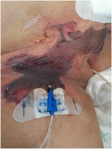
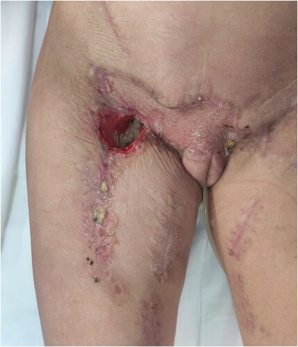
Due to hypocalcemia, calcium gluconate was injected through a central venous access in the right femoral vein. The central vascular catheter was regularly monitored. Despite this, there was a calcium extravasation that caused a large hyperemic area with blood-serum blisters on the pubis involving the labia majora and the superior part of the right thigh. The central vascular catheter was immediately removed and the infusion was stopped, cold compresses and vaseline gauzes was applied ().
Figure 1. Necrosis of the pubic area on day 1.
The abdomen magnetic resonance imaging angiography highlighted deposits of calcium from the anterior pelvis to the proximal third of the anterior side of the right thigh.
The surgical treatment of extravasation was not prioritised due to the patient’s serious overall condition and the wound was managed with topical dressings.
Necrosis began to appear during the next few days. On the fifteenth day, the necrotic area was removed, and Negative Pressure Wound Therapy (NPWT) was started. The patient underwent hyperbaric oxygen therapy in the meantime.
NPWT was applied and the device was changed weekly. 30 days after calcium extravasation and 13 days after the application of NPWT thrombosis of the femoral artery and femoral vein occurred and surgical revascularization of the right lower leg was carried out through an extra anatomical bypass and a biological prosthesis.
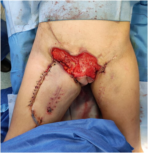
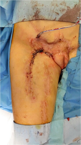
After three weeks of NPWT the necrotic area was excised (). At this stage we performed an advancement flap from the thigh to cover the femoral vessels exposed and NPWT was reapplied on the pubic area ().
Figure 2. The pubic area after excision necrosis and 20 days of NPWT.
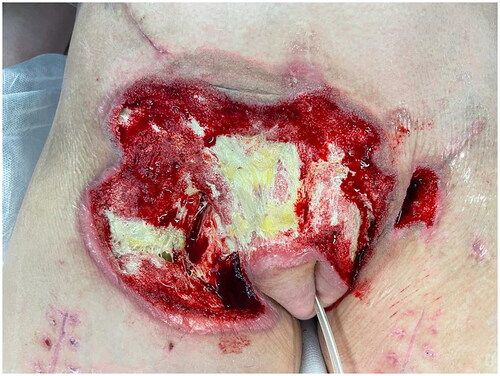
Figure 3. Appearance of the thigh after the advancement flap.
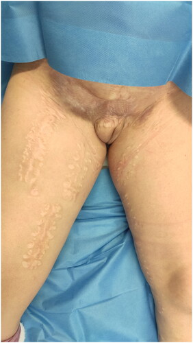
After three more weeks a skin graft was taken from the contralateral thigh to cover the pubic area and NPWT was reapplied for 7 days (). The graft was almost completely attached within 10 days, except for a 2 × 2 cm area close to the groin at the most cranial portion of the advancement flap in continuity with the lower portion of the skin graft where there was a residual seroma ().
Figure 4. Appearance of the thigh after the skin graft.
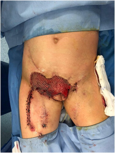
Figure 5. The residual seroma at the right inguinal level.
Skin swabs revealed growth of Citrobacter freundii and Pseudomonas aeruginosa, accordingly treated with ciprofloxacin antibiotic therapy.
After two weeks of NPWT and surgical removal of necrotic and fibrotic tissue () a new advancement thigh flap was realized to cover the residual exposed area ().
Figure 6. Application of NPWT.
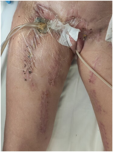
Figure 7. Appearance of the thigh after removal of thrombosed portion of the femoral artery and femoral vein and the new advancement thigh flap.
At the follow up consultation 9 months later, the skin graft was healed without significant adhesion or retraction. The patient received rehabilitation care with intensive physiotherapy and a prosthesis of the lower limb was packed to allow the patient to resume walking with aids ().
Figure 8. Appearance of the thigh after 9 months of follow-up.
3. Discussion
Calcium gluconate is used to correct hypocalcemia, especially in premature births in intensive care units [Citation1].
Calcium gluconate extravasation can cause different kind of injuries. According to the literature, the two most common injuries after extravasation are erythema and swelling followed by indurated skin, yellow-white plaques or papules [Citation6,Citation7]. However, it can cause serious lesions, such as skin necrosis and deposits of calcium phosphate crystal in soft tissues visible on the X-ray, described as calcinosis cutis [Citation7–9].
The lesion progression ranges from an acute tissue injury to a delayed clinical manifestation. In a bibliographical review by Francisco Javier Pacheco Compaña et al. edema and erythema appear at an early stage, during the first 7 days after the injury, while plaques and necrosis usually are seen during the subsequent 3 weeks [Citation3]. The necrosis induced by the cationic agent can be severe due to the infiltration of the solution under the fascia and the muscle layers. The most common location of lesions is on the back of the hand and/or the wrist, followed by the upper limb and the lower limb [Citation3].
Currently, there is not a common strategy for the wound management after calcium gluconate extravasation in the literature.
In most cases patients are managed conservatively. The conservative treatment is based on stopping the infusion to eliminate the infiltrated drug as much as possible. Heat or cold compresses, tulle dressing and local corticosteroids can be applied as initial treatment. Arm or limb elevation can be useful to promote fluid drainage, although evidence for this technique remains weak [Citation3,Citation10,Citation11].
The necrotic area appears between 7 to 15 days after extravasation and surgical debridement is required. Surgical skin coverage technique depends on the loss of surface area and the quality of the subcutaneous plane: split or full thickness skin grafts and local flaps are possible options [Citation3,Citation5].
Although calcium gluconate extravasation may result in serious damage, very few studies discuss the use of antidotes. Various local antidotes have been proposed, such as hyaluronidase, sodium thiosulphate, triamcinolone acetonide and physiologic solution in experimental study in mice. Francisco Javier et al. have proven that sodium thiosulfate infiltration prevents the development of calcium deposits and skin lesions in mice, while local hyaluronidase infiltration reduces only the presence of calcium deposits [Citation12].
Triamcinolone acetonide has an anti-inflammatory, antiproliferative and immunosuppressive effect. It has been used in an experimental study in rabbits with calcinosis cutis, intralesional injection of triamcinolone facilitates the resorption of calcium in the tissue and healing in a shorter period [Citation13] although no standardised protocol has yet been adopted.
The complexity of our case lies not only in the extension and depth of the calcium extravasation but also in the difficult management of hypoperfused tissues in a patient with labile general conditions.
Due to the complexity of the lesion, multiple therapeutic strategies have been implemented: dressings, negative pressure wound therapy, hyperbaric oxygen therapy, grafts and flaps.
The NPWT has been used in 2016 by Xu [Citation4] in a case of calcium chloride extravasation in 83-year-old man and in 2019 by P. Girard [Citation5] in two cases of calcium gluconate extravasation respectively in a 13-year-old child and in a premature male newborn to reduce edema and to enhance the quality of subcutaneous tissues.
In our case NPWT allowed a significant reduction of local edema and exudate and has promoted the formation of granulation tissue, improving vascularization and graft integration.
Furthermore, hyperbaric oxygen therapy has been used to promote oxygen tissue diffusion, to improve blood perfusion in semi-obstructed vascular districts, to reduce edema, to increase bactericidal leukocyte function and to promote neovascularization.
In complex clinical situations like this one, multidisciplinary management and the application of various therapeutic strategies are essential to improve patients’ clinical condition and lead to local and general recovery.
4. Conclusion
Extravasated lesions can significantly worsen patients’ health and increase hospitalisation. Recognition of high-risk patients, proper cannulation technique and close surveillance of venous access are the milestones to prevent extravasation lesions. Healthcare personnel should be conscious and skilled in extravasation management, so that as soon as it occurs, prompt measures and procedures could be adopted.
In complicated clinical cases like ours, multiple therapeutic strategies and a multidisciplinary approach are crucial to improve general and local conditions.
In our experience, hyperbaric oxygen therapy and NPWT are fundamental steps for wound management and surgical reconstruction since they improve general and local perfusion in order to make the damaged tissue more suitable for surgery and promote faster healing.
Disclosure statement
No potential conflict of interest was reported by the author(s).
References
- Ly C. The care of skin lesions caused by extravasation of intravenous fluids in peripheral venous perfusion. Arch Pediatr. 2017;24(9):884–893. doi:10.1016/j.arcped.2017.06.011.
- Paquette V, McGloin R, Northway T, et al. Describing intravenous extravasation in children (DIVE study). Can J Hosp Pharm. 2011;64(5):340–345. doi:10.4212/cjhp.v64i5.1069.
- Pacheco Compaña FJ, Midón Míguez J, de Toro Santos FJ. Lesions associated With calcium gluconate extravasation: presentation of 5 clinical cases and analysis of cases published. Ann Plast Surg. 2017;79(5):444–449. doi:10.1097/SAP.0000000000001110.
- Xu C, Turner A, Yeoh TM, et al. Management of severe calcium chloride extravasation injury: a case report. ANZ J Surg. 2016;86(5):421–422. doi:10.1111/ans.13437.
- Girard P, Plancq MC, Tourneux P, et al. Extravasation of calcium solution in the child: value of negative-pressure wound therapy. Arch Pediatr. 2019;26(7):407–410. doi:10.1016/j.arcped.2019.09.011.
- Millam DA. Managing complications of i.v. therapy (continuing education credit). Nursing. 1988;18(3):34–43. doi:10.1097/00152193-198803000-00020.
- Rumancik BE, Rahnama-Moghadam S. Severe iatrogenic calcinosis cutis from extravasated calcium gluconate. Cureus. 2020;12(8):e9712. doi:10.7759/cureus.9712.
- Ahn KH, Park ES. A rare case report of neonatal calcinosis cutis induced by distant and delayed extravasation of intravenous calcium gluconate. Arch Plast Surg. 2021;48(6):641–645. doi:10.5999/aps.2020.01942.
- Domínguez-Fernández I, Goíriz R, Pérez-Gala S, et al. Calcinosis cutis following extravasation of calcium salts. J Eur Acad Dermatol Venereol. 2008;22(4):505–506. doi:10.1111/j.1468-3083.2007.02369.x.
- Reynolds PM, MacLaren R, Mueller SW, et al. Management of extravasation injuries: a focused evaluation of noncytotoxic medications. Pharmacotherapy. 2014;34(6):617–632. doi:10.1002/phar.1396.
- Casanova D, Bardot J, Magalon G. Emergency treatment of accidental infusion leakage in the newborn: report of 14 cases. Br J Plast Surg. 2001;54(5):396–399. doi:10.1054/bjps.2001.3593.
- Pacheco Compaña FJ, Midón Míguez J, de Toro Santos FJ, et al. The use of antidotes for calcium gluconate extravasation: an experimental study in mice. Plast Reconstr Surg. 2018;142(3):699–707. doi:10.1097/PRS.0000000000004640.
- Ahn SK, Kim KT, Lee SH, et al. The efficacy of treatment with triamcinolone acetonide in calcinosis cutis following extravasation of calcium gluconate: a preliminary study. Pediatr Dermatol. 1997;14(2):103–109. doi:10.1111/j.1525-1470.1997.tb00214.x.