
Abstract
Severe infections can cause difficult neurologic sequelae, sensorineural hearing loss included. In this case report, we describe a 58-year-old female, who had lost hearing in her right ear due to pneumococcal sepsis and meningitis 10 years earlier. More recently, urinary tract infection resulting in Escherichia coli urosepsis, completed the profound hearing disability by means of a total left ear hearing loss. After receiving a cochlear implant on her left ear and uneventful recuperation period, the implant was activated four weeks after the surgery with immediate speech reception and patient satisfaction. First postoperative audiometry two months after the surgery yielded an 84% speech reception.
Introduction
Meningitis is a well-known risk factor for acute sensorineural hearing loss (HL), even deafness [Citation1]. Pneumococcal meningitis, in particular, results relatively often in hearing loss and other neurologic sequelae [Citation1]. Following sepsis, acute sensorineural HL is far less common [Citation2] compared to meningitis-induced HL and to our knowledge, the role of Escherichia coli has not been specifically reported previously. Should the conventional hearing aid rehabilitation prove inadequate in the treatment of HL, cochlear implantation is potentially a viable treatment option for these patients. Indeed, cochlear implantation has gained a solid position as a means to treat patients with severe to profound hearing loss [Citation3,Citation4]. In addition to the established guidelines for cochlear implantation, there is also an increasing trend for off-label and nontraditional indication consideration, asymmetrical HL included [Citation5].
Case report
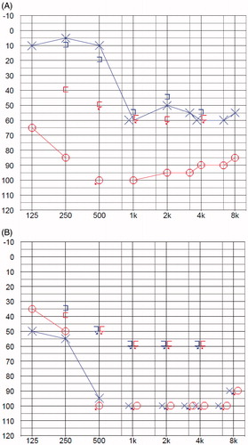
A 58-year-old woman had a number of long-term illnesses: asthma, obstructive sleep apnea, obesity, hypertension, hyperlipidemia, esophageal reflux, depression and anxiety disorder, and she was using a total of 18 different drugs daily. In 2008, she had suffered a pneumococcal meningitis and septichaemia, treated with vancomycin and levofloxacin, and consequently had lost hearing in the right ear (Figure ). During the course of the disease, the patient initially developed fever and had trouble breathing. She was diagnosed with right upper lobe pneumonia and blood culture was positive for gram-positive cocci. She was treated in intensive care unit for 17 days, with a total hospitalization of 26 days. The left ear retained a speech reception threshold of 32 dB with a speech recognition score of 72%, and the patient benefited from a conventional behind the ear hearing aid in her left ear.
Figure 1. (A) Pure tone audiogram of the patient after the first septic meningitis. (B) Preoperative pure tone audiogram of the patient, having suffered second sepsis.
During the autumn of 2018, the patient got a urinary tract infection leading eventually into E. coli sepsis, treated with ceftriaxone and subsequently with sulfadiazine-trimethoprim. Initial symptoms included only fever and vomiting and during her ER visit, the patient had a blood pressure of 114/95 mmHg with a heart rate of 125 bpm and a 78–82% oxygen saturation. A standard urinalysis revealed the presence of erythrocytes, protein, nitrites and leukocytes. In arterial blood gas analysis the partial pressure of oxygen was decreased (6.55 kPa), the partial pressure of carbon dioxide was increased (7.53 kPa), and pH was decreased (7.22). Later, blood culture and urine culture were positive for E. coli. Whereas during the previous pneumococcal infection C-reactive protein values reached 500 mg/L, highest value of 361 mg/L was noticed during urosepsis. Leukocytosis was moderate, up to 10.9 × 10⁹/L, and initial creatinine value of 347 µmol/L decreased to a normal level of 90 µmol/L in 5 days. Apart from the renal failure, no other organ failures were seen and the patient was transferred from the intensive care unit after three days. There was no need for intubation, but the patient received ventilation support by means of a continuous positive airway pressure mask treatment.
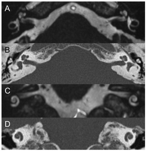
During and after the infection, the patient felt the hearing in her left ear become increasingly worse, and was referred to her local ENT-department. During the visit, the patient was only able to communicate via reading and was essentially deaf with no measurable speech reception (Figure ). This increased the anxiety of the patient considerably, as she was forced to rely solely on written communication with her husband and other people. Although with a quite heavy illness burden, the possibility of cochlear implantation was considered, and since computed tomography and magnetic resonance imaging (Figure ) and consultation with a psychologist lead to no obvious contraindication, a decision was made to treat the patient with a cochlear implant. A prominent sigmoid sinus, a relatively poor aeriation of the mastoidal air cells and a longer history of deafness in the right ear directed cochlear implantation decision in favor of the left ear. The left ear was operated on using a Cochlear Nucleus CI532-implant (Cochlear Ltd., Sydney, Australia). In addition to the availability in the department, a thin electrode design in CI532 was considered advantageous in order to minimize insertion trauma [Citation6]. Since intracutaneous absorbable sutures were used for closure of the standard retroauricular incision, a first postoperative follow-up visit, with implant activation, was agreed 4 weeks after the surgery. The recuperation period was uneventful and the patient had mild vertigo only in the morning of the first postoperative day. Vestibular function was not specifically evaluated, however, during this symptom or before the surgery. On the day of the implant activation the patient already was coping quite adequately without the help of a writing interpreter. Following the routine protocol of the ENT department in the University Hospital of Turku, Finland, a second postoperative control two months after the operation included also a free sound field hearing examination, where the patient reached a speech recognition score of 84%. In hearing in noise test (Oldenburg Hearing in noise test, Finnish version [Citation7]), a 50% threshold of –2.1 dB was observed. Six months after the operation, values of 88% and –4.0 dB were measured, respectively.
Figure 2. Preoperative MRI (A, axial; C, coronal) and CT (B, axial; D, coronal) showed normal cochlear and vestibular fluid spaces, with no imaging evidence of labyrinthitis. Specifically, MRI showed no abnormal obliteration, signal change, or contrast enhancement of fluid spaces, and CT did not show ossification.
Discussion
Acute sensorineural hearing loss, leading to deafness and possibly into hearing rehabilitation via cochlear implant, can occur through several mechanisms. Severe infections and head trauma are among reported aetiologies [Citation8–12]. The ramifications of such life-threatening infections are not only dire personal crises leading to often permanent disabilities, but also a major financial burden to the society in general [Citation13].
The mechanisms of acute sensorineural HL in the event of meningitis or sepsis are not thoroughly understood. In case of meningitis, a possible route of bacterial and bacterial toxin penetration is via internal auditory canal, which could lead into infection of vestibulocochlear nerve and cochlea [Citation12]. Otitis media spreading into the cochlea is a possibility, as are sepsis-derived vessel emboli with following hypoxia or anoxia of cochlea or auditory neural pathways [Citation12]. Interestingly, our patient did not have any abnormal cochlear obliteration or ossification after her pneumococcal meningitis, even in her right ear, which had lost hearing 10 years earlier. In addition, after her more recent E. coli urosepsis infection and acute HL of the left ear, no imaging evidence of acute labyrinthitis was seen. It is possible that meningitis resulted in infectious damage of the eight cranial nerves, with potentially ototoxic drug, namely vancomycin, having some role in previous hearing loss. Septic emboli, on the other hand, could in this case have contributed to the HL of the left ear. It is important to note, however, that the exact mechanism of HL remains unidentified. Until now, there are no reports of acute HL caused by septic E. coli infection.
Cochlear implantation has been long accepted as a cost-effective method of improving the quality of life of patients with profound deafness [Citation3]. In general, bilateral implantation seems to produce more favorable hearing results as far as sound localization and speech reception in noise are concerned [Citation14], and bilateral implantation is currently increasingly considered. In terms of a specific hearing rehabilitation circumstance, namely sudden deafness caused by meningitis, a prompt cochlear implantation decision is advisable in order to gain a full, uncomplicated electrode insertion before marked ossification of the cochlea takes place [Citation15,Citation16]. If this patient had not wanted CI treatment currently, the possibility of a placeholder electrode surgery could have been brought up in order to maintain CI option available in the future [Citation17]. It might be possible to meet the challenges related to CI in ossified cochlea, if there is only oval window ossification or partial ossification of the cochlea [Citation18]. In a case of a postmeningitis HL with a total bilateral ossification of cochlea, on the other hand, an auditory brainstem implant might be an option, albeit prognosis for serviceable hearing after the surgery is considerably worse compared to conventional cochlear implantation [Citation19].
In the adult population in Finland the incidence of severe sepsis requiring intensive care in a study by Karlsson et al. [Citation20] was 0.38/1000 (95% confidence interval 0.31–0.41). Although rare, severe sepsis is related to considerable mortality, 1-year mortality being over 40% in the same nationwide study [Citation20]. The patient in our case report survived two severe infections, but lost serviceable hearing after urosepsis caused by E. coli – an occurrence we did not come across in the previous literature. After consideration, unilateral cochlear implantation was agreed. Despite many illnesses and significant medication, the patient has been delighted to be able to hear again, has regained her former social activity and is able to participate in conversations, and will continue her hearing rehabilitation protocol.
Ethical considerations
The patient has given her permission to publish this case report.
Disclosure statement
No potential conflict of interest was reported by the authors.
References
- Edmond K, Clark A, Korczak VS, et al. Global and regional risk of disabling sequelae from bacterial meningitis: a systematic review and meta-analysis. Lancet Infect Dis. 2010;10(5):317–328.
- Molyneux EM, Dube Q, Banda FM, et al. The treatment of possible severe infection in infants: an open randomized safety trial of parenteral benzylpenicillin and gentamicin versus ceftriaxone in infants <60 days of age in Malawi. Pediatr Infect Dis J. 2017;36(12):e328–e333.
- Cheng AK, Niparko JK. Cost-utility of the cochlear implant in adults: a meta-analysis. Arch Otolaryngol Head Neck Surg. 1999;125(11):1214–1218.
- Brodie A, Smith B, Ray J. The impact of rehabilitation on quality of life after hearing loss: a systematic review. Eur Arch Otorhinolaryngol. 2018;275(10):2435–2440.
- Carlson ML, Sladen DP, Gurgel RK, et al. Survey of the American neurotology society on cochlear implantation: part 1, candidacy assessment and expanding indications. Otol Neurotol. 2018;39(1):e12–e19.
- Cuda D, Murri A. Cochlear implantation with the nucleus slim modiolar electrode (CI532): a preliminary experience. Eur Arch Otorhinolaryngol. 2017;274(12):4141–4148.
- Dietz A, Buschermöhle M, Aarnisalo AA, et al. The development and evaluation of the Finnish matrix sentence test for speech intelligibility assessment. Acta Otolaryngol. 2014;134(7):728–737.
- Arslan F, Karagöz E, Beköz HS, et al. Epstein-Barr virus-associated haemophagocytic lymphohistiocytosis presenting with acute sensorineural hearing loss: a case report and review of the literature. Infez Med. 2017;25(3):277–280.
- Birkner L. Lemierre's syndrome associated with mechanical ventilation and profound deafness. Case Rep Infect Dis. 2017;2017:4261429.
- Lachowska M, Lukaszewicz-Moszynska Z, Pastuszka A, et al. Hearing restoration with cochlear implantation in patients deafened after blunt head trauma. Int Adv Otol. 2019;14(3):347–352.
- Haas LE, van der Ploeg RS, Quak JJ, et al. A young man with severe and disabling complications of septic shock. Am J Crit Care. 2015;24(5):450–452.
- Kenna MA. Acquired hearing loss in children. Otolaryngol Clin North Am. 2015;48(6):933–953.
- Karlsson S, Ruokonen E, Varpula T, Finnsepsis Study Group, et al. Long-term outcome and quality-adjusted life years after severe sepsis. Crit Care Med. 2009;37(4):1268–1274.
- van Schoonhoven J, Sparreboom M, van Zanten BG, et al. The effectiveness of bilateral cochlear implants for severe-to-profound deafness in adults: a systematic review. Otol Neurotol. 2013;34(2):190–198.
- Liu CC, Sweeney M, Booth TN, et al. The impact of postmeningitic labyrinthitis ossificans on speech performance after pediatric cochlear implantation. Otol Neurotol. 2015;36(10):1633–1637.
- Tokat T, Catli T, Bayrak F, et al. Cochlear implantation in postmeningitic deafness. J Craniofac Surg. 2018;29(3):e245–e248.
- Hill FCE, Grenness A, Withers S, et al. Cochlear patency after translabyrinthine vestibular schwannoma surgery. Otol Neurotol. 2018;39(7):e575–e578.
- Wang L, Zhang D. Surgical methods and postoperative results of cochlear implantation in 79 cases of ossified cochlea. Acta Otolaryngol. 2014;134(12):1219–1224.
- Malerbi A, Goffi-Gomez MVS, Tsuji RK, et al. Auditory brainstem implant in postmeningitis totally ossified cochleae. Acta Otolaryngol. 2018;138(8):722–726.
- Karlsson S, Varpula M, Ruokonen E, et al. Incidence, treatment, and outcome of severe sepsis in ICU-treated adults in Finland: the Finnsepsis study. Intensive Care Med. 2007;33(3):435–443.