
Abstract
Phleboliths are calcified thrombi that occur in vascular channels associated with venous malformations. When phleboliths are located in the salivary gland, they are difficult to differentiate from sialolithiasis due to the similarities in their clinical and radiological features. We present the case of a patient who complained of left parotid pain and was found to have multiple calcified bodies, interpreted as sialolithiasis, in the parotid gland on plain computed tomography (CT). T2-weighted magnetic resonance imaging (MRI) showed a hyper-intense signal region in and around the parotid gland, and this finding suggested the possibility of venous malformations. A superficial parotidectomy was performed, and the histological diagnosis was venous malformations containing phleboliths. MRI was useful in differentiating between the aforementioned diseases. We describe the clinical course and review the literature.
Introduction
Pathologic calcification of soft tissues occurs when calcium phosphate is deposited in soft tissues in association with aging and in a variety of diseases. Causes of calcified lesions in the parotid gland include sialolithiasis, phleboliths, neoplasms, and tuberculous lymphadenitis [Citation1,Citation2]. It is important to differentiate between these conditions as treatment strategies for them differ. Open surgery is the mainstay of treatment for neoplasms, while sialoendoscopic surgery is an effective treatment of choice for sialolithiasis. We had the opportunity to treat a case of venous malformation with multiple phleboliths located in the parotid gland. While cases of venous malformation with phleboliths in the parotid gland have been previously reported, this disease is rare. Phleboliths resemble sialolithiasis not only radiologically but also clinically in terms of presentation with pain in the salivary gland. The similarities in clinical and radiological properties of phleboliths and sialolithiasis often leave clinicians at a loss for a diagnosis. Herein, we report on the present case, and the related literature is reviewed.
Patient information
The patient was a 16-year-old adolescent male. His primary complaint was left parotid pain. He had no specific family history and no past medical history other than allergic rhinitis.
Clinical findings and history of present illness
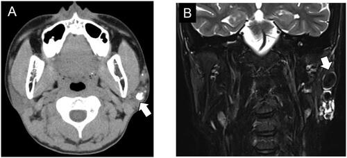
The patient visited another hospital as an emergency outpatient complaining of left parotid pain after dinner. The pain occurred intermittently and recurrently, without swelling of the parotid gland during eating. Workup with plain computed tomography (CT) scan demonstrated multiple calcifications of variable size in his left parotid gland (Figure ), which was interpreted as sialolithiasis. He was referred to our hospital for treatment. A few soybean-sized, hard masses were palpable under the skin at the left parotid region. The angle of the mandible appeared to be tinged faintly with a pale bluish-purple coloration, which was present from the time of his birth.
Figure 1. Preoperative findings. (A) Plain axial CT image shows calcified materials of various sizes (white arrow) in the left parotid gland. (B) Coronal T2-weighted MRI shows a hyperintense, lobulated lesion with multiple hypointense areas (white arrow) consistent with phleboliths in the left parotid gland.
Diagnostic assessment
Considering vascular malformations with phleboliths as a differential diagnosis, several imaging examinations were additionally performed. A contrast-enhanced CT scan of the parotid gland area revealed at least nine calcifications, the surrounding tissue of which was contrasted well by contrast agent. T2-weighted magnetic resonance imaging (MRI) showed a poorly-defined hyper-intense signal region in and around the left parotid gland, along with round, well-defined hypo-intense areas surrounded with a membranous hyper-intense signal (Figure ). These round masses, which correspond to calcifications on CT, manifested as hypo-intense areas on T1-weighted MRI and their circumferences were highly contrasted by gadolinium.
Therapeutic intervention
With the presumed diagnosis of sialolithiasis or vascular malformations containing phleboliths, a superficial parotidectomy was performed. When the skin flap was elevated, a hemorrhagic vascular lesion was observed in the parotid gland. This vascular lesion mainly existed in the parotid gland, and a portion of the lesion advanced behind the facial nerve and contacted the masseter muscle. After resecting this lesion with preservation of the facial nerve, the remnant parotid gland tissue was covered with a rotation flap of the sternocleidomastoid muscle in order to avoid Frey’s syndrome. The operative time was 3 h 26 min and the blood loss was 30 mL.
Follow-up and outcome
The patient’s facial movement was normal following surgery and the postoperative course was uneventful. Complications such as Frey’s syndrome have not been observed. No recurrence of phleboliths on contrast-enhanced CT was found at 2 years 4 months postoperatively.
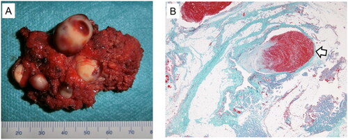
The resected parotid lesion contained multiple stone-like objects, which were round, red-white, and had a smooth surface (Figure ). The microscopic features showed that most of the parotid gland was replaced by fat tissue and collagenous fibrous tissue. Blood vessels in the lesion were dilated and thin-walled, and they contained thrombi and phleboliths within the lumen (Figure ). Endothelial cells lining the lumen of the vascular vessels were CD31-positive, partially positive for CD34, and D2-40-negative. The lesion consisted of abnormal venous components and thus it was diagnosed as venous malformations.
Figure 2. Excised specimen. (A) Macroscopic findings of the resected specimen are shown. Grossly, the parotid lesion is 54 mm in maximum diameter with multiple white stone-like objects. The largest object is 18 mm in size. (B) Elastic-Masson trichrome staining reveals nodular, irregular smooth muscle growth and disconnected elastic fibers in the abnormal vascular wall. Phleboliths (white arrow) are observed in the vascular lumen.
Discussion
Vascular malformations are differentiated from vascular tumors, so-called hemangiomas, according to the International Society for the Study of Vascular Anomalies (ISSVA) classification. The source of the ISSVA classification dates back to the report of Mulliken and Glowack in 1982 [Citation3]. They described that hemangioma and vascular malformation were different pathological conditions, focusing on vascular endothelial cell proliferation. Hemangiomas are vascular tumors that are rarely apparent at birth, grow rapidly during the first 6 months of life, involute with time, and do not necessarily infiltrate but can sometimes be destructive [Citation4]. On the other hand, vascular malformations are irregular vascular networks, where blood vessels grow abnormally. Vascular malformations are classified into capillary malformations, venous malformations, lymphatic malformations, and arteriovenous malformations based on their main constituent vessels. Venous malformations are a type of vascular malformation, which is the most common type of congenital vascular malformation, with an incidence of 1 to 2 in 10,000 and a prevalence of 1% [Citation5]. Venous malformations are not noticeable during birth, and they appear to develop in adults because they tend to worsen due to puberty, pregnancy, or traumatic injury [Citation4]. Unlike hemangiomas, venous malformations never regress and continue to expand with time. Altered blood flow dynamics within venous malformations can lead to thrombus formation and phleboliths. The pathogenesis of phleboliths is thought to involve an organized thrombus produced when the peripheral blood flow slows [Citation2,Citation6,Citation7]. Achache et al. [Citation8] reported that vascular malformations were identified in 10 cases (1.6%) out of 614 parotidectomies performed at their institution. The authors of that study noted that the frequency has been previously reported as 0.5% by Bears et al. [Citation9], who reported 760 parotid tumors, and as 0.6% by Byars et al. [Citation10], who reported 460 parotid tumors. In the parotid gland, venous malformations may arise from the gland itself, or by the invasion of subcutaneous blood vessels into the gland structure [Citation11]. The symptoms including pain and swelling are related to phleboliths and venous stasis, which masquerade as sialolithiasis.
Both phleboliths and sialoliths are observed as calcified lesions on CT. When phleboliths are located in the parotid gland, they appear radiologically similar to sialoliths. It is therefore difficult to distinguish between sialoliths and phleboliths by plain CT alone. If the calcified lesion on CT is suspected to be something other than a sialolith, other imaging examinations such as MRI and ultrasound should be performed additionally. MRI can visualize the enlarged vessels of vascular malformations which manifest with the appearance of a lobulated bunch of grapes with hyperintensity on T2-weighted images [Citation5]. Phleboliths are identified as hypo-intense nodular or punctate structures in all sequences on MRI [Citation2]. Magnetic resonance (MR) sialography, which produces images of the salivary duct system, is useful for the diagnosis of chronic obstructive sialadenitis, including sialolithiasis and stenosis [Citation12,Citation13]. Sialoliths tend to be located in the salivary duct system, whereas phleboliths are located outside the salivary duct system; thus, MR sialography may be able to distinguish these different characteristics. Ultrasound (US) is another useful imaging tool for evaluating vascular malformations. US evaluation of venous malformations shows a heterogeneous hypoechoic pattern with sinusoidal spaces, where slow flow is seen as back-and-forth movement of low/intermediate echo intensity [Citation14]. Doppler ultrasound may also detect vascular lesions with increased blood flow [Citation2,Citation15]. Phleboliths can be detected as hyperechoic areas producing acoustic shadowing [Citation14]. In the present case, the peculiar skin color at his mandibular angle made us suspect that the lesion might be a venous malformation with phleboliths, although a definitive diagnosis could not be achieved preoperatively. Skin color often appears bluish-purple when a venous malformation extends subcutaneously. Not only image inspection but also clinical characteristics like skin appearance are important for making a diagnosis.
The conventional treatment methods for vascular malformations have included surgical removal, sclerotherapy, embolization, as well as combinations of these methods. Sclerotherapy is an attractive treatment for venous malformations. The most commonly used sclerosing agents for the treatment of venous malformations are ethanol and sodium tetradecyl sulphate [Citation16,Citation17]. These agents come into direct contact with the vessel wall, causing endothelial damage, inflammation, thrombosis, fibrosis, and ultimately occlusion of the vascular lumen. Su et al. [Citation16] reported that sclerotherapy under the guidance of venography was highly effective for cases of venous malformations in the face and neck region. Sclerotherapy has the advantage of being effective and less invasive, but on the other hand, there is a risk of complications such as skin necrosis and nerve injury. Surgery is another treatment option and may be curative for localized venous malformations. Achache et al. [Citation8] reported that their treatment for vascular malformations of the parotid area was mainly surgical because it allows for definitive diagnosis via histological examination. Venous malformations often show unclear boundaries and snarled shapes on MRI, so confirming a pathological diagnosis through surgery is effective in terms of differentiation from malignant neoplasms. Hu et al. [Citation18] reported that phleboliths are a risk factor associated with painful venous malformations of the extremities. Phleboliths can be removed through surgery, so surgery should be considered in cases of accompanying pain, although phleboliths cause less pain in the head and neck region compared with the extremities [Citation18,Citation19]. In the present case, the patient suffered from parotid pain caused by plural phleboliths. Given the difficulty of preoperative diagnosis and the fact that the patient complained of pain, the choice of surgery for our patient was thought to be reasonable. As Achache et al. [Citation8] note, dissection and resection of vascular malformations is not a particularly complex procedure compared with that for other parotid tumors. However, careful attention should be paid to controlling bleeding and preserving the facial nerve as in other parotid surgeries. Frey’s syndrome is one of the most common complications of parotid surgery, known as gustatory sweating. It is caused by reattachment of the auriculotemporal nerve to the sweat glands of the skin instead of the parotid gland during the healing process. In the present case, we attempted to suture the remaining parotid fascia after removal of the lesion, but the parotid fascia defect was too large to suture. Therefore, we made a rotation flap using the sternocleidomastoid muscle and filled the defect site with the flap. Filling in the defect site in this manner is one of the techniques for preventing Frey’s syndrome, as it creates a barrier that hinders aberrant nerve regeneration between the remaining parotid gland and the sweat glands.
Conclusion
Venous malformation with phleboliths in the parotid gland is a rare entity. It is sometimes difficult to distinguish phleboliths from sialolithiasis because of the similarities in their clinical and radiological features. If the calcified lesion is suspected to be something other than a sialolith, additional imaging examinations such as MRI and ultrasound should be performed. If a diagnosis of phleboliths is suspected, then venous malformation ought also to be considered to exist around them. Thus, awareness of this disease entity is important for producing an accurate diagnosis. The conventional treatment methods for vascular malformations of the parotid gland mainly include surgical removal and sclerotherapy. Surgery should be considered if the lesion is localized and the patient complains of pain due to phleboliths.
Informed consent
The patient’s legal guardian gave informed consent for the publication of this case report.
Acknowledgements
The authors thank FORTE Science Communications (https://www.forte-science.co.jp/) for English language editing.
Disclosure statement
The authors report no conflict of interest.
References
- Tagnon BB, Theate I, Weynand B, et al. Long-standing mucosa-associated lymphoid tissue lymphoma of the parotid gland: CT and MR imaging findings. AJR Am J Roentgenol. 2002;178(6):1563–1565. doi: 10.2214/ajr.178.6.1781563.
- Kato H, Ota Y, Sasaki M, et al. A phlebolith in the anterior portion of the masseter muscle. Tokai J Exp Clin Med. 2012;37:25–29.
- Mulliken JB, Glowacki J. Hemangiomas and vascular malformations in infants and children: a classification based on endothelial characteristics. Plast Reconstr Surg. 1982;69(3):412–422. doi: 10.1097/00006534-198203000-00002.
- Richter GT, Friedman AB. Hemangiomas and vascular malformations: current theory and management. Int J Pediatr. 2012;2012:645678–645610. doi: 10.1155/2012/645678.
- Behravesh S, Yakes W, Gupta N, et al. Venous malformations: clinical diagnosis and treatment. Cardiovasc Diagn Ther. 2016;6(6):557–569. doi: 10.21037/cdt.2016.11.10.
- O’Riordan B. Phleboliths and salivary calculi. Br J Oral Surg. 1974;12:119–131. doi: 10.1016/0007-117x(74)90120-6.
- Raymond AK, Batsakis JG. Angiolithiasis and sialolithiasis in the head and neck. Ann Otol Rhinol Laryngol. 1992;101(5):455–457. doi: 10.1177/000348949210100514.
- Achache M, Fakhry N, Varoquaux A, et al. Management of vascular malformations of parotid area. Eur Ann Otorhinolaryngol Head Neck Dis. 2013;130(2):55–60. doi: 10.1016/j.anorl.2011.11.004.
- Beahrs OH, Woolner LB, Carveth SW, et al. Surgical management of parotid lesions. Review of seven hundred sixty cases. Arch Surg. 1960;80:890–904. doi: 10.1001/archsurg.1960.01290230008002.
- Byars LT, Ackerman LV, Peacock E. Tumors of salivary gland origin in children: a clinical pathologic appraisal of 24 cases. Ann Surg. 1957;146(1):40–51. doi: 10.1097/00000658-195707000-00005.
- Chuang CC, Lin HC, Huang CW. Submandibular cavernous hemangiomas with multiple phleboliths masquerading as sialolithiasis. J Chin Med Assoc. 2005;68(9):441–443. doi: 10.1016/S1726-4901(09)70162-5.
- Sánchez Barrueco Á, Santillán Coello JM, González Galán F, et al. Epidemiologic, radiologic, and sialendoscopic aspects in chronic obstructive sialadenitis. Eur Arch Otorhinolaryngol. 2022;279(12):5813–5820. doi: 10.1007/s00405-022-07473-w.
- Sobrino-Guijarro B, Cascarini L, Lingam RK. Advances in imaging of obstructed salivary glands can improve diagnostic outcomes. Oral Maxillofac Surg. 2013;17(1):11–19. doi: 10.1007/s10006-012-0327-8.
- Flis CM, Connor SE. Imaging of head and neck venous malformations. Eur Radiol. 2005;15(10):2185–2193. doi: 10.1007/s00330-005-2828-4.
- Su Y-X, Liao G-Q, Wang L, et al. Sialoliths or phleboliths? Laryngoscope. 2009;119(7):1344–1347. doi: 10.1002/lary.20514.
- Su L, Fan X, Zheng L, et al. Absolute ethanol sclerotherapy for venous malformations in the face and neck. J Oral Maxillofac Surg. 2010;68(7):1622–1627. doi: 10.1016/j.joms.2009.07.094.
- Siniluoto TM, Svendsen PA, Wikholm GM, et al. Percutaneous sclerotherapy of venous malformations of the head and neck using sodium tetradecyl sulphate (sotradecol). Scand J Plast Reconstr Surg Hand Surg. 1997;31(2):145–150. doi: 10.3109/02844319709085481.
- Hu L, Chen H, Yang X, et al. Risk factors associated with pain in patients with venous malformations of the extremities. Vasc Med. 2019;24(1):56–62. doi: 10.1177/1358863X18802007.
- Eivazi B, Fasunla AJ, Güldner C, et al. Phleboliths from venous malformations of the head and neck. Phlebology. 2013;28(2):86–92. doi: 10.1258/phleb.2011.011029.