
Abstract
Amyloidosis is a condition characterised by extracellular tissue deposition of fibrils causing a wide range of clinical manifestations. This protein deposition can occur in any tissue, most commonly in the kidney, heart, skin, peripheral nervous system, and gastrointestinal tract. However, the deposition of amyloid fibrils in the synovium is seldom reported. Musculoskeletal manifestations are subtle, subclinical and rarely the patient presents with symptoms that resemble rheumatic diseases. Here, we describe a 55-year-old man with AL (amyloid light chain) amyloidosis who presented with inflammatory polyarthritis with significant morning stiffness, inflammatory low back pain, and painful thickened tongue. The patient had anaemia, macroglossia with lateral scalloping of the tongue, papules, and plaques in the periocular, perioral and perinasal area, bilateral carpal tunnel syndrome, localised soft tissue swelling over the joints, restricted movement in different joints with flexion contractures in some joints. Rheumatoid factor and ACPA were negative and the X-ray of the sacroiliac joints was normal. We confirmed amyloidosis by biopsy of an affected skin lesion. In the urine, no Bence Jones protein was found and bone marrow study and x-ray of the skull were normal. Plasma immuno-electrophoresis and serum free light chain (FLC) assay confirmed lambda light chain type monoclonal gammopathy. Take home message: Although AL amyloidosis is a rare condition, it should be considered while evaluating atypical symptoms in patients presenting with rheumatic complaints. A high index of suspicion is necessary for proper diagnosis as delay in diagnosis will yield a poorer treatment outcome.
Keywords:
Introduction
Polyarthritis encompasses a wide range of differential diagnoses. Rheumatoid arthritis and spondyloarthritis (e.g. reactive arthritis, psoriatic arthritis) are the most common forms of inflammatory polyarthritis. But, there are other systemic autoimmune diseases (e.g. systemic lupus erythematosus, primary Sjogren syndrome, vasculitis, etc.) that may cause joint pain. Diagnosing polyarthritis depends upon an appropriate medical history, a thorough physical examination, and judiciously selected laboratory investigations. There are some rare systemic disorders like amyloidosis, lymphoma, multiple myeloma, POEMS syndrome (polyneuropathy, organomegaly, endocrinopathy, myeloma protein, and skin change) that may present with inflammatory arthritis [Citation1]. Amyloidosis is a rare localised as well as systemic disorder that results from the extracellular deposition of a variety of fibrillar proteins leading to changes in tissue architecture or function.
There are several types of amyloidosis depending upon their precursor proteins. Immunoglobulin light chain (AL) amyloidosis is the most common form of systemic amyloidosis [Citation2]. The signs and symptoms of amyloidosis depend upon the type of amyloid protein deposition and organs that are affected. The most frequently affected organs are the heart, kidney, liver, skin, gastrointestinal tract (GIT), autonomic, and peripheral nervous system. Amyloid protein may deposit in the joints, joint capsules, and in the articular cartilage. Articular manifestations of amyloidosis are collectively known as amyloid arthropathy. Amyloid arthropathy is a relatively uncommon manifestation of amyloidosis [Citation3]. Rarely this may be the only presenting feature of systemic amyloidosis. Most patients with this disorder may experience pain and swelling in different joints with morning stiffness that may mimic rheumatoid diseases [Citation4]. Polyarthropathy preferentially occurs in β2-microglobulin-derived amyloidosis, which complicates chronic renal failure with haemodialysis but is uncommon in amyloid light-chain (AL) amyloidosis [Citation5].
Here we describe a case of amyloid arthropathy who presented with inflammatory polyarthritis and eventually diagnosed as a case of AL amyloidosis. We followed the Care guidelines for case reporting [Citation6].
Case report
A 55- year-old man was admitted to our rheumatology department with complaints of pain in multiple joints for two years, for which he had been treated by several physicians outside our hospital, with insufficient effect The pain was insidious in onset, inflammatory in nature with significant morning stiffness. The pain initially started in both wrists, then gradually involved small and large joints of both upper and lower limbs, predominantly the large joints. The joint involvement was symmetrical. He also noticed low back pain for one and a half years which was insidious in onset, non-radiating, mild to moderate in intensity, and inflammatory. Gradually the pain became severe to hamper his mobility and daily activities. For his joint pain, he was prescribed different NSAIDs including naproxen and sulindac, painkillers like paracetamol and tramadol hydrochloride, in different dosages and duration, glucocorticoid (deflazacort 12 mg), methotrexate (MTX) 20 mg weekly during one year, and tofacitinib (10 mg) for eight months without any significant improvement. From the last one year, the patient experienced an enlarged and painful tongue, with continuously dull and aching pain, aggravated by chewing food. He also noticed fatigue and a significant weight loss of five kg in three months. After admission, he was found to have chronic kidney disease evidenced by persistently raised serum creatinine of 1.9 mg/dl and 1.6 mg/dl three months before, though being normotensive and non-diabetic. He had paresthaesia in both hands. A nerve conduction study established bilateral carpal tunnel syndrome (CTS). A carpal tunnel release surgery gave some improvement, but after 5–6 months, his symptoms (e.g. paresthaesia) reappeared. He denied any history of painful red eye, erythematous scaly skin lesions, preceding diarrhoea or dysentery, any alteration of bowel habit, photosensitivity, fever or cough, and had no contact with tuberculosis patient. He had no family history of such type of illness. He didn’t have any clinical features of cardiac problems, no lower extremity edoema, no elevated jugular venous pressure, ascites, or dyspnoea. He had no history of syncope or presyncope. On admission, he complained of severe pain (VAS 9/10).
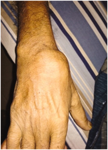
On physical examination, the patient was found moderately pale looking, all vital signs were normal. His pulse rate was 80/min, regular with normal volume, blood pressure 120/70 mm of Hg, respiratory rate 18/min, there was no lower limb edoema or lymphadenopathy. There were papules and plaques over the periocular, perinasal, and perioral area, macroglossia with indentation of the tongue, pinch purpura in the oral cavity ( and ). Nail dystrophy was present in some of his fingers. There was no organomegaly, apex beat was situated in the 5th intercostal space along the midclavicular line. Musculoskeletal examination revealed localised, mildly tender, soft tissue swelling of variable size and shape (largest one was 5Х3 cm, over the wrist) over flexor and extensor aspects of wrists and back of knees. Both shoulders were swollen (shoulder pad sign positive, ), tenderness was present over MCPs, wrists, elbows, and shoulders. He had an antalgic gait. Active and passive movements of wrists and shoulders were painful and restricted. Flexion contracture (30 degrees) was present in the left elbow.
Figure 1. Lateral scalloping with macroglossia.
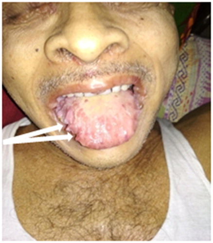
Figure 2. Papules and plaque over periocular, perinasal and perioral area.
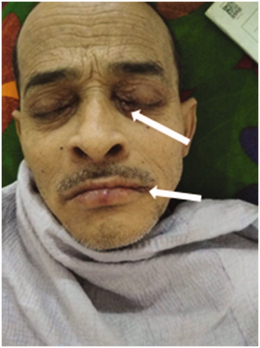
Figure 3. Anterior shoulder pad sign.
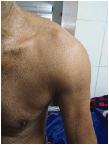
Figure 4. Soft tissue swelling over the wrist due to amyloid deposit.
The movement of the cervical spine was restricted in all directions. There was a loss of lumbar lordosis, with restricted forward flexion (positive Schober test of 13 cm) of the lumbar spine. Movements were restricted in both hips (positive Thomas test) with fixed flexion deformity. Both knees were tender. Nervous system examination revealed wasting of the thenar muscles of both hands and post-surgical scars with weakness of abduction and apposition of the thumbs. Phalen sign was positive in both wrists with an otherwise normal neurological examination. The examination of other systems was unremarkable.
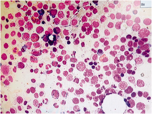
Laboratory investigations showed normocytic normochromic anaemia (hemoglobin 8.2 mg/dl (normal 15 ± 2 mg/dl.)) with normal white blood cells and platelet counts, erythrocyte sedimentation rate (ESR) 55 mm in the first hour (reference range 0–10), C-Reactive protein of 97.9 mg/L (reference range <5), TSAT (transferrin saturation) 58%, serum ferritin 358 µgm/dl (reference range 250–450), serum albumin 33.57 gm/L (reference range 38–54), serum total protein 47 gm/L (reference range 54–83). The liver function tests, calcium, phosphate, and thyroid function were normal. Serum creatinine was 1.9 mg/dl and creatinine clearance was 37 ml/min. Urine analysis revealed mild proteinuria, his 24-hour urinary total protein was 0.82 gm/day (reference range <0.2 gm/day) with no abnormal urinary sediment. Bence-Jones protein was absent in the urine. Rheumatoid factor, anti-CCP antibody, and HLA B27 were negative. The X-ray of the sacroiliac joints was normal. The heart was normal at echocardiography. Ultrasound of the right wrist joint revealed well-defined extensor tendons surrounded by a hypoechoic and inhomogeneous mass above the wrist joint. There was mild synovial thickening of the wrist joint but the power Doppler signal was absent and joint erosion was not observed. . A transverse scan of the palmar side of the wrist revealed enlarged and swollen median nerve (sectional area of 15 mm2), hypoechoic mass around flexor tendons and median nerve. A biopsy from an affected skin lesion was performed (plaque of lower lip margin) showing apple-green birefringence with Congo red stain confirmed the diagnosis of amyloidosis (). The bone marrow was normocellular with appropriate trilinear haematopoiesis. The plasma cell percentage was four percent, no immature or anaplastic plasma cells were seen. Prussian blue (iron) stained bone marrow shows normal iron deposits in macrophages ().
Figure 5. (a) and (b) Plaque biopsy shows amyloid deposit confirmed by Congo red staining with apple green birefringence.
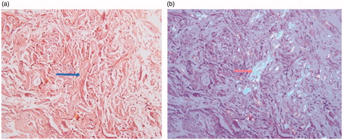
Figure 6. High-power appearance of the spread bone marrow aspirate showing normal bone particles of bone marrow at the end of cellular trails.
The serum protein electrophoresis showed alpha 1,2 was slightly raised and there was no ‘M-spike’. The serum immunoglobulin levels showed almost normal values of all three lines: IgG 950 mg/dl (reference range 700–1600 mg/dl), IgA 210 mg/dl (reference range 70–400 mg/dl) and IgM 235 mg/dl (reference range 40–230 mg/dl). Flow cytometry was not done as this is not available in our centre. Immunoelectrophoresis revealed lambda light chain type monoclonal gammopathy in serum (). The serum-free light chain (FLC) assay showed a greatly enhanced free lambda light chain with alternation of free kappa/lambda ratio ().
Figure 7. Increased accumulation of Lambda light chain in the Lambda zone on serum IFE.
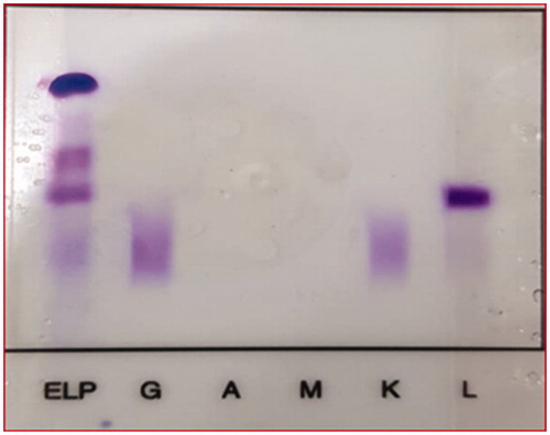
Table 1. The serum free light chain (FLC) assay.
The treatment of AL amyloidosis grossly has two parts: haematopoietic cell transplantation (HCT) eligible and not eligible for HCT. High dose melphalan followed stem cell transplant is preferred. Otherwise, bortezomib based therapy like cyclophosphamide, bortezomib, and dexamethasone (CyBorD) or bortezomib, melphalan, and dexamethasone regimen are used. He was eligible for high dose melphalan with autologous haematopoietic cell transplantation (HCT) support. HCT was not offered because of financial constraints, so the bortezomib-based triple regimen, i.e. bortezomib, cyclophosphamide, and dexamethasone (CyBorD) was started [Citation7].
The patient developed diarrhoea after the second dose of bortezomib (after 13 days of 1st dose of bortezomib) which delayed the schedule of the next cycle. In the meantime, a slight reduction in the size of the papules and macules of face and lips and the soft tissue swelling over wrists was observed. Initially, the patient experienced the worst pain possible level 09 as measured by Visual Analogue Scale(VAS) 0–10; after initiation of treatment, the pain intensity was reduced to a severity level 06 as measured by VAS. Before giving the third dose of bortezomib, the patient succumbed due to massive haematemesis leading to hypovolemic shock.
Discussion
Our patient presented with typical inflammatory polyarthritis with subcutaneous nodules over the wrists mimicking rheumatoid arthritis that preceded the features of amyloidosis. Presence of macroglossia with lateral indentation of the tongue, shoulder pad sign, perioral and periocular papules, bilateral carpal tunnel syndrome, renal impairment, biopsy evidence of amyloid deposition distinguished it from rheumatoid arthritis. Most of the case reports reported polyarthritis in AL amyloidosis are associated with multiple myeloma [Citation8,Citation9]. The prevalence of a dominant soft tissue and bone involvement is 3.9–4.3 times higher in patients with AL amyloidosis with multiple myeloma than in patients with AL amyloidosis without multiple myeloma [Citation10]. We had excluded multiple myeloma in our case with the help of radiological skeletal survey, the study of bone marrow, and serum protein electrophoresis.
Amyloidosis should be suspected in any patient with unexplained nephropathy, cardiac failure, peripheral or autonomic neuropathy, carpal tunnel syndrome, hepatic or splenic enlargement, and/or dysfunction [Citation11]. Biopsy should be done from the affected organ or abdominal subcutaneous fat in all patients suspected to be a case of amyloidosis. The diagnosis is based upon finding amorphous extracellular Congo red positive deposits, which displays characteristic apple-green birefringence under polarised microscopy. Type of amyloidosis should be sought by mass spectrometry, immunohistochemical staining, and search for monoclonal protein in serum or urine or a bone marrow study for monoclonal plasma cell [Citation12].
Diagnostic criteria for AL amyloidosis have been developed by the Mayo Clinic and the International Myeloma working group and require the presence of all the four criteria [Citation13] (Appendix 1). The current patient fulfilled the criteria of this working group. We used immunohistochemical staining that can determine the type of amyloid [Citation14] instead of spectrometry-based proteomic analysis or immunoelectron microscopy as these methods were not available in our country.
Amyloidosis is a heterogeneous group of disorders associated with extracellular tissue deposits of amyloid fibril protein sharing a similar structure. There are several types of amyloidosis. The principal types of amyloidosis are AL (primary) and AA (secondary) types. Other major types are dialysis-related amyloidosis, heritable amyloidosis, age-related systemic amyloidosis, and organ-specific amyloid [Citation15]. The nomenclature for amyloid subunit proteins formulated in the Nomenclature Committee of the International Society of Amyloidosis (ISA) in 2014 was based on the protein precursor of amyloid fibrils such as AL, AA, ATTR, Aß2M, etc [Citation16]. Immunoglobulin light chain (AL) amyloidosis (historically referred to as primary amyloidosis) is the most common and most severe form of systemic amyloidosis in the developed countries [Citation2]. It occurs more in males than in females, and the average age at presentation is 64 years [Citation15]. Immunoglobulin kappa or lambda chain act as a precursor protein in AL amyloidosis. Primary or AL amyloidosis can occur alone or in association with multiple myeloma, Waldenstrom's macroglobulinaemia, or non-Hodgkin's lymphoma. It affects 8–12 per million patients per year [Citation17]. AA amyloidosis (previously known as reactive or secondary amyloidosis) is more common in developing countries. It is associated with rheumatic diseases (rheumatoid arthritis, ankylosing spondylitis, psoriatic arthritis, juvenile idiopathic arthritis, systemic lupus erythematosus, primary Sjögren's syndrome (SS) or secondary SS concomitant with rheumatoid arthritis, etc.), infectious diseases (bronchiectasis, tuberculosis, leprosy), inherited diseases (familial Mediterranean fever, tumour necrosis factor receptor-associated periodic fever syndrome), idiopathic diseases (sarcoidosis) and malignant tumours (Hodgkin’s lymphoma, renal cell carcinoma, mesothelioma) [Citation7,Citation18]. AA derived from serum protein precursor SAA, an acute phase reactant, is typically upregulated in chronic inflammatory processes. There were no indications for any of these diseases in our patients. There. were no clinical signs or symptoms of Sjogren’s syndrome-like dry eyes or xerostomia in our patient. In association with SS usually, AA amyloidosis rather than AL amyloidosis occurs.
Amyloid protein can deposit in various organs/tissues. Clinical features depend upon the type of amyloid protein and organ/tissue where deposited. Systemic AL amyloidosis can involve any organ except the central nervous system [Citation2]. An updated definition of organ involvement in AL amyloidosis has been made in a meeting of the International Society of Amyloidosis held in 2010 in Rome [Citation19,Citation20]. Amyloid arthropathy is a relatively uncommon presentation (approximately 5%) of systemic amyloidosis [Citation21]. It is low grade, subacute, progressive, and symmetric with a predilection for shoulders, knees, wrists, MCPs, PIPs, and less commonly elbows and hips. There may be pain, swelling, and morning stiffness that may mimic chronic inflammatory arthropathies [Citation1]. Though amyloid arthropathy is rather uncommon as a presenting feature of amyloidosis, when present, it is regarded as a disease-specific finding in AL amyloidosis [Citation21]. It may also cause nodular hypertrophy of synovium due to amyloid deposits. Rarely erosive arthritis has been reported in patients with AL amyloidosis coexisting with monoclonal gammopathy of uncertain significance [Citation22]. Swelling may be particularly prominent around glenohumeral joints resulting in a characteristic “shoulder pad” sign. It is pathognomonic for AL amyloidosis [Citation23].
On ultrasound of the right wrist joint, we observed well-defined extensor tendons surrounded by a hypoechoic and inhomogeneous mass above the wrist joint. In Amyloidosis related to chronic dialysis usually, a hyperechoic area is found [Citation24]. The picture we found was not compatible with the findings in rheumatoid arthritis, as there was no joint erosion or power Doppler signal.
Amyloid fibrils may also deposit in the muscle causing muscular weakness (myopathy) and enlargement (pseudohypertrophy). Muscle involvement is generally associated with systemic involvement. A large tongue (macroglossia) due to amyloid infiltration may occur, and when present, it is almost always a part of systemic AL amyloidosis, of which it is a pathognomonic sign [Citation25]. Cutaneous involvement is also a common finding in AL amyloidosis. Cutaneous amyloidosis often shows yellowish or brownish macules, papules, plaque, nodules, or rarely blisters and periorbital haematoma ("raccoon-eyes" sign) in different parts of the body, mostly localised to face and trunk [Citation2]. Fingernail dystrophy may also occur in AL amyloidosis.
Peripheral neuropathy in AL amyloidosis is manifested as mixed sensory and motor neuropathy (20% resembling diabetic neuropathy) and/or autonomic neuropathy (15%) [Citation26]. Carpal tunnel syndrome is typically bilateral and symmetric. Renal involvement is the most frequent organ involvement of AL amyloidosis affecting about two-thirds of patients. Haematuria and hypertension are uncommon [Citation2]. About 75 percent of patients present with proteinuria varying from mild to massive (>20 gram/day) form like it is seen in multiple myeloma [Citation27,Citation28]. Mild renal impairment is common, but rapidly progressive renal failure is rare. The presented patient has moderate renal impairment (S creatinine 1.9 mg/dl) with mild proteinuria (0.82 gram/day). The amyloid deposits are primarily limited to the vessels leading to narrowing of the vascular lumens [Citation29].
Bortezomib is the first proteasome inhibitor approved by the USFDA for the treatment of multiple myeloma. Besides multiple myeloma, it is now being used in various hematological malignancies like mantle cell lymphoma, cutaneous or peripheral T cell lymphoma, follicular lymphoma, Waldenström's macroglobulinemia, etc. For the treatment of immunoglobulin light chain (AL) amyloidosis autologous haematopoietic cell transplantation (HCT) is the preferred option for the eligible patients. A bortezomib-based triplet regimen such as bortezomib, cyclophosphamide, dexamethasone (CyBorD) or bortezomib, melphalan, and dexamethasone (BMD) is the choice of therapy for transplant non-eligible patients. Peripheral neuropathy is the most common adverse reaction followed by thrombocytopenia and diarrhoea. Diarrhoea is may occur in 19 to 52% of patients [Citation30]. Our patient developed diarrhoea after the second dose of bortezomib. We excluded the infectious causes of diarrhoea.
The patient also experienced massive gastrointestinal bleeding in the form of haematemesis, which lead to the development of profound shock and death. Amyloidosis may directly be associated with bleeding disorders. Possible mechanisms responsible are factor X deficiency due to selective binding to amyloid deposits in the perivascular tissues of the spleen and also acquired von Willebrand disease [Citation31,Citation32].
Treatment of primary amyloidosis depends upon patients’ age, comorbidity, organ involvement, renal or hepatic function. The preferred option is melphalan followed by autologous stem cell transplant. If the patient is not eligible for a transplant (presence of major comorbidities, the involvement of three or more organs, poor performance status, and advanced cardiac amyloidosis) or cannot afford, the alternative is bortezomib based therapy (bortezomib, cyclophosphamide, and dexamethasone-CyBorD or bortezomib, melphalan, and dexamethasone-BMD). A melphalan based therapy (melphalan, dexamethasone) could be another alternate one for those who are not candidates for bortezomib. Though there is no head to head trial between CyBorD and BMD regimens, an international trial that used BMD versus melphalan and dexamethasone revealed a superior response rate in the BMD group [Citation33]. The hematological response should be checked by serial measurement of serum-free light chain assay [Citation19]. Alternative systemic therapy should be chosen if there is haematologic or organ progression at any time during treatment, relapsed/refractory cases, if there is <50 percent reduction in the difference between the involved and uninvolved free light chain levels (dFLC) after two cycles of chemotherapy; or if dFLC is ≥40 mg/L after four to six cycles of chemotherapy or on day 100 after transplant [Citation7]. The amyloid deposition may decrease quickly when the underlying disorder is successfully treated [Citation8]. The prognosis of AL amyloidosis depends upon the extent, nature, and number of organ involvement as well as the stage of the disease. The presence and severity of cardiac involvement and the concurrent presence of myeloma greatly regulate the outcome of myeloma [Citation34]. The major causes of death are cardiac, hepatic, or renal failure, and infection [Citation35].
A strength of this case report is that it is one of the rare descriptions of polyarthritis in AL amyloidosis not associated with multiple myeloma. The deposition of amyloid fibrils in the synovium is seldom reported and patients rarely present as rheumatoid arthritis. We were able to describe the clinical history, physical symptoms of the patient as well as pathology. We gave a review of current therapeutic possibilities.
A limitation of this case report is that we could not observe the full response to treatment as the patient expired prior to the third dose of chemotherapy.
Take home message: Although AL amyloidosis is a rare condition, it should be considered while evaluating atypical symptoms in patients presenting with rheumatic complaints. A high index of suspicion is necessary for proper diagnosis as delay will result in a poorer treatment outcome.
Patient consent
Written permission was obtained from the patient for taking the photographs and writing this article for publication.
Ethical approval
Not applicable.
Acknowledgment
The authors are grateful to Dr. Md. Moniruzzaman, Dr. Ahammad Shafiq Sikder Adel, Dr. MF Ullah, Dr. Sadi Masud Al Turab, and Dr. TQM Ashraful Haque for their cordial help in different aspects.
Conflicts of interest
None.
References
- Duna GF, Cash JM. Rheumatic manifestations of dysproteinemias and lymphoproliferative disorders. Rheum Dis Clin North Am. 1996;22(1):39–51.
- Desport E, Bridoux F, Sirac C, et al. AL Amyloidosis. Orphanet J Rare Dis. 2012;7:54.
- Glenne G. Amyloid deposits and amyloidosis: the beta-fibrilloses (first ot two parts). N Engl J Med. 1980;302:1283–1292.
- Wiernik PH. Amyloid joint disease. Medicine (Baltimore). 1972;51(6):465–479.
- Katoh N, Tazawa KI, Ishii W, et al. Systemic AL amyloidosis mimicking rheumatoid arthritis. Intern Med. 2008;47(12):1133–1138.
- Gagnier JJ, Kienle G, Altman DG, et al. The CARE guidelines: consensus-based clinical case reporting guideline development. J Med Case Rep. 2013;7:223.
- Mayo Stratification of Myeloma and Risk-Adapted Therapy (mSMART) Consensus on AL Amyloidosis: Diagnosis and Treatment (v6, Feb 2017). [accessed 2020 Feb 19]. Available from: https://www.msmart.org/treating-al.
- Alpay N, Artim-Esen B, KamalI S, et al. Amyloid arthropathy mimicking seronegative rheumatoid arthritis in multiple myeloma: case reports and review of the literature. Amyloid. 2009;16(4):226–231.
- Schoninger S, Homsi Y, Kreps A, et al. A case of multiple myeloma misdiagnosed as seronegative rheumatoid arthritis and review of relevant literature. Case Rep Rheumatol. 2018;2018:9746241.
- Prokaeva T, Spencer B, Kaut M, et al. Soft tissue, joint, and bone manifestations of AL amyloidosis: clinical presentation, molecular features, and survival. Arthritis Rheum. 2007;56(11):3858–3868.
- Merlini G, Westermark P. The systemic amyloidoses: clearer understanding of the molecular mechanisms offers hope for more effective therapies. J Intern Med. 2004;255(2):159–178.
- Mahmood S, Palladini G, Sanchorawala V, et al. Update on treatment of light chain amyloidosis. haematologica. 2014;99(2):209–221.
- Rajkumar SV, Dimopoulos MA, Palumbo A, et al. International Myeloma Working Group updated criteria for the diagnosis of multiple myeloma. Lancet Oncol. 2014;15(12):e538-48–e548.
- Brambilla F, Lavatelli F, Di Silvestre D, et al. Reliable typing of systemic amyloidoses through proteomic analysis of subcutaneous adipose tissue. Blood. 2012;119(8):1844–1847.
- Gorevic P. ‘Overview of amyloidosis.’ UpTodate; 2020 [accessed 2020 Jan 23]. Available from: https://www.uptodate.com/contents/overview-of-amyloidosis
- Sipe JD, Benson MD, Buxbaum JN, et al. Nomenclature 2014: amyloid fibril proteins and clinical classification of the amyloidosis. Amyloid. 2014;21(4):221–224.
- McCausland KL, White MK, Guthrie SD, et al. Light chain (AL) amyloidosis: the journey to diagnosis. Patient. 2018;11(2):207–216.
- Röcken C, Shakespeare A. Pathology, diagnosis and pathogenesis of AA amyloidosis. Virchows Arch. 2002;440(2):111–122.
- Gertz MA, Comenzo R, Falk RH, et al. Definition of organ involvement and treatment response in immunoglobulin light chain amyloidosis (AL): a consensus opinion from the 10th International Symposium on Amyloid and Amyloidosis, Tours, France, 18–22 April 2004. Am J Hematol. 2005;79(4):319–328.
- Gertz MA. Definition of organ involvement and response to treatment in AL amyloidosis: an updated consensus opinion. Amyloid. 2010;17:48–49.
- Cho YJ, Chun YS, Rhyu KH, et al. Amyloid arthropathy of the hip joint associated with multiple myeloma: a case report. Hip Pelvis. 2016;28(2):127–131.
- Vitali C, Baglioni P, Vivaldi I, et al. Erosive arthritis in monoclonal gammopathy of uncertain significance: report of four cases. Arthritis Rheum. 1991;34(12):1600–1605.
- Katz GA, Peter JB, Pearson CM, et al. The shoulder-pad sign-a diagnostic feature of amyloid arthropathy. N Engl J Med. 1973;288(7):354–355.
- Cardinal E, Buckwalter KA, Braunstein EM, et al. Amyloidosis of the shoulder in patients on chronic hemodialysis: sonographic findings. AJR Am J Roentgenol. 1996;166(1):153–156.
- Pepys MB. Amyloidosis. Annu Rev Med. 2006;57:223–241.
- Dispenzieri A. ‘Clinical presentation, laboratory manifestations, and diagnosis of immunoglobulin light chain (AL) amyloidosis’. UpTodate; 2020 [accessed 2020 Mar 23]. Available from: https://www.uptodate.com/contents/clinical-presentation-laboratory-manifestations-and-diagnosis-of-immunoglobulin-light-chain-al-amyloidosis.
- Palladini G, Merlini G. Current treatment of AL amyloidosis. Haematologica. 2009;94(8):1044–1048.
- Wechalekar AD, Hawkins PN, Gillmore JD. Perspectives in treatment of AL amyloidosis. Br J Haematol. 2008;140(4):365–377.
- Falck HM, Törnroth T, Wegelius O. Predominantly vascular amyloid deposition in the kidney in patients with minimal or no proteinuria. Clin Nephrol. 1983;19(3):137–142. Mar
- ‘Bortezomib: Drug information’. Uptodate. [Accessed 2020 Mar 24]. Available from: https://www.uptodate.com/contents/bortezomib-drug-information.
- Yood RA, Skinner M, Rubinow A, Talarico L, et al. Bleeding manifestations in 100 patients with amyloidosis. JAMA. 1983;249(10):1322–1324.
- Mumford AD, O'Donnell J, Gillmore JD, et al. Bleeding symptoms and coagulation abnormalities in 337 patients with AL-amyloidosis. Br J Haematol. 2000;110(2):454–460.
- Kastritis E, Leleu X, Arnulf B, et al. Bortezomib, melphalan, and dexamethasone for light-chain amyloidosis. J Clin Oncol. 2020;38(28):JCO2001285.
- Dispenzieri A. Treatment and prognosis of immunoglobulin light chain (AL) amyloidosis and light and heavy chain deposition diseases. UpTodate; 2020 [accessed 2020 Aug 23]. Available from: https://www.uptodate.com/contents/treatment-and-prognosis-of-immunoglobulin-light-chain-al-amyloidosis-and-light-and-heavy-chain-deposition-diseases.
- Kyle RA, Gertz MA, Greipp PR, et al. A trial of three regimens for primary amyloidosis: colchicine alone, melphalan and prednisone, and melphalan, prednisone, and colchicine. N Engl J Med. 1997;336(17):1202–1207.
Appendix 1 International Myeloma Working Group diagnostic criteria for systemic AL amyloidosis
Diagnosis of systemic AL amyloidosis requires all of the following (14):
Presence of an amyloid-related systemic syndrome (eg, renal, liver, heart, gastrointestinal tract, or peripheral nerve involvement)
Positive amyloid staining by Congo red in any tissue (eg, fat aspirate, bone marrow, or organ biopsy)
Evidence that amyloid is light-chain-related established by direct examination of the amyloid using mass spectrometry-based proteomic analysis, or immunoelectron microscopy, and
Evidence of a monoclonal plasma cell proliferative disorder (serum or urine monoclonal protein, abnormal free light-chain ratio, or clonal plasma cells in the bone marrow).