
ABSTRACT
A 23-year-old man presented to the emergency department after ingesting an unknown amount of an immediate-release preparation of carbamazepine. Thirty minutes after presentation, he became obtunded and required intubation for airway protection. Initial serum carbamazepine concentration was 59 μg/mL and peaked at 120 μg/mL 16 hours later. High-flux hemodialysis (HFHD) was performed, followed by continuous venovenous hemodiafiltration (CVVH). The patient remained comatose as his hospital course was remarkable for prolonged serum carbamazepine toxicity. On day 12, esophagogastroduodenoscopy (EGD) was performed and a 5 cm carbamazepine bezoar weighing 9.9 g was recovered from his stomach using an esophageal overtube and a Roth Net to facilitate extraction. Following endoscopic intervention, serum carbamazepine concentrations rapidly declined and his mental status improved with no permanent neurological deficit or other sequelae.
KEYWORDS:
Introduction
Bezoars are concretions of foreign material formed in the gastrointestinal tract, most often in the stomach [Citation1]. The formation of pharmacobezoars, which contain pharmaceutical products, depends on physiochemical (e.g. hygroscopicity, tablet dissolution), pharmacokinetic (e.g. rate and fraction of absorption) and pharmacodynamic (e.g. gastrointestinal motility, pylorospasm) factors [Citation2].
Carbamazepine is an antiepileptic agent with anti-muscarinic properties. Bezoar formation from sustained-release carbamazepine is a well-described entity [Citation3]. Here, we present a 23-year-old male who presented with a severe immediate release carbamazepine overdose complicated by carbamazepine bezoar.
Case presentation
A 23-year-old male with a history of paranoid schizophrenia presented to the Emergency Department after ingesting over fifty 100 mg carbamazepine immediate release tablets, obtained from his mother's medications. His prescribed medications included benztropine 0.5 mg BID, clonazepam 1 mg BID, fluphenazine 25 mg intramuscular every three weeks, and fluphenazine 2.5 mg at bedtime. On arrival, he was conscious, agitated and vomiting. His vital signs were a blood pressure of 134/88 mmHg, heart rate 105/minute, respiratory rate 18/min, temperature 36.2 °C, and oxygen saturation 94% on room air. On physical examination, he was non-diaphoretic and his pupils were 3 mm in diameter, equal, and reactive to light. There were audible rhonchi in both lung fields. Within half an hour of presentation, he was in coma and required endotracheal intubation for airway protection. On repeat examination, he had developed dry skin and mucous membranes, widely dilated pupils that were unresponsive to light, and markedly diminished bowel sounds. Laboratory tests revealed a white blood cell count of 15.7 × 10³/µL, but were otherwise unremarkable. Within the first six hours following admission, he suffered a generalized tonic-clonic seizure that resolved following administration of 12 mg of I.V. lorazepam. An electroencephalogram the following day showed generalized cerebral dysfunction without further evidence of seizure activity.
His initial serum concentration of carbamazepine was 59 µg/mL (therapeutic level 4–10 µg/mL) but rose to a peak concentration of 120 µg/mL 16 hours after admission. High flux hemodialysis (HFHD) was employed to enhance elimination of carbamazepine. After 24 hours of HFHD, the serum carbamazepine concentration was 37.4 µg/mL. HFHD was stopped and a single oral dose of charcoal was administered. Repeat laboratory testing revealed rising serum concentrations of carbamazepine. Continuous venovenous hemodiafiltration (CVVH) was started and continued for two days (). On day five, HFHD was performed for three hours but resulted in only a slight drop in plasma drug concentrations. Over the next several days, carbamazepine concentrations remained elevated suggesting the existence of either ineffective drug removal, laboratory error, or ongoing drug absorption.
Figure 1. Carbamazepine concentrations and interventions. Initial serum carbamazepine concentration was 59 μg/mL and peaked at 120 μg/mL 16 hours later.
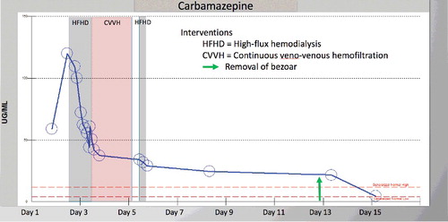
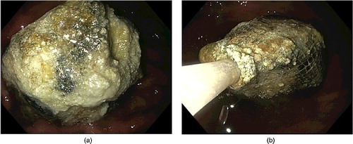
On day 12 after admission, the patient was still in coma with persistently high serum concentrations of carbamazepine. An esophagogastroduodenoscopy (EGD) was performed and revealed an encrusted, crateriform pharmacobezoar 5 cm in diameter, in the gastric antrum. Using a Roth Net and an esophageal overtube (), the bezoar was extracted, and on laboratory analysis was shown to contain 9.9 g of carbamazepine. Following bezoar removal, serum concentrations of carbamazepine declined rapidly with recovery of consciousness. He was discharged to a mental health facility 23 days after admission, without any permanent neurological deficit or other sequelae.
Figure 2. (a) Carbamazepine bezoar seen on esophagogastroduodenoscopy in situ measuring 5 cm in diameter, 9.9 g by weight on laboratory analysis. (b) The pharmacobezoar was fragmented and captured using a Roth Net. Three passes of the endoscope were required and an esophageal overtube was used to facilitate the complete retrieval of pharmacobezoar.
Discussion
Establishing the presence of a pharmacobezoar requires a high index of suspicion. In most cases, there are no physical findings or imaging modalities that reliably detect the presence of a pharmacological bezoar [Citation2]. On rare occasions, bezoars may cause gastrointestinal complications including gastric outlet obstruction, pneumatosis intestinalis, pancreatitis, bleeding and perforation [Citation2,Citation3]. Treatment is highly dependent on the specific drug that is involved [Citation3]. Consideration should be given to the toxicity of the ingested agent, the size and location of the bezoar, patient-specific factors () and the degree of illness.
Table 1. Patient-specific factors associated with bezoar formation [Citation2,Citation3,Citation10–12].
There is no antidote available for carbamazepine overdose. Basic medical management consists of supportive care, prevention of further absorption and extracorporeal elimination [Citation4]. The gastrointestinal absorption rate and plasma half-life are highly unpredictable [Citation5]. In life-threatening carbamazepine overdose, oral administration of multiple doses of activated charcoal is recommended and has been shown to decrease absorption, reduce plasma half-life, and improve clinical outcomes [Citation6]. Extracorporeal elimination can achieve rapid and substantial removal of carbamazepine, and is usually recommended in the setting of overdose unresponsive to conventional treatment [Citation6]. Charcoal hemoperfusion is effective in severe overdose and, when combined with hemodialysis, may result in a 50% reduction in the serum concentrations [Citation7]. When charcoal hemoperfusion is not available, HFHD is an effective extracorporeal elimination alternative [Citation8].
Gastrointestinal endoscopy is indicated if bezoar formation is suspected after an ingested toxin or medication overdose is not effectively treated by less invasive means. Endoscopy allows for direct visualization of a bezoar and for diagnostic certainty by obtaining samples to determine its chemical composition. In addition, endoscopy may shorten hospital stay and reduce the need for repeated laboratory testing [Citation3]. Endoscopic retrieval is limited to the upper and lower GI tract and may be accomplished by direct endoscopic retrieval, retrieval following mechanical fragmentation, or retrieval following chemical dissolution [Citation2]. The immediate risks include gastrointestinal hemorrhage and perforation. Bezoar fragmentation may be accomplished by use of water jets, direct suction, compression by forceps, snares, neodymium–yttrium–aluminum garnet lasers, or bezotriptors [Citation2]. Clearing of large solid fragments may be accomplished using a Roth Net or a Dormia basket, which preserves the integrity of the sample. Safe retrieval at EGD may be facilitated by the use of an overtube which reduces the risk of esophageal injury and aspiration. Allowing a large fragmented pharmacobezoar to pass through the gastrointestinal tract allows ongoing drug absorption and is undesirable. To date, there are no reports of using a single- or double-balloon enteroscope for the retrieval of pharmacobezoars in the jejunum or ileum.
The clinical persistence of signs of toxic pharmacological effects, with sustained, high, rebounding, or rising serum concentrations of drug, should raise suspicion of the presence of clumps of medication or bezoar formation in the bowel, especially when these signs persist despite enhanced elimination treatment [Citation9]. In such situations, endoscopic investigation is minimally invasive, carries little risk, and may be warranted to assess for the presence of pharmacobezoar.
Acknowledgments
The authors thank Denis M. McCarthy, MD, Larry Crocket, MD, Christopher Bustos, Joshua Jones, Lorraine Kronowit, and Christine Molina, TriCore Reference Laboratory.
Disclosure statement
None of the authors have any conflicts of interest to disclose.
References
- Williams R. The fascinating history of bezoars. Med J Aust. 1985;145:613–614.
- Eng K, Kay M. Review: gastrointestinal bezoars: history and current treatment paradigms. Gastroenterol Hepatol. 2012;8:776–778.
- Höjer J, Personne M. Endoscopic removal of slow release clomipramine bezoars in two cases of acute poisoning. Clin Toxicol. 2008;46:317–319.
- Ghannoum M, Yates C, Galvao TF, et al. Extracorporeal treatment for carbamazepine poisoning: systematic review and recommendations from the EXTRIP workgroup. Clin Toxicol. 2014;52:993–1004.
- Graudins A, Peden G, Dowsett R. Massive overdose with controlled‐release carbamazepine resulting in delayed peak serum concentrations and life‐threatening toxicity. Emerg Med. 2002;14:89–94.
- Brahmi N, Kouraichi N, Thabet H, et al. Influence of activated charcoal on the pharmacokinetics and the clinical features of carbamazepine poisoning. Am J Emerg Med. 2006;24:440–443.
- Bock E, Keller F, Heitz J, et al. Treatment of carbamazepine poisoning by combined hemodialysis/hemoperfusion. Int J Clin Pharmacol Ther Toxicol. 1989;27:490–492.
- Sikma MA, van den Broek MPH, Meulenbelt J. Increased unbound drug fraction in acute carbamazepine intoxication: suitability and effectiveness of high-flux haemodialysis. Intensive Care Med. 2012;38:916–917.
- Sethna M, Solomon G, Cedarbaum J, et al. Successful treatment of massive carbamazepine overdose. Epilepsia. 1989;30:71–73.
- Dimarino AJ, Benjamin SB, editors. Gastrointestinal disease: an endoscopic approach. Thorofare (NJ): Slack Incorporated; 2002. p. 555–556.
- Sanders M. Bezoars: from mystical charms to medical and nutritional management. Pract Gastroenterol. 2004;28:37–50.
- Kement M, Ozlem N, Colak E, et al. Synergistic effect of multiple predisposing risk factors on the development of bezoars. World J Gastroenterol. 2012;18:960–964.