
Abstract
To date, increasing numbers of myositis-specific autoantibodies (MSAs) have been reported and their clinical significance has been elucidated. Anti-aminoacyl-tRNA synthetase (ARS) and anti-melanoma-differentiation associated gene 5 (MDA5) are strongly associated with interstitial lung disease (ILD); however, the clinical course of ILD is different depending on which autoantibody is present. Anti-ARS is associated with chronic and repetitive ILD and anti-MDA5 is associated with rapidly progressive ILD. Anti-MDA5, anti-transcriptional intermediary factor (TIF) 1-γ, anti-nuclear matrix protein (NXP) 2 and anti-Mi-2 antibodies are dermatomyositis specific. Anti-TIF1-γ and anti-NXP-2 antibodies are associated with malignancy, and anti-signal recognition particle (SRP) and anti-3-hydroxy-3-methylglutaryl-coenzyme A reductase (HMGCR) antibodies are associated with immune-mediated necrotizing myopathy (IMNM). Prognosis is also different among MSA-positive patient groups. Thus, MSAs are of great use for predicting disease course, prognosis, and determining therapeutic strategy as well as the diagnosis of idiopathic inflammatory myopathy (IIM) patients. Investigation of the pathogenic role of MSAs and their corresponding autoantigens will help us to understand the pathophysiology of IIM and identify new therapeutic targets.
1. Introduction
Idiopathic inflammatory myopathies (IIMs) are a heterogeneous group of autoimmune diseases that affect skeletal muscle and various systemic organs, including the skin, lung, heart and joints. The subcategorization of IIMs was formed on the basis of clinical phenotype or histopathological character consisting of polymyositis (PM), dermatomyositis (DM), amyopathic dermatomyositis (ADM), inclusion body myositis (IBM) and immune-mediated necrotizing myopathy (IMNM). However, there are a variety of phenotypes, organ involvement and disease courses even within the subcategories, suggesting the heterogeneity of pathophysiology. To date, various autoantibodies have been detected in IIMs, termed myositis-specific autoantibodies (MSAs) and myositis-associated autoantibodies (MAAs), and their clinical significance in terms of clinical symptoms, complications, reactivity to treatment and prognosis has been elucidated. Thus, MSAs/MAAs can help subcategorize IIM patients, determine their prognosis and their clinical management.
2. Myositis-specific autoantibodies
Myositis-specific autoantibodies and their corresponding autoantigens, frequency, and clinical significance are listed in . MSAs are useful for identifying patients at risk of interstitial lung disease (ILD), malignancy and necrotizing myopathy (). Anti-aminoacyl-tRNA synthetase (ARS) and anti-melanoma-differentiation associated gene 5 (MDA5) are strongly associated with ILD. Anti-transcriptional intermediary factor (TIF) 1-γ and anti-nuclear matrix protein (NXP) 2 are associated with malignancy. Anti-signal recognition particle (SRP) and anti-3-hydroxy-3-methylglutaryl-coenzyme A reductase (HMGCR) are associated with IMNM. MSAs are detected in 45–85% of adult IIM patients and in 50–70% of juvenile IIM (JIIM) patients [Citation1–8]. In adult patients, anti-ARSs are most frequently detected (20–40% of IIM cases) followed by anti-MDA5 (8–20%), anti-SRP (3–10%), anti-TIF1-γ (5–7%), anti-Mi-2 (3–8%) and anti-NXP2 (5–8%) [Citation2,Citation9]. The most frequent MSAs in JIIM are anti-NXP2 (15–23%), anti-TIF1-γ (15–35%) and anti-MDA5 (7–28%); in contrast, anti-ARSs are relatively rare (2–8%) [Citation5].
Figure 1. Schematic subcategorization of patients with different myositis-specific autoantibodies (MSAs). Because IIM patients with different MSAs have common clinical characteristics, each MSA-positive patient can be determined to be a subset of IIM. The horizontal axis shows the spectrum of skin disease and muscle involvement, and the vertical axis shows the acute or chronic type of ILD.
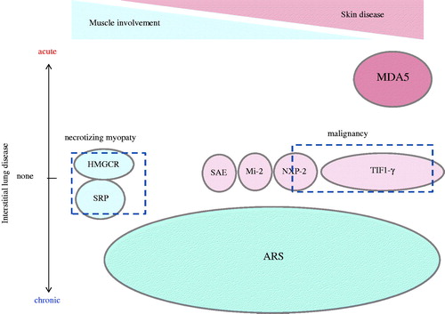
Table 1. The frequency and significance of myositis-specific autoantibodies in adult IIM patients.
2.1. Anti-ARS
Anti-aminoacyl-tRNA synthetase are enzymes that catalyze the binding of amino acids to corresponding transfer RNAs to form aminoacyl-tRNAs in an energy-dependent manner. Eight different ARSs have been identified as autoantigens of MSAs, including anti-Jo-1 (histidyl-tRNA synthetase) [Citation10,Citation11], anti-PL-7 (threonyl) [Citation12], anti-PL-12 (alanyl) [Citation13], anti-EJ (glycyl) [Citation14], anti-OJ (isoleucyl) [Citation15], anti-KS (asparaginyl) [Citation16], anti-tyrosyl [Citation17] and anti-Zo (phenylalanyl) tRNA synthetase antibodies [Citation18]. Among them, anti-Jo-1 is the most common antibody, found in 15–25% of PM/DM patients, whereas all other anti-ARSs are found in 0–7% of cases [Citation19–21]. Anti-ARS-positive patients develop common clinical manifestations, characterized by myositis, ILD, arthritis, fever, Raynaud’s phenomenon and mechanic’s hand, which is called ‘anti-synthetase syndrome (ASS)’ [Citation22]. Anti-ARSs have a strong association with ILD because 70–95% of anti-ARS-positive patients [Citation21,Citation23,Citation24] have ILD and 40–55% of PM/DM patients with ILD are positive for anti-ARSs [Citation2,Citation25]. Anti-ARS-positive ILD tends to be diagnosed at the same time or even before the development of myositis [Citation22]. Indeed, anti-ARSs are detected in 5–10% of idiopathic interstitial pneumonia (IIP) patients [Citation24,Citation26]. Although anti-ARS-positive patients develop clinical manifestations similar to ASS, some differences in clinical manifestations among patients with different anti-ARS antibodies have been described. Anti-PL-12, -OJ and -KS antibodies are associated with ILD rather than myositis [Citation16,Citation21,Citation27]. Other studies showed that arthritis and myopathy were more frequent in patients with anti-Jo-1 antibodies rather than with anti-non-Jo-1-ARS antibodies [Citation23,Citation28,Citation29]. In addition, survival is longer for anti-Jo-1 patients compared with anti-non-Jo-1-ARS patients [Citation28,Citation29]. ILD in anti-ARS-positive patients tends to be chronic progressive. The most frequent pattern of high-resolution computed tomography (HRCT) findings is nonspecific interstitial pneumonia (NSIP), whereas usual interstitial pneumonia (UIP) patterns with honeycombing are rare [Citation24,Citation30]. Glucocorticoids (GC) are the empirical first-line therapy because both myositis and ILD in anti-ARS-positive patients respond well to initial treatment with GC [Citation22,Citation30]. However, additional immunosuppressive agents are often necessary because both myositis and ILD recur when GC is tapered and repetitive recurrence is associated with the chronic deterioration of muscle strength and pulmonary function [Citation22,Citation30,Citation31].
2.2. Anti-MDA5
Anti-MDA5, first reported to be a specific autoantibody for clinically ADM (CADM), and named as anti-CADM-140 in 2005 [Citation32]. Subsequently, the target autoantigen was identified as melanoma differentiation-associated gene 5 (MDA5), also known as interferon-induced with helicase C domain protein 1 (IFIH1) [Citation33,Citation34]. MDA5 is a cytoplasmic retinoic acid-inducible gene-I (RIG-I)-like receptor that upon the recognition of viral RNA induces the expression of type 1 interferon and other inflammatory cytokines [Citation35]. Anti-MDA5 is a DM-specific autoantibody detected in 20–50% of Asian adult DM patients (including CADM) and is associated with relatively lower creatine kinase (CK) levels, a high frequency of ILD (90–95%) especially rapidly progressive ILD (RP-ILD) (50–80%), and poor prognosis because of respiratory failure [Citation32,Citation34,Citation36–38]. However, these characteristics are not the same (lower frequency of the antibody and RP-ILD) in American and European cohorts [Citation39–42]. Such clinical discrepancies may be explained by differences in ethnicity or environmental background [Citation43–45]. Anti-MDA5 antibody was also reported in juvenile DM (JDM) cohorts and the clinical characteristics were similar to those of adult patients with anti-MDA5 in Japan [Citation46,Citation47]. However, in a European cohort, the frequency and characteristics of anti-MDA5-positive JDM was different from Japanese JDM cohort as seen in adult cohorts [Citation48]. Anti-MDA5-positive patients have distinctive features related to physical manifestations as well as blood test and radiological findings. Elevated hepatobiliary enzymes and serum ferritin levels are often seen in anti-MDA5-positive DM patients compared with anti-MDA5-negative DM patients. Moreover, serum ferritin levels correlate with the activity of ILD [Citation34,Citation49]. Several reports suggested that anti-MDA5 titers could be used to monitor disease activity and were good predictors of relapse in anti-MDA5-positive DM [Citation50,Citation51]. Because anti-MDA5 is strongly associated with RP-ILD, patients with anti-MDA5 have the poorest prognosis in IIM (). High levels of initial serum ferritin, alveolar-arterial oxygen gradient (P[A-a]O2), and score of ground-glass opacity (GGO) involving the right middle lobe were reported to be poor prognostic factors in anti-MDA5-positive patients [Citation52]. Increased serum IL-6, IL-8 and IL-10 levels were associated with RP-ILD in PM/DM, and high concentrations of IFN-α and soluble CD163, and the upregulation of IFN-inducible genes were reported in anti-MDA5-positive patients [Citation53–57]. These findings suggest the upregulation of the type 1 IFN system via activation of monocytes, macrophages, or other immunocompetent cells in the pathophysiology of anti-MDA5-positive dermatomyositis with RP-ILD, although further investigation is needed. Recently, the efficacy of intensive immunosuppressive therapy combining several immunosuppressants, including GC, calcineurin inhibitors and intravenous cyclophosphamide, from the early stage of the disease were reported [Citation2]. However, some anti-MDA5-positive patients are resistant to intensive immunosuppressive treatments. Therefore, further investigation and the establishment of other therapeutic strategies for anti-MDA5-positive patients are required.
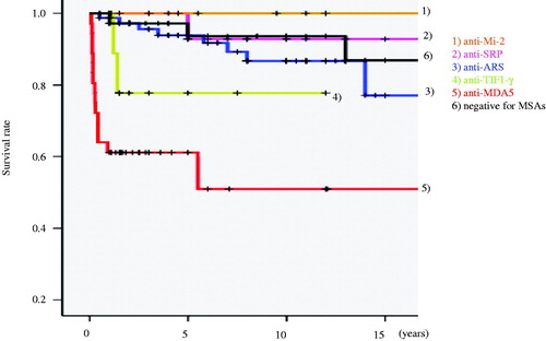
Figure 2. The survival rate of patient groups with different myositis-specific autoantibodies (MSAs). Overall, 245 Japanese IIM patients who visited our department were divided into groups, myositis-specific autoantibody-positive subsets (anti-Mi-2, anti-SRP, anti-ARS, anti-TIF1-γ, anti-MDA5 and negative for MSAs), and their survival rate was plotted.
2.3. Anti-Mi-2
Anti-Mi-2 is a dermatomyositis-specific autoantibody. Its target antigens are Mi-2α (240 kDa) and Mi-2β (218 kDa), which form a protein complex with histone deacetylases, termed nucleosome remodeling deacetylase complex. Anti-Mi-2 is associated with relatively mild myositis, fewer complications in ILD or cardiac disease, and a fair response to treatment with a favorable prognosis [Citation58–60]. There are several interesting reports on the relationship between exposure to ultraviolet (UV) light and anti-Mi-2-positive dermatomyositis. Love et al. [Citation61] suggested that UV radiation intensity was associated with the incidence of DM and anti-Mi-2-positivity. Burd et al. [Citation62] suggested that UV light-induced the upregulation of Mi-2 in human keratinocytes. Because anti-Mi-2 have an association with HLA-DR7, genetic background, as well as environmental factors, may influence the development of anti-Mi-2-positive DM [Citation19].
2.4. Anti-TIF1
Anti-TIF1 autoantibodies were first reported as anti-p155 by Targoff et al. and as anti-155/140 by Kaji et al. These studies suggested that the antibodies immunoprecipitated a 155 kDa protein often with a 140 kDa protein by 35S-methionine-labeled protein immunoprecipitation using HeLa cells or K562 cells. Subsequently, the 155 kDa autoantigen was identified as transcription intermediary factor 1 (TIF1) γ. Moreover, Fujimoto et al. showed the 140 kDa antigen was TIF1α, and that anti-TIF1β (120 kDa protein) was present in some DM patients with or without anti-TIF1 α/γ. Anti-TIF1 autoantibodies are specifically found in DM (15–20% of adult DM and 20% of JDM cases) [Citation5,Citation8,Citation63,Citation64]. Anti-TIF1γ has a positive association with malignancy, especially when it appears with anti-TIF1α, and a negative association with ILD [Citation64]. A meta-analysis based on six studies including 312 adult DM patients suggested that the pooled sensitivity of anti-TIF1γ for diagnosing cancer-associated DM was 78% (95% CI 45–94%), specificity was 89% (95% CI 82–93%), and the diagnostic OR was 27.26 (95% CI 6.59–112.82%) [Citation65]. However, Fujimoto et al. showed there was no malignancy in anti-TIF1-positive patients under 40 years of age although anti-TIF1γ/α was significantly associated with malignancy in patients aged over 40 years [Citation64], suggesting a lower risk of malignancy in JDM and young adults. Another clinical characteristic of anti-TIF1γ is the high risk of dysphagia [Citation66,Citation67]. Mugii et al. [Citation67] reported that 13 out of 92 Japanese adult DM patients showed dysphagia and 11 (84.6%) were positive for anti-TIF1γ. Multivariate analysis showed that significant risk factors for the development of dysphagia in DM were internal malignancy (odds ratio 22.2), anti-TIF-1γ Ab (odds ratio 11.8) and MMT score of sternomastoid muscle (odds ratio 0.46) [Citation67]. In another study, high-resolution manometry showed that Jackhammer esophagus could be seen in 30% of anti-TIF1γ-positive IIM patients, suggesting dysphagia may be caused by skeletal muscle weakness in the neck and by esophageal dysmotility [Citation66].
2.5. Anti-NXP2
Anti-NXP2 was originally reported as anti-MJ, which targeted a 140 kDa protein in a US cohort of JDM patients [Citation68]. Anti-NXP2 is also specific for DM (6–30% in adult DM and about 20% in JDM) [Citation5,Citation8,Citation68–70]. Espada et al. [Citation71] suggested an association between anti-NXP2 and a significant compromise of functional status characterized by muscle contractures and atrophy in an Argentine pediatric cohort. Two other independent cohorts in France and the UK showed that anti-NXP2 was associated with a severe subtype of JDM and developed a severe muscular disease with a lower remission rate [Citation72,Citation73]. Another significant clinical factor of anti-NXP2 in JDM is a high frequency of calcinosis [Citation73,Citation74]. Tansley et al. [Citation73] demonstrated that anti-NXP2 substantially increased the risk of calcinosis across all ages of JDM although younger age at disease onset was associated with an increased risk of calcinosis.
Anti-NXP2 in adult patients was first reported in a UK cohort [Citation70], where anti-NXP-2-positive patients had frequent systemic involvement including weight loss or fever and ILD as well as typical skin lesions of DM, but rarely showed calcinosis. However, an Italian adult cohort suggested that anti-NXP-2-positive patients showed calcinosis more often and heart or lung involvement more rarely than anti-NXP-2-negative PM/DM patients [Citation69]. In a Japanese adult cohort, anti-NXP-2-positive patients had no association with calcinosis or ILD [Citation75,Citation76]. Recent reports suggested an association between anti-NPX-2 and malignancy in adults [Citation75,Citation77]. Ichimura et al. [Citation75] demonstrated that 3 (38%) of 8 anti-NXP-2-positive IIM patients had internal malignancies within 3 years of the diagnosis of IIM. Fiorentino et al. [Citation77] further confirmed an association between anti-NXP-2 and malignancy demonstrating that 9 (31%) of 29 cancer-associated dermatomyositis patients were positive for anti-NXP-2 and had an increased frequency of cancer in anti-NXP-2-positive DM patients of 24% (9 of 37) compared with DM patients without anti-NXP-2 or anti-TIF1-γ of 5% (5 of 96).
2.6. Anti-SAE
Anti-SAE was originally described by Betteridge et al. [Citation78] in a Caucasian European myositis cohort. The corresponding autoantigen is a small ubiquitin-like modifier activating enzyme [Citation78]. This autoantibody is specific for DM, occurring in 6–8% of DM cases in a European cohort [Citation78–80]. The frequency is lower in Asian cohorts, at 1.5–3% of adult DM patients [Citation81,Citation82]. Anti-SAE-positive patients often present as CADM initially and then progress to develop myositis with a high frequency of systemic features, including dysphagia that occurs in 30–78% of anti-SAE-positive patients [Citation79,Citation81,Citation82]. In European cohorts, anti-SAE had no relationship with ILD [Citation79], but was more common in Asian cohorts (60–70%) [Citation81,Citation82]. Fujimoto et al. [Citation81] suggested that ILD of anti-SAE-positive patients in a Japanese cohort was generally mild and responded well to therapy. These differences in frequency and phenotype of patients may be related to ethnicity. Regarding genetic background, the HLA-DQB1*03 haplotype was suggested to be a risk factor for anti-SAE-positivity [Citation79].
2.7. Anti-SRP
Anti-SRP antibody was described by Reeves et al. in 1986 and Okada et al. in 1987 [Citation83,Citation84]. SRP is a cytoplasmic small-RNA-protein complex involved in regulating the translocation of newly synthesized proteins across the endoplasmic reticulum membrane. Anti-SRP is detected in 4–15% of adult IIM [Citation85–87], and is associated with PM, especially severe muscle weakness resulting in marked disability, dysphagia and highly elevated levels of serum creatinine kinase, with relatively acute onset [Citation85,Citation87,Citation88]. Moreover, anti-SRP was associated with a low prevalence of ILD, arthritis and Raynaud’s phenomenon [Citation85]. Anti-SRP-positive IIM patients are usually resistant to standard treatment with corticosteroids (CS) and show frequent exacerbation [Citation85–87]. Several studies demonstrated the association of anti-SRP with a histological subset of IMNM [Citation88–90]. The prevalence of necrotizing myopathy in anti-SRP-positive IIM ranges between 70–100% [Citation88–90] and 13–54% of necrotizing myopathy cases are positive for anti-SRP [Citation91,Citation92]. The difference in the frequency of anti-SRP may be related to genetic or environmental factors, selection bias of patients, or different detection systems (immunoprecipitation, enzyme-linked immunosorbent assay or line immunoassay). In JIIM, anti-SRP was detected in 2–4% of cases, which is lower than that observed for adult patients [Citation5,Citation8]. According to a literature review [Citation93], anti-SRP-positive JIIM patients were associated with severe proximal weakness in the absence of typical DM skin lesions. Each of Raynaud’s phenomena, including arthritis and dysphagia, were reported in 38% of cases, and histologically, muscle necrosis without lymphocytic infiltrate was common (63%). The tendency for a poor response to standard initial treatment with CS with or without methotrexate and a generally poor outcome resulting in disability was reported in anti-SRP-positive JIIM patients as well as in adult IIM patients [Citation93].
2.8. Anti-HMGCR
Anti-HMGCR was originally reported as an anti-200/100-kDa antibody specific for IMNM [Citation94], and subsequently, its corresponding autoantigen of approximately 100 kDa was identified as HMGCR [Citation95]. The approximately 200 kDa antigen was suggested to be a dimer or an associated protein of HMGC R [Citation95]. Anti-HMCGR-positive patients are characterized by proximal weakness, high creatine kinase (CK) levels, irritable myopathy on electromyography (EMG), and prominent degenerating, regenerating and/or necrotic fibers without significant inflammatory infiltrates. Notably, exposure to statins prior to the onset of weakness was found in 38–63% of anti–HMGCR-positive patients [Citation94,Citation96,Citation97], and this was more pronounced in older patients [Citation95]. Anti-HMGCR titers were associated with CK levels before and after treatment and were inversely associated with muscle strength [Citation98]. Regarding prognosis, Tiniakou et al. [Citation98] reported that anti-HMGCR-positive patients found it difficult to recover the full strength of muscles with immunosuppressive therapy and that over 50% of those who recovered full strength of their muscles continued to have CK levels in excess of 500 IU/L. Moreover, younger patients had more a severe disease and worse prognosis compared with older patients [Citation98].
Recently, the pathogenic role of autoantibodies in IMNM was suggested. Arouche-Delaperche et al. [Citation99] showed that anti-SRP and anti-HMGCR-induced muscle fiber atrophy and impairment of muscle regeneration in vitro. Indeed, the expression of SRP and HMGCR proteins was observed on regenerating muscle fibers in vitro [Citation100] and in vivo [Citation95,Citation101]. Allenbach et al. [Citation101] demonstrated that SRP and HMGCR proteins were present on the sarcolemma of regenerating fibers from autoantibody-positive patients and that anti-SRP and anti-HMGCR from patients recognized their cognate antigens on the sarcolemma. In addition, sarcolemma C5b-9 deposits were suggested to correlate with muscle fiber necrosis and activation of the classical complement pathway [Citation101]. Thus, anti-SRP and anti-HMGCR may have pathogenic roles in IMNM via activation of the classical complement pathway.
2.9. Other autoantibodies
Anti-cytosolic 5′ nucleotidase 1A (cN1A) was described in 2011 by Salajegheh et al. [Citation102] as an autoantibody against a 43 kDa protein associated with inclusion body myositis (IBM). The corresponding autoantigen was identified as cN1A expressed in skeletal muscle [Citation103,Citation104]. This autoantigen has a role in the hydrolysis of adenosine monophosphate leading to the physiological homeostasis of energy, metabolic regulation and cell replication. Anti-cN1A is present in about 33–34% of IBM, 4–5% of PM and 3–4% of DM cases [Citation103,Citation104]. Because Herbert et al. [Citation105] showed that this autoantibody was present in 36% of Sjogren’s syndrome and 20% of SLE cases, the specificity of this autoantibody for myositis is probably low. However, it might help differentiate between myositis subgroups. In a recent study, the pathogenic role of anti-cN1A in IBM was demonstrated both in vivo and in an in vitro passive immunization model suggesting that the autoantibody might affect protein degradation in myofibers [Citation106].
Anti-DNA mismatch repair enzymes (MMREs) were first described in 2001 [Citation107]. Humans have seven MMREs, MSH2, MSH3, MSH6, MLH1, MLH3, PMS1 and PMS2. Anti-MMREs were found in 6% of IIM cases in a Japanese cohort, but were restricted to MLH1, PMS1, MSH2 and PMS2 with simultaneous positivity for more than one autoantibody occurring in some patients [Citation108]. To date, specific characteristics for individual clinical IIM subtypes have not been determined in anti-MMREs-positive IIM patients, but their association with a relatively good prognosis has been suggested [Citation108].
In 2017, Hosono et al. [Citation109] reported a new autoantibody against splicing factor proline/glutamine-rich (SFPQ), which was only found in 57% of anti-MDA5-positive patients. Interestingly, anti-SFPQ was detected at diagnosis in some cases but in other cases, it appeared during the disease course or even after intensive immunosuppressive treatment. Compared with anti-MDA5-positive patients, anti-SFPQ was associated with older age at onset, high frequency of mechanic’s hand, and low frequency of arthritis. Moreover, the maximum KL-6 levels of anti-SFPQ-positive patients were significantly higher than those of anti-SFPQ-negative patients, although no significant difference was found in prognosis between the two groups. There seemed to be seasonality in the disease onset according to the detection timing of anti-SFPQ [Citation109].
3. Conclusion
In 2017, a new classification criterion for adult and juvenile IIM was established by the European League Against Rheumatism (EULAR)/American College of Rheumatology (ACR) and its high performance in classifying IIM patients was reported [Citation110]. However, among MSAs, only anti-Jo-1 antibody was included as a variable in the criteria [Citation110]. Indeed, MSAs may not be necessary in the classification of IIM, but they are useful for the subclassification of IIM, predicting prognosis, and determining the management of classified IIM patients. Investigation of the pathophysiologic role of MSAs and their corresponding autoantigens may help us to elucidate the pathogenicity of IIM and potential therapeutic targets.
Disclosure statement
R.N. collaborated with Medical Biological Laboratories Co., Ltd. to develop detection systems for myositis-specific autoantibodies.
References
- McHugh NJ, Tansley SL. Autoantibodies in myositis. Nat Rev Rheumatol. 2018;14:290–302.
- Nakashima R, Hosono Y, Mimori T. Clinical significance and new detection system of autoantibodies in myositis with interstitial lung disease. Lupus. 2016;25:925–933.
- Yang H, Peng Q, Yin L, et al. Identification of multiple cancer-associated myositis-specific autoantibodies in idiopathic inflammatory myopathies: a large longitudinal cohort study. Arthritis Res Ther. 2017;19:259.
- Rider LG, Shah M, Mamyrova G, et al. The myositis autoantibody phenotypes of the juvenile idiopathic inflammatory myopathies. Medicine (Baltimore). 2013;92:223–243.
- Ueki M, Kobayashi I, Takezaki S, et al. Myositis-specific autoantibodies in Japanese patients with juvenile idiopathic inflammatory myopathies. Mod Rheumatol. 2018. DOI:10.1080/14397595.2018.1452353
- Ceribelli A, Isailovic N, De Santis M, et al. Myositis-specific autoantibodies and their association with malignancy in Italian patients with polymyositis and dermatomyositis. Clin Rheumatol. 2017;36:469–475.
- Srivastava P, Dwivedi S, Misra R. Myositis-specific and myositis-associated autoantibodies in Indian patients with inflammatory myositis. Rheumatol Int. 2016;36:935–943.
- Tansley SL, Simou S, Shaddick G, et al. Autoantibodies in juvenile-onset myositis: their diagnostic value and associated clinical phenotype in a large UK cohort. J Autoimmun. 2017;84:55–64.
- Cavazzana I, Fredi M, Ceribelli A, et al. Testing for myositis specific autoantibodies: comparison between line blot and immunoprecipitation assays in 57 myositis sera. J Immunol Methods. 2016;433:1–5.
- Nishikai M, Reichlin M. Heterogeneity of precipitating antibodies in polymyositis and dermatomyositis. Characterization of the Jo-1 antibody system. Arthritis Rheum. 1980;23:881–888.
- Mathews MB, Bernstein RM. Myositis autoantibody inhibits histidyl-tRNA synthetase: a model for autoimmunity. Nature. 1983;304:177–179.
- Mathews MB, Reichlin M, Hughes GR, et al. Anti-threonyl-tRNA synthetase, a second myositis-related autoantibody. J Exp Med. 1984;160:420–434.
- Bunn CC, Bernstein RM, Mathews MB. Autoantibodies against alanyl-tRNA synthetase and tRNAAla coexist and are associated with myositis. J Exp Med. 1986;163:1281–1291.
- Targoff IN, Trieu EP, Plotz PH, et al. Antibodies to glycyl-transfer RNA synthetase in patients with myositis and interstitial lung disease. Arthritis Rheum. 1992;35:821–830.
- Targoff IN, Trieu EP, Miller FW. Reaction of anti-OJ autoantibodies with components of the multi-enzyme complex of aminoacyl-tRNA synthetases in addition to isoleucyl-tRNA synthetase. J Clin Invest. 1993;91:2556–2564.
- Hirakata M, Suwa A, Nagai S, et al. Anti-KS: identification of autoantibodies to asparaginyl-transfer RNA synthetase associated with interstitial lung disease. J Immunol. 1999;162:2315–2320.
- Betteridge Z, Gunawardena H, North J, et al. Anti-synthetase syndrome: a new autoantibody to phenylalanyl transfer RNA synthetase (anti-Zo) associated with polymyositis and interstitial pneumonia. Rheumatology (Oxford). 2007;46:1005–1008.
- Hashish LTE, Sadanandan P, Targoff IN. Igentification of autoantibodies to tyrosyl-tRNA synthetase in dermatomyositis with features consistent with antisynthetase syndrome [abstract]. Arthritis Rheum. 2005;52:S312.
- Love LA, Leff RL, Fraser DD, et al. A new approach to the classification of idiopathic inflammatory myopathy: myositis-specific autoantibodies define useful homogeneous patient groups. Medicine (Baltimore). 1991;70:360–374.
- Targoff IN. Update on myositis-specific and myositis-associated autoantibodies. Curr Opin Rheumatol. 2000;12:475–481.
- Hamaguchi Y, Fujimoto M, Matsushita T, et al. Common and distinct clinical features in adult patients with anti-aminoacyl-tRNA synthetase antibodies: heterogeneity within the syndrome. PLoS One. 2013;8:e60442.
- Yoshifuji H, Fujii T, Kobayashi S, et al. Anti-aminoacyl-tRNA synthetase antibodies in clinical course prediction of interstitial lung disease complicated with idiopathic inflammatory myopathies. Autoimmunity. 2006;39:233–241.
- Lega JC, Fabien N, Reynaud Q, et al. The clinical phenotype associated with myositis-specific and associated autoantibodies: a meta-analysis revisiting the so-called antisynthetase syndrome. Autoimmun Rev. 2014;13:883–891.
- Nakashima R, Imura Y, Hosono Y, et al. The multicenter study of a new assay for simultaneous detection of multiple anti-aminoacyl-tRNA synthetases in myositis and interstitial pneumonia. PLoS One. 2014;9:e85062.
- Hozumi H, Fujisawa T, Nakashima R, et al. Comprehensive assessment of myositis-specific autoantibodies in polymyositis/dermatomyositis-associated interstitial lung disease. Respir Med. 2016;121:91–99.
- Watanabe K, Handa T, Tanizawa K, et al. Detection of antisynthetase syndrome in patients with idiopathic interstitial pneumonias. Respir Med. 2011;105:1238–1247.
- Friedman AW, Targoff IN, Arnett FC. Interstitial lung disease with autoantibodies against aminoacyl-tRNA synthetases in the absence of clinically apparent myositis. Semin Arthritis Rheum. 1996;26:459–467.
- Aggarwal R, Cassidy E, Fertig N, et al. Patients with non-Jo-1 anti-tRNA-synthetase autoantibodies have worse survival than Jo-1 positive patients. Ann Rheum Dis. 2014;73:227–232.
- Hervier B, Devilliers H, Stanciu R, et al. Hierarchical cluster and survival analyses of antisynthetase syndrome: phenotype and outcome are correlated with anti-tRNA synthetase antibody specificity. Autoimmun Rev. 2012;12:210–217.
- Hozumi H, Enomoto N, Kono M, et al. Prognostic significance of anti-aminoacyl-tRNA synthetase antibodies in polymyositis/dermatomyositis-associated interstitial lung disease: a retrospective case control study. PLoS One. 2015;10:e0120313.
- Takato H, Waseda Y, Watanabe S, et al. Pulmonary manifestations of anti-ARS antibody positive interstitial-pneumonia – with or without PM/DM. Respir Med. 2013;107:128–133.
- Sato S, Hirakata M, Kuwana M, et al. Autoantibodies to a 140-kd polypeptide, CADM-140, in Japanese patients with clinically amyopathic dermatomyositis. Arthritis Rheum. 2005;52:1571–1576.
- Sato S, Hoshino K, Satoh T, et al. RNA helicase encoded by melanoma differentiation-associated gene 5 is a major autoantigen in patients with clinically amyopathic dermatomyositis: association with rapidly progressive interstitial lung disease. Arthritis Rheum. 2009;60:2193–2200.
- Nakashima R, Imura Y, Kobayashi S, et al. The RIG-I-like receptor IFIH1/MDA5 is a dermatomyositis-specific autoantigen identified by the anti-CADM-140 antibody. Rheumatology (Oxford). 2010;49:433–440.
- Kato H, Takeuchi O, Sato S, et al. Differential roles of MDA5 and RIG-I helicases in the recognition of RNA viruses. Nature. 2006;441:101–105.
- Hoshino K, Muro Y, Sugiura K, et al. Anti-MDA5 and anti-TIF1-gamma antibodies have clinical significance for patients with dermatomyositis. Rheumatology (Oxford). 2010;49:1726–1733.
- Chen F, Wang D, Shu X, et al. Anti-MDA5 antibody is associated with A/SIP and decreased T cells in peripheral blood and predicts poor prognosis of ILD in Chinese patients with dermatomyositis. Rheumatol Int. 2012;32:3909–3915.
- Koga T, Fujikawa K, Horai Y, et al. The diagnostic utility of anti-melanoma differentiation-associated gene 5 antibody testing for predicting the prognosis of Japanese patients with DM. Rheumatology (Oxford). 2012;51:1278–1284.
- Fiorentino D, Chung L, Zwerner J, et al. The mucocutaneous and systemic phenotype of dermatomyositis patients with antibodies to MDA5 (CADM-140): a retrospective study. J Am Acad Dermatol. 2011;65:25–34.
- Ceribelli A, Fredi M, Taraborelli M, et al. Prevalence and clinical significance of anti-MDA5 antibodies in European patients with polymyositis/dermatomyositis. Clin Exp Rheumatol. 2014;32:891–897.
- Labrador-Horrillo M, Martinez MA, Selva-O'Callaghan A, et al. Anti-MDA5 antibodies in a large Mediterranean population of adults with dermatomyositis. J Immunol Res. 2014;2014:1.
- Hall JC, Casciola-Rosen L, Samedy LA, et al. Anti-melanoma differentiation-associated protein 5-associated dermatomyositis: expanding the clinical spectrum. Arthritis Care Res (Hoboken). 2013;65:1307–1315.
- Gono T, Kawaguchi Y, Kuwana M, et al. Brief report: association of HLA-DRB1*0101/*0405 with susceptibility to anti-melanoma differentiation-associated gene 5 antibody-positive dermatomyositis in the Japanese population. Arthritis Rheum. 2012;64:3736–3740.
- Muro Y, Sugiura K, Hoshino K, et al. Epidemiologic study of clinically amyopathic dermatomyositis and anti-melanoma differentiation-associated gene 5 antibodies in central Japan. Arthritis Res Ther. 2011;13:R214.
- Hosono Y, Nakashima R, Imura Y, et al. The onsets of myositis with myositis-specific autoantibodies (MSAs) are associated with the seasons. Ann Rheum Dis. 2013;72:656.
- Kobayashi I, Okura Y, Yamada M, et al. Anti-melanoma differentiation-associated gene 5 antibody is a diagnostic and predictive marker for interstitial lung diseases associated with juvenile dermatomyositis. J Pediatr. 2011;158:675–677.
- Ikeda S, Arita M, Morita M, et al. Interstitial lung disease in clinically amyopathic dermatomyositis with and without anti-MDA-5 antibody: to lump or split?. BMC Pulm Med. 2015;15:159.
- Tansley SL, Betteridge ZE, Gunawardena H, et al. Anti-MDA5 autoantibodies in juvenile dermatomyositis identify a distinct clinical phenotype: a prospective cohort study. Arthritis Res Ther. 2014;16:R138.
- Gono T, Kawaguchi Y, Ozeki E, et al. Serum ferritin correlates with activity of anti-MDA5 antibody-associated acute interstitial lung disease as a complication of dermatomyositis. Mod Rheumatol. 2011;21:223–227.
- Matsushita T, Mizumaki K, Kano M, et al. Antimelanoma differentiation-associated protein 5 antibody level is a novel tool for monitoring disease activity in rapidly progressive interstitial lung disease with dermatomyositis. Br J Dermatol. 2017;176:395–402.
- Sato S, Kuwana M, Fujita T, et al. Anti-CADM-140/MDA5 autoantibody titer correlates with disease activity and predicts disease outcome in patients with dermatomyositis and rapidly progressive interstitial lung disease. Mod Rheumatol. 2013;23:496–502.
- Fujiki Y, Kotani T, Isoda K, et al. Evaluation of clinical prognostic factors for interstitial pneumonia in anti-MDA5 antibody-positive dermatomyositis patients. Mod Rheumatol. 2018;28:133–140.
- Gono T, Kaneko H, Kawaguchi Y, et al. Cytokine profiles in polymyositis and dermatomyositis complicated by rapidly progressive or chronic interstitial lung disease. Rheumatology (Oxford). 2014;53:2196–2203.
- Horai Y, Koga T, Fujikawa K, et al. Serum interferon-alpha is a useful biomarker in patients with anti-melanoma differentiation-associated gene 5 (MDA5) antibody-positive dermatomyositis. Mod Rheumatol. 2015;25:85–89.
- Kawasumi H, Gono T, Kawaguchi Y, et al. IL-6, IL-8, and IL-10 are associated with hyperferritinemia in rapidly progressive interstitial lung disease with polymyositis/dermatomyositis. Biomed Res Int. 2014;2014:1.
- Zhang SH, Zhao Y, Xie QB, et al. Aberrant activation of type I interferon system may contribute to the pathogenesis of anti-MDA5 dermatomyositis. Br J Dermatol. 2018. DOI:10.1111/bjd.16917
- Kawasumi H, Katsumata Y, Nishino A, et al. Association of serum soluble CD163 with polymyositis and dermatomyositis, especially in anti-MDA5 antibody-positive cases. J Rheumatol. 2018;45:947–955.
- Targoff IN, Reichlin M. The association between Mi-2 antibodies and dermatomyositis. Arthritis Rheum. 1985;28:796–803.
- Hengstman GJ, Vree Egberts WT, Seelig HP, et al. Clinical characteristics of patients with myositis and autoantibodies to different fragments of the Mi-2 beta antigen. Ann Rheum Dis. 2006;65:242–245.
- Komura K, Fujimoto M, Matsushita T, et al. Prevalence and clinical characteristics of anti-Mi-2 antibodies in Japanese patients with dermatomyositis. J Dermatol Sci. 2005;40:215–217.
- Love LA, Weinberg CR, McConnaughey DR, et al. Ultraviolet radiation intensity predicts the relative distribution of dermatomyositis and anti-Mi-2 autoantibodies in women. Arthritis Rheum. 2009;60:2499–2504.
- Burd CJ, Kinyamu HK, Miller FW, et al. UV radiation regulates Mi-2 through protein translation and stability. J Biol Chem. 2008;283:34976–34982.
- Targoff IN, Mamyrova G, Trieu EP, et al. A novel autoantibody to a 155-kd protein is associated with dermatomyositis. Arthritis Rheum. 2006;54:3682–3689.
- Fujimoto M, Hamaguchi Y, Kaji K, et al. Myositis-specific anti-155/140 autoantibodies target transcription intermediary factor 1 family proteins. Arthritis Rheum. 2012;64:513–522.
- Trallero-Araguas E, Rodrigo-Pendas JA, Selva-O'Callaghan A, et al. Usefulness of anti-p155 autoantibody for diagnosing cancer-associated dermatomyositis: a systematic review and meta-analysis. Arthritis Rheum. 2012;64:523–532.
- Casal-Dominguez M, Pinal-Fernandez I, Mego M, et al. High-resolution manometry in patients with idiopathic inflammatory myopathy: elevated prevalence of esophageal involvement and differences according to autoantibody status and clinical subset. Muscle Nerve. 2017;56:386–392.
- Mugii N, Hasegawa M, Matsushita T, et al. Oropharyngeal dysphagia in dermatomyositis: associations with clinical and laboratory features including autoantibodies. PLoS One. 2016;11:e0154746.
- Oddis CVFN, Goel A, Espada G, et al. Clinical and serological characterization of the anti-MJ antibody in childhood myositis [abstract]. Arthritis Rheum. 1997;40:S139.
- Ceribelli A, Fredi M, Taraborelli M, et al. Anti-MJ/NXP-2 autoantibody specificity in a cohort of adult Italian patients with polymyositis/dermatomyositis. Arthritis Res Ther. 2012;14:R97.
- Betteridge ZEGH, Chinoy H, et al. Autoantibodies to the p140 autoantigen NXP-2 in adult dermatomyositis. Arthritis Rheum. 2009;60:S304.
- Espada G, Maldonado Cocco JA, Fertig N, et al. Clinical and serologic characterization of an Argentine pediatric myositis cohort: identification of a novel autoantibody (anti-MJ) to a 142-kDa protein. J Rheumatol. 2009;36:2547–2551.
- Aouizerate J, De Antonio M, Bader-Meunier B, et al. Muscle ischaemia associated with NXP2 autoantibodies: a severe subtype of juvenile dermatomyositis. Rheumatology (Oxford). 2018;57:873–879.
- Tansley SL, Betteridge ZE, Shaddick G, et al. Calcinosis in juvenile dermatomyositis is influenced by both anti-NXP2 autoantibody status and age at disease onset. Rheumatology (Oxford). 2014;53:2204–2208.
- Gunawardena H, Wedderburn LR, Chinoy H, et al. Autoantibodies to a 140-kd protein in juvenile dermatomyositis are associated with calcinosis. Arthritis Rheum. 2009;60:1807–1814.
- Ichimura Y, Matsushita T, Hamaguchi Y, et al. Anti-NXP2 autoantibodies in adult patients with idiopathic inflammatory myopathies: possible association with malignancy. Ann Rheum Dis. 2012;71:710–713.
- Ishikawa A, Muro Y, Sugiura K, et al. Development of an ELISA for detection of autoantibodies to nuclear matrix protein 2. Rheumatology (Oxford)). 2012;51:1181.
- Fiorentino DF, Chung LS, Christopher-Stine L, et al. Most patients with cancer-associated dermatomyositis have antibodies to nuclear matrix protein NXP-2 or transcription intermediary factor 1γ. Arthritis Rheum. 2013;65:2954–2962.
- Betteridge Z, Gunawardena H, North J, et al. Identification of a novel autoantibody directed against small ubiquitin-like modifier activating enzyme in dermatomyositis. Arthritis Rheum. 2007;56:3132–3137.
- Betteridge ZE, Gunawardena H, Chinoy H, et al. Clinical and human leucocyte antigen class II haplotype associations of autoantibodies to small ubiquitin-like modifier enzyme, a dermatomyositis-specific autoantigen target, in UK Caucasian adult-onset myositis. Ann Rheum Dis. 2009;68:1621–1625.
- Bodoki L, Nagy-Vincze M, Griger Z, et al. Four dermatomyositis-specific autoantibodies-anti-TIF1gamma, anti-NXP2, anti-SAE and anti-MDA5-in adult and juvenile patients with idiopathic inflammatory myopathies in a Hungarian cohort. Autoimmun Rev. 2014;13:1211–1219.
- Fujimoto M, Matsushita T, Hamaguchi Y, et al. Autoantibodies to small ubiquitin-like modifier activating enzymes in Japanese patients with dermatomyositis: comparison with a UK Caucasian cohort. Ann Rheum Dis. 2013;72:151–153.
- Ge Y, Lu X, Shu X, et al. Clinical characteristics of anti-SAE antibodies in Chinese patients with dermatomyositis in comparison with different patient cohorts. Sci Rep. 2017;7:188.
- Reeves WH, Nigam SK, Blobel G. Human autoantibodies reactive with the signal-recognition particle. Proc Natl Acad Sci USA. 1986;83:9507–9511.
- Okada N, Mimori T, Mukai R, et al. Characterization of human autoantibodies that selectively precipitate the 7SL RNA component of the signal recognition particle. J Immunol. 1987;138:3219–3223.
- Targoff IN, Johnson AE, Miller FW. Antibody to signal recognition particle in polymyositis. Arthritis Rheum. 1990;33:1361–1370.
- Hirakata M, Mimori T, Akizuki M, et al. Autoantibodies to small nuclear and cytoplasmic ribonucleoproteins in Japanese patients with inflammatory muscle disease. Arthritis Rheum. 1992;35:449–456.
- Watanabe Y, Uruha A, Suzuki S, et al. Clinical features and prognosis in anti-SRP and anti-HMGCR necrotising myopathy. J Neurol Neurosurg Psychiatry. 2016;87:1038–1044.
- Hengstman GJ, ter Laak HJ, Vree Egberts WT, et al. Anti-signal recognition particle autoantibodies: marker of a necrotising myopathy. Ann Rheum Dis. 2006;65:1635–1638.
- Miller T, Al LMT, Lopate G, et al. Myopathy with antibodies to the signal recognition particle: clinical and pathological features. J Neurol Neurosurg Psychiatry. 2002;73:420–428.
- Takada T, Hirakata M, Suwa A, et al. Clinical and histopathological features of myopathies in Japanese patients with anti-SRP autoantibodies. Mod Rheumatol. 2009;19:165.
- Suzuki S, Yonekawa T, Kuwana M, et al. Clinical and histological findings associated with autoantibodies detected by RNA immunoprecipitation in inflammatory myopathies. J Neuroimmunol. 2014;274:202–208.
- Wang L, Liu L, Hao H, et al. Myopathy with anti-signal recognition particle antibodies: clinical and histopathological features in Chinese patients. Neuromuscul Disord. 2014;24:335–341.
- Binns EL, Moraitis E, Maillard S, et al. Effective induction therapy for anti-SRP associated myositis in childhood: a small case series and review of the literature. Pediatr Rheumatol Online J. 2017;15:77.
- Christopher-Stine L, Casciola-Rosen LA, Hong G, et al. A novel autoantibody recognizing 200-kd and 100-kd proteins is associated with an immune-mediated necrotizing myopathy. Arthritis Rheum. 2010;62:2757–2766.
- Mammen AL, Chung T, Christopher-Stine L, et al. Autoantibodies against 3-hydroxy-3-methylglutaryl-coenzyme A reductase in patients with statin-associated autoimmune myopathy. Arthritis Rheum. 2011;63:713–721.
- Watanabe Y, Suzuki S, Nishimura H, et al. Statins and myotoxic effects associated with anti-3-hydroxy-3-methylglutaryl-coenzyme A reductase autoantibodies: an observational study in Japan. Medicine (Baltimore). 2015;94:e416.
- Musset L, Allenbach Y, Benveniste O, et al. Anti-HMGCR antibodies as a biomarker for immune-mediated necrotizing myopathies: a history of statins and experience from a large international multi-center study. Autoimmun Rev. 2016;15:983–993.
- Tiniakou E, Pinal-Fernandez I, Lloyd TE, et al. More severe disease and slower recovery in younger patients with anti-3-hydroxy-3-methylglutaryl-coenzyme A reductase-associated autoimmune myopathy. Rheumatology (Oxford). 2017;56:787–794.
- Arouche-Delaperche L, Allenbach Y, Amelin D, et al. Pathogenic role of anti-signal recognition protein and anti-3-Hydroxy-3-methylglutaryl-CoA reductase antibodies in necrotizing myopathies: myofiber atrophy and impairment of muscle regeneration in necrotizing autoimmune myopathies. Ann Neurol. 2017;81:538–548.
- Rojana-Udomsart A, Mitrpant C, Bundell C, et al. Complement-mediated muscle cell lysis: a possible mechanism of myonecrosis in anti-SRP associated necrotizing myopathy (ASANM). J Neuroimmunol. 2013;264:65–70.
- Allenbach Y, Arouche-Delaperche L, Preusse C, et al. Necrosis in anti-SRP + and anti-HMGCR + myopathies: role of autoantibodies and complement. Neurology. 2018;90:e507–e517.
- Salajegheh M, Lam T, Greenberg SA. Autoantibodies against a 43 kDa muscle protein in inclusion body myositis. PLoS One. 2011;6:e20266.
- Larman HB, Salajegheh M, Nazareno R, et al. Cytosolic 5'-nucleotidase 1A autoimmunity in sporadic inclusion body myositis. Ann Neurol. 2013;73:408–418.
- Pluk H, van Hoeve BJ, van Dooren SH, et al. Autoantibodies to cytosolic 5'-nucleotidase 1A in inclusion body myositis. Ann Neurol. 2013;73:397–407.
- Herbert MK, Stammen-Vogelzangs J, Verbeek MM, et al. Disease specificity of autoantibodies to cytosolic 5'-nucleotidase 1A in sporadic inclusion body myositis versus known autoimmune diseases. Ann Rheum Dis. 2016;75:696–701.
- Tawara N, Yamashita S, Zhang X, et al. Pathomechanisms of anti-cytosolic 5'-nucleotidase 1A autoantibodies in sporadic inclusion body myositis. Ann Neurol. 2017;81:512–525.
- Casciola-Rosen LA, Pluta AF, Plotz PH, et al. The DNA mismatch repair enzyme PMS1 is a myositis-specific autoantigen. Arthritis Rheum. 2001;44:389–396.
- Muro Y, Nakashima R, Hosono Y, et al. Autoantibodies to DNA mismatch repair enzymes in polymyositis/dermatomyositis and other autoimmune diseases: a possible marker of favorable prognosis. Arthritis Rheumatol. 2014;66:3457–3462.
- Hosono Y, Nakashima R, Serada S, et al. Splicing factor proline/glutamine-rich is a novel autoantigen of dermatomyositis and associated with anti-melanoma differentiation-associated gene 5 antibody. J Autoimmun. 2017;77:116–122.
- Lundberg IE, Tjarnlund A, Bottai M, et al. 2017 European League against Rheumatism/American College of Rheumatology classification criteria for adult and juvenile idiopathic inflammatory myopathies and their major subgroups. Arthritis Rheumatol. 2017;69:2271–2282.