
Abstract
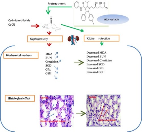
Cadmium contributes to nephrotoxicity linked with oxidative stress in humans and animals. This study used Atorvastatin to examine its effect on cadmium chloride-induced nephrotoxicity in a rat model using biochemical and histological methodologies. Experiments were performed on 56 adult male Wistar rats (200 ± 20 g), randomly assigned to eight groups. Rats in Group A received physiologic saline. Group B was treated with a dosage of 20 mg/ kg body weight/day AT for 15 days. Groups C, D, and E received CdCl2 with dosages of 1, 2, and 3 mg/kg body weight, respectively. Groups F, G, and H were pretreated with Atorvastatin, 30 min prior to the administration of CdCl2. Rats received intra-gastric Atorvastatin for 15 days during which cadmium chloride was given from days 8 to 15. On day 16, blood samples were collected, and kidneys were excised to evaluate the biochemical and histopathological changes. Cadmium chloride significantly increased malondialdehyde (MDA), serum creatinine (Cr), blood urea nitrogen (BUN), and decreased superoxide dismutase (SOD), glutathione (GSH), and glutathione peroxidase (GPx) levels. Administration of Atorvastatin (20 mg/kg) significantly decreased lipid peroxidation, BUN and Cr, while it significantly increased glutathione and antioxidant enzymes activity. Atorvastatin improved the histological changes and all biochemical markers and shed light on its protecting role against cadmium chloride-induced oxidative stress in the kidney.
GRAPHICAL ABSTRACT
Introduction
Cadmium (Cd) is frequently used in various industrial processes and is considered as one of the toxic metals to the environment. Occupational and environmental exposures to Cd are linked to industrial activities (Mezynska and Brzóska Citation2018). The kidney is known as the main target organ for chronic Cd exposure (Rana et al. Citation2018). The biological half-life of Cd in humans reported 10–30 years in the kidney cortex which may account for a higher occurrence of nephrotoxicity (Moitra et al. Citation2014). In the kidney, the accumulation of 50 μg Cd/g, wet tissue weight resulted in renal dysfunction (Satarug et al. Citation2000).
Evidence suggests that Cd absorption may result in oxidative stress. Cd-induced oxidative injury includes a difference between the production and removal of reactive oxygen species (ROS) in the kidney. Furthermore, the production and accumulation of ROS are associated with hydrogen peroxide (H2O2), hydroxyl radical (OH−), superoxide anions, singlet oxygen, lipid hydroperoxides, and phospholipid hydroperoxides (Patra et al. Citation2011) causing inflammation and injury in the cell membrane, enzymatic pathways, and connective tissue structures (Gabr et al. Citation2019). Cadmium chloride (CdCl2)-induced renal dysfunction is most likely the result of an oxidant/antioxidant imbalance in the kidney since it was parallel with elevated LPO and NO levels, which could be considered as signals of increased generation of reactive oxygen species (ROS) and reactive nitrogen species (RNS) (Almeer et al. Citation2019). Moreover, a decrease in antioxidant activity may have a pathological role in biological reactions and Cd-induced nephrotoxicity (Şişman et al. Citation2003). Cd is filtered by the glomeruli and is then reabsorbed by the epithelial cells of the proximal tubule, possessing a potent toxic effect on the kidney (Yang and Shu Citation2015; Rana et al. Citation2018).
Cd generates physiological and biochemical alterations in human and animal kidneys (Renugadevi and Prabu Citation2009; Patra et al. Citation2011; Bernhoft Citation2013). Cd administration in rats resulted in a significant decrease of antioxidant indicators such as superoxide dismutase (SOD), glutathione peroxidase (GPx), catalase (CAT), and glutathione (GSH), and a rise in malondialdehyde (MDA) peroxidation marker in kidney and pancreas (Hormozi et al. Citation2018; Andjelkovic et al. Citation2019; Aja et al. Citation2020; Aja et al. Citation2020; Evcimen et al. Citation2020). Chronic exposure to cadmium chloride may attribute to some morphological and metabolic alterations, causing an increased dose-dependent in blood urea nitrogen (BUN) and Creatinine in rats (Renugadevi and Prabu Citation2009). Meanwhile, Long-term Cd exposure can develop tissue necrosis in rats (Erboga et al. Citation2016; Kim et al. Citation2018).
Atorvastatin (AT), with the chemical formula: C33H35FN2O5 and molecular weight: of 558.6, is the leading antioxidant of the class of statins and has been shown to possess anti-inflammatory, antioxidant (10 mg/kg, for 10 days) (Pal et al. Citation2015), antithrombotic (10 mg/kg/day) (Ozbek et al. Citation2009), antiplatelet, immune-modulatory (40 mg for 4 weeks) (Fuentes-Orozco et al. Citation2018), and antitumor effects in experimental animal models (Garjani et al. Citation2012). Doses of Statin compounds at 20 mg/day have reduced inflammation by HMG-CoA reductase inhibitors, lowered low-density lipoprotein (LDL) cholesterol in plasma, and promoted antioxidant effects by suppressing oxidation pathways (Goodarzi et al. Citation2016; Wang et al. Citation2017). AT is a potent HMG-CoA reductase inhibitor and is effective in lipid profiles, lipid peroxidation, and antioxidant systems (Yan et al. Citation2020). AT has ameliorated renal dysfunction (El-Moselhy and El-Sheikh Citation2014), morphological changes (Yan et al. Citation2020), and oxidative stress injuries in rats (Wu et al. Citation2014).
Overall, the widespread emission of Cd has increased exposure in the working and general population and demanded further research to prevent health-related hazards. Although researchers have indicated the antioxidant potential of AT, but few experimental studies have investigated and reported the protective role of Atorvastatin on toxic metals in an animal model with renal failure. As per our knowledge, the protective role of AT on Cadmium-induced nephrotoxicity in rats has not been demonstrated. Therefore, the main goal of the current experiment was to investigate in vivo biochemical and histological changes induced by CdCl2 on the kidney of an adult rat model. We evaluated the possible role of Atorvastatin in protecting CdCl2-induced renal toxicity in rats and in ameliorating the renal oxidative stress biomarkers and histological changes in rats. Confirming the hypothesis that oxidative stress contributes to Cd intoxication and alteration in kidney tissue, AT may shed light on the prevention of Cd poisoning.
Materials and methods
Experimental animals
We used adult Wistar male rats (n = 56) weighing between 200 and 220 g. The animals were kept in metal cages under hygienic conditions and maintained at 22 ± 2°C and 12-h light-dark cycles with free access to food and water. Rats were acclimatized for one week before treatment commenced.
Drug and chemicals
AT was purchased from Tehran Chemie pharmaceutical Co. Cadmium chloride (CdCl2) was obtained from Merck (Darmstadt, Germany). Biochemical markers were assessed using Rat SOD, GPx, MDA, and GSH ELISA Kits from ZellBio, GmbH, (Germany) as instructed by the manufacturer. BUN and Creatinine in the kidney tissues were detected as instructed by kits obtained from Pars Azemoon, Tehran, (Iran). Purchased Chemicals were of standard grade and purity for performing experimental tests.
Ethics
Prior to the experiment, we reviewed and ensured that the protocols were in accordance with the guidelines of animal acts proposed by the institutional Ethical Review Board of Semnan University of Medical Sciences (IR.SEMUMS.REC.1395.177).
Animal groupings and treatments
Rats were pretreated with AT (20-mg/kg body weight) dissolved in saline solution and gastric gavage at a volume of 4 ml/kg. The given dose of AT in our experiment was based on previous reports of the antioxidant effects of the drug (Sun et al. Citation2009; Goodarzi et al. Citation2020). In a dose-response experiment, rats received doses of CdCl2 at 1, 2, and 3 mg/kg dissolved in distilled water at an intra-peritoneal volume of 2-ml/kg produced a dose-related increase in the frequency of histological and biochemical alterations in kidneys (Golbaghi et al. Citation2019). From the first day through the fifteenth day, rats received intra-gastric gavage of AT. In addition, from day 8 to day 15 rats received intraperitoneal injections of CdCl2. Then, on the 16th day, biochemical and histological analyzes were performed. The timeline of experimental treatments is shown in Figure .
Figure 1. The time schedule for experimental rat treatments.
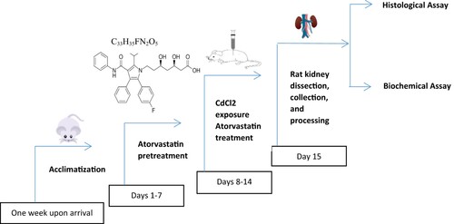
The Wistar rats were randomly assigned into eight groups of seven rats in each group. Group A of rats received physiologic saline. Group B was treated with oral gavages a dosage of 20 mg/ kg body weight/day AT for 15 days. Groups C, D, and E received intra-peritoneal (i.p.) CdCl2 with doses of 1, 2 and 3 mg/kg, respectively. Groups F, G, and H were pretreated with oral gavages containing 20 mg AT/kg body weight, 30 min prior to the intra-peritoneal (i.p.) administration of CdCl2 at 1, 2, and 3 mg/kg. All animals received CdCl2 from day 8 to day 15. After 24 h of the last administration, blood samples were taken from the heart under anesthesia with sodium pentobarbital (50 mg/kg). Excised Kidneys were washed thoroughly with saline solution. Obtained parts of kidney tissues were fixed and examined by light microscopy, using the hematoxylin and eosin (H&E) staining technique and 0.5 × 0.5-cm2 slices of additional half were cut for the measurement of GSH, SOD, MDA, and GPx.
Biochemical analyzes
The samples of kidney tissue were washed with saline. Then, the right kidneys were homogenized with phosphate buffered solution for the determination of tissue oxidative and anti-oxidative markers. The supernatants were prepared by centrifuging the homogenates at 800× g for 10 min at 4°C and preserving the samples at −80°C. The activities of GSH, MDA, GPx, and SOD were measured in the obtained supernatants (Yang and Shu Citation2015). Serum was separated from blood samples by centrifugation at 3000 rpm for 20 min and stored at −20°C until the measurement of parameters. Serum levels of BUN, and CR levels and were assessed by using standard assay kits. These parameters were measured using a colorimetric kit (Pars Azemoon, Tehran, Iran) as prescribed in the manufacturer’s instructions provided with commercial kits. The serum level of Cr was measured by Jaffe’s method (Brouwers et al. Citation2013).
Thiobarbituric acid reaction with MDA Assay kit estimated the level of lipid peroxidation. This results in the formation of a red complex product that was read with a NanoDrope Spectrophotometer at 532 nm. The detection limit of MDA was 0.1 µM (Pirmoradi et al. Citation2019). An assay kit (Zellbio Co) was used to measure the activity of SOD in kidney tissue. Measurement of SOD was based on an enzyme reacting with superoxide anion to produce oxygen and hydrogen peroxide. GSH and GPx were quantified by colorimetric method at 412 nm, using chemical assay kits ZellBio GmbH, (Ulm, Germany) with a 0.1 mM detection limit (Sheikh Citation2016). The content of renal Gpx was assessed by measuring NADH catalyzed one micromole GSH per minute to Oxidized glutathione (GSSG). The protein content of supernatant was assayed according to the method described by the Bradford method using standard bovine serum albumin at 560 nm (Verdi et al. Citation2005).
Histological analyzes
The left kidneys were fixed in buffered formalin (10%) for 48 h and embedded in paraffin wax. Slices of 10 μm were cut and placed on glass slides. After making and drying kidney tissue slides, they were stained with the hematoxylin and eosin (H&E) method. Kidney cell injury was examined based on dilated nuclei, loss of staining capacity, and swelling of kidney tubular cells. Five fields of each slide were randomly selected and photographed under a magnification of ×400 and evaluated by a pathologist and a histologist.
Statistical analyzes
Statistical analyzes were performed using the Graphpad Prism 8 software (San Diego, CA, USA). Experimental data were processed to present the Standard Error Means. Data were analyzed by one-way analysis of variance (ANOVA), and Tukey’s multiple comparisons tests were performed to compare the means of all groups to the mean of every other group. Differences were statistically considered significant at P < .05.
Results
Effects of CdCl2 and Atorvastatin on kidney MDA level
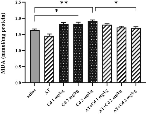
MDA level in the rat kidney homogenates increased significantly in the group treated with CdCl2 compared to the control group A (Group E: CdCl2, 3 mg/ kg/day, **P = .0011, and Group D: CdCl2, 2 mg/kg/ day *P = .0428). AT administered at a dosage of 20 mg/kg/day significantly decreased MDA level in CdCl2 treated rats (Group H: CdCl2, 3 mg/kg/day + AT 20 mg/kg/day, *P = .0268) as illustrated in Figure .
Figure 2. Effects of AT on MDA levels in kidney tissues of rats treated with CdCl2 at the dosages of 1, 2, and 3 mg/kg. Administration of CdCl2 (2 and 3 mg/kg) significantly increased levels of MDA in serum compared to the rats received saline. Pretreatment with AT decreased the effect of CdCl2 (3 mg/kg). Presented figures are mean ± S.E.M (n = 7) 0.05999. *P < .05, **P < .01.
Effects of treatments on antioxidant enzymes
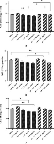
The activities of non-enzymatic antioxidants (GSH) and, enzymatic (SOD and GPx) in the kidney of rats are shown in Figures (a–c).
Figure 3. (a). Effects of AT on GSH levels in kidney of rats received CdCl2 (1, 2, and 3 mg/kg). Administration of CdCl2 (3 mg/kg) significantly lowered the content of GSH. Pretreatment of AT significantly suppressed the CdCl2-induced effects of 2 and 3 mg/kg doses. Values are mean ± S.E.M (n = 7) 0.7959. *P < .05, **P < .01. (b). Effects of AT on SOD activity in the kidney tissues of rats received CdCl2 (1, 2, and 3 mg/kg). CdCl2 (3 mg/kg) significantly decreased the content of SOD compared to the saline-treated rats. AT pretreatment improved SOD at the level of the control group and significantly suppressed the effect of CdCl2 (1 mg/kg). Results are mean ± S.E.M (n = 7) 0.4352. *P < .05, **P < .01.3.2.3. Glutathione peroxidase (GPx). (c). Effects of AT on GPx activity in renal tissues of rats received CdCl2 with doses of 1, 2, and 3 mg/kg. Data showed no significant difference in GPx levels between rats pretreated with AT and the rats received the various doses of CdCl2. Results are mean ± S.E.M (n = 7) 0.7233. **P = .0078, *P = .0138.
Glutathione GSH
The effect of CdCl2 on GSH levels and treatment with a combination of CdCl2 plus AT on GSH concentration of rat renal tissues is depicted in Figure (a). The administration of CdCl2 significantly decreased GSH antioxidant compared to control values in kidney (Group E: CdCl2, 3 mg/kg/day, **P = .0078). AT in combination with CdCl2 significantly increased kidney GSH content compared to CdCl2 treated rats (Group G: CdCl2, 2 mg/kg/day + AT 20 mg/kg/day, *P = .0401 and Group H: CdCl2, 3 mg/kg/day + AT 20 mg/kg/day, *P = .0390).
Superoxide dismutase (SOD)
In rats given CdCl2 at a dose of 3 mg/kg, SOD activity significantly lowered compared to the control group (**P = .0056). Equally, pretreatment of AT (20 mg/kg/day) significantly increased SOD enzymatic activity (*P = .0323) compared to CdCl2-treated rats (Group C: 1 mg/kg/day) as depicted in Figure (b).
The effect of CdCl2 on rat kidney tissue GPx activity and treatment with a combination of CdCl2 plus AT on the rat tissue GPx activity is depicted in Figure (c). Administration of CdCl2 significantly reduced GPx concentration in kidney compared to control values (Group D: Cd 2 mg/kg/day, *P = .0138, and Group E: Cd 3 mg/kg/day, **P < .0078). The combination of CdCl2 plus AT increased kidney GPx content compared to CdCl2 treated rats, but the difference was not significant.
Effects of treatments on the serum level of BUN
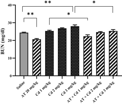
A dosage of 3-mg/kg CdCl2 significantly increased BUN compared to the rats treated with saline (**P < .01) but not significantly at a dose of 2 mg as shown in Figure . Pretreatment with AT (20 mg/kg/day) significantly decreased BUN level in Cd-induced changes of 1 and 3 mg/kg (*P < .05) and control group (***P < .01) but not significantly at Cd dosage of 2 mg/kg.
Figure 4. Effects of AT on BUN concentrations in the rats exposed to CdCl2 with doses 1,2, and 3 mg/kg. Administration of CdCl2 (3 mg/kg) significantly increased the level of BUN. AT pretreatment significantly decreased BUN compared to saline and CdCl2-treated rats (1 and, 3 mg/kg). Figures are mean ± S.E.M (n = 7) (0.8043). Saline and AT ***P = .0009, CdCl2 3 mg/kg and saline **P = .0015, CdCl2 1 mg/kg and AT + CdCl2 1 mg/kg *P = .0123, CdCl2 3 mg/kg and AT + CdCl2 3 mg/kg *P = .0378.
Effects of CdCl2 and AT on the serum level of creatinine
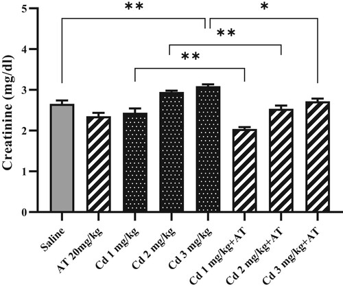
Figure shows the status of creatinine serum levels of control group and experimental group. CdCl2 administration (Group E: 3 mg/kg/day) significantly increased creatinine in the serum as compared to control rats (**P = .0029). Increased levels of creatinine due to CdCl2 challenge were significantly decreased at doses of 1, 2, and 3 mg/kg/day upon the pre-treatment with Atorvastatin 20 mg/kg/day (Group F: CdCl2 1 mg/kg + AT **P = .0072, Group G: CdCl2 2 mg/kg + AT **P = .0048, Group H: CdCl2 3 mg/kg + AT *P = .0155).
Figure 5. Effects of AT on creatinine in rat kidney tissues exposed to CdCl2. Administration of CdCl2 (3 mg/kg), induced significant increase in creatinine level compared to the rats treated with saline and AT pretreatment significantly decreased creatinine and the effect of CdCl2 (1, 2 and 3 mg/kg) . Results are presented as mean ± S.E.M (n = 7) 0.1018. *P < .02, **P < .01.
Histological changes in the rat kidney
The Histological examination on the renal tissues of the control rats demonstrated normal architecture and regularly arranged kidney tissue cells in both lateral cortex and medullary segments, respectively. In the central part of the tissue, normal epithelial cells were arranged in the collecting duct. The appearance of epithelial cells and glomerular size were observed normally in the proximal convoluted tubule and the distal convoluted tubule (Figure (a)).
Figure 6. (a). Light microscopy of rat renal tissue of the control group under normal saline treatment illustrating the healthy architecture of Bowman’s capsule (black arrow), glomeruli, distal tubules, and collector duct. (b). Light microscopy of rat kidney received CdCl2 (3 mg/kg) displaying renal damages: degeneration of glomeruli (G), hemorrhage (H), deposited epithelial cells in collector duct (C). (c). Light microscopy of rat kidney structure following pretreatment of AT (20 mg/kg) 30 min before administration of CdCl2. AT protective effect on CdCl2- induced kidney damage, exhibiting normal kidney tissue structure with glomeruli, renal distal convoluted, and collecting duct.
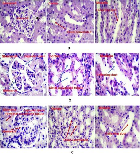
The kidney tissue of the CdCl2-treated (does of 3 mg/kg) rats group demonstrated severe histological damages, reduced renal glomerular size, and induced hemorrhage inside the Bowman's capsule. Furthermore, lymphocytic cells increased in the renal tissues, and the death of epithelial cells in the wall of Bowman's capsule was observed in glomeruli. Disrupted epithelial cells were deposited in the collector duct. In the central part of the tissue, the number of renal cells in the proximal and distal convoluted tubules was indistinguishable, indicating histopathological alterations in the cellular structure. Further, in rats treated with CdCl2 (3 mg/kg), the aggregation of interstitial lymphocyte infiltration was increased in renal tubular cells, and vacuolated cytoplasm was observed, which ultimately led to cellular death (Figure (b)). In the central part of the tissue, abnormal epithelial cells were placed in the collection duct.
CdCl2 – induced histopathological changes were reduced in the kidney of rats following AT (20 mg/kg/day) pretreatment. The results of this study in the rat groups treated with CdCl2 and AT 20 mg/kg/day pretreatment showed that Bowman's capsule wall epithelial cells in the kidney glomeruli had a normal appearance. Bowman space is slightly larger in the distance between the epithelial cells and the vessel wall thicker than in the control groups. The percentage of lymphocyte cells in tissue was normal and interstitial hemorrhage was not observed. In the cortical tissue, the entire complex tubes were seen around and near the epithelial wall and the cells were aligned. The epithelial cells near the convoluted tubules and the surrounding tubules were normal (Figure (c)).
Discussion
This in-vivo experimental study examined the potential effects of AT on CdCl2-induced kidney toxicity in rats. Our results showed that administration of CdCl2 significantly increased MDA levels, and decreased the enzymatic and non-enzymatic antioxidants in the kidney of rats. Correspondingly, Renugadevi and Prabu (Citation2009) suggested oxidative stress as the main mechanism of acute Cd toxicity. Prior studies reported significantly increased MDA levels in the kidney from CdCl2 -administered rats (Gabr et al. Citation2019; Mohammed and Hashem Citation2019). In addition, Mohammed and Hashem (Citation2019) reported that CdCl2 (5 mg/kg/day) orally for 30 days significantly increased MDA, lowered activity of GSH, and histopathological changes in the rat. Our results suggest that CdCl2 administration resulted in the reduction of renal GSH concentration as compared to the control group, which is in agreement with the previous results obtained by Xiao et al. (Citation2002) and Messaoudi et al. (Citation2009). These findings agreed with previous study on cadmium in male jewelry manufacturing workers, indicating decrease in plasma antioxidant enzymes, and an increase in MDA and erythrocyte instability (Moitra et al. Citation2014).
Our results showed the administration of CdCl2 significantly decreased GPx (doses of 2 and3 mg/kg), SOD, and GSH (dose of 3 mg/kg) activities in the kidney compared to a control group and resulted in oxidative stress in the rat kidney that was reflected by the renal histopathological and biochemical changes. Our results were in agreement with a rat experimental study by Adi et al. (Citation2016) concluded that Cd exposure (20 mg/kg body weight for 30 days) meaningfully reduced CAT, GR, SOD, and GPx activities and improved LPx and GST activities. Also, Hormozi et al. (Citation2018) demonstrated that concurrent workplace exposure to lead and cadmium in the tile industry might result in a remarkable increase in lipid peroxidation, and alter antioxidant enzymes (CAT, SOD, and GPx) and oxidative stress.
In our study, the functional nephrotoxicity was indexed through BUN and creatinine levels, which were increased in CdCl2-treated rats as matched to control rats. Our results confirm the study by Wallin et al. (Citation2014) revealed a link between Cd levels in urine and blood with exposure to Cd and chronic kidney disease. Our findings also correlated with the results of Andjelkovic et al. (Citation2019) that suggested Cd (15 and 30 mg/kg BW) significantly increased BUN and Cr compared to a control group CdCl2- induced oxidative stress in rat kidneys. Another study showed that BUN and serum creatinine levels significantly reduced following administration of CdCl2 (25 mg/kg, orally for 7 days) in rats (Kim et al. Citation2018). Previous studies have shown severe histological changes in the kidney of CdCl2-treated rats (Gabr et al. Citation2019; Mohammed and Hashem Citation2019). Besides, in our study CdCl2 exposure induced toxic histophatological injuries to the renal and declined glomerular function and progressive renal failure in the kidney, which were consistent with the findings reported by Gabr et al. (Citation2019), Renugadevi and Prabu (Citation2009), and Mohammed and Hashem (Citation2019).
In our experiment, the pretreatment of AT before CdCl2 poisoning improved biochemical parameters. Previous study showed a protective effect of AT on antioxidant enzymes and inhibits the reduction of endogenous antioxidant enzymes (Ozbek et al. Citation2009). In this study, taking AT (20 mg/kg/day) plus CdCl2 produced much biochemical and pathological impact in rat kidneys. However, conflicting results have been reported regarding the effect of AT on renal tissue. Some studies find the effect toxic and others find it useful and supportive (Mehrzadi et al. Citation2016; Nasri et al. Citation2016). Mehany et al. (Citation2013) reported the effect of pretreatment with AT and vitamin E on rat kidneys. They suggested that in potassium dichromate-induced nephrotoxicity (15 mg/kg) in rats, pretreatment with vitamin E (200 mg/k) and Atorvastatin (10 mg/kg/day for 14 days) resulted in lowered toxicity and as well as improvement of kidney histopathological changes. In another study, Talebpour Amiri et al. (Citation2018) reported that AT administration produced a significant protective effect against radiation-induced nephrotoxicity.
A study reported opposite results showed that administration of AT (30 mg/kg/day for 8 weeks) induced adverse effects in renal tissues and a post-treatment of Arjunolic acid (20 mg/kg for 4 days) and vitamin C might protect kidney from AT-induced severe tissue toxicity (Pal et al. Citation2015). Three major differences between our findings and the results of Pal et al. (Citation2015) could be due to the discrepancy between dose, exposure time, and animals. In the current study, rats were exposed to 20 mg/kg AT for 15 days, while in Pal et al. (Citation2015) study mice were given 30 mg/kg/day AT for 8 weeks.
Our results highlighted that the administration of CdCl2 in the rat model may lead to nephrotoxicity. Histological examination showed many alterations in renal tissue structure following exposure to CdCl2 (3 mg/kg) (Figure (b)). These results are consistent with those of Mohammed and Hashem (Citation2019) who described CdCl2 (5 mg/kg b.w, orally for 4 weeks) induced glomerular injury, acute dilatation of Bauman’s capsule, congestion of the renal blood vessels, and injury to glomerular epithelial in rat.
A previous report concluded that CdCl2 administration (5 mg/kg for 30 days) indicated an adverse effect on cortical blood flow and renal parenchyma replacement with numerous lymphocytes infiltrates, and dilation of glomeruli in rats (Gabr et al. Citation2019). Also, El-Sokkary et al. (Citation2010) reported that CdCl2 (5-mg /kg b.w for 22 days) is associated with spaces separating the cortical tissue, attributed to the cellular degeneration and interstitial edema, mesangial cell proliferative glomerulonephritis in rat, and dilation of Bowman's spaces.
According to our study, CdCl2 administration (1, 2 mg/kg) did not show histopathological disturbance in the kidney tissue (figures not shown).
Our results indicated that cadmium-induced nephrotoxicity pretreatment of AT (20 mg/kg/day) significantly improved concentrations of SOD, GSH, and GPx in rat kidneys and significantly decreased MDA, BUN, and Creatinine contents. The results of this study confirm previous studies, suggesting the antioxidant effect of AT (Ozbek et al. Citation2009; Ghelani et al. Citation2019). Additionally, our findings suggest that the administration of AT might reduce cadmium-induced tissue damage. Similar to our findings El-Moselhy and El-Sheikh (Citation2014) showed AT intake (10 mg/kg for 10 days) produced a renal protective effect against doxorubicin (15 mg/kg for 5 days) induced nephrotoxicity.
Conclusion
This study indicated that varying doses of CdCl2 caused oxidative stress and accounted for decreasing enzymatic activity in neutralizing free radicals and histological changes. Our results highlighted AT might protect rats against CdCl2-induced oxidative stress. Workers must be informed about the potential health effects related to Cd exposure. We proposed that AT might be clinically relevant to CdCl2-induced renal disorders due to its potential therapeutic use in industrial workers.
Institutional Review Board Statement
The study was conducted in accordance with the animal study protocol approved by the Ethics Committee of Semnan University of Medical Sciences (Ethics approval code IR.SEMUMS.REC.1395.177).
Authors contributions
AD, EK, ZG contributed to conception, design, drafting, and revising of the study. AG, ARB, and SY conducted experiments, and AD, EK, ZG, AG, and ARB performed data analyzes and interpretations. AD, EK, and ZG contributed to writing and revising the manuscript. AD supervised the study and final approval of the version to be published. The authors declare that all data were generated in-house and that no paper mill was used.
Disclosure statement
No potential conflict of interest was reported by the author(s).
Data availability statement
Data supporting reported results in this study can be found at https://data.mendeley.com/datasets/fknzy9twbz/5.
Additional information
Funding
References
- Adi PJ, Burra SP, Vataparti AR, Matcha B. 2016. Calcium, zinc and vitamin E ameliorate cadmium-induced renal oxidative damage in albino Wistar rats. Toxicol Rep. 3:591–597. doi:10.1016/j.toxrep.2016.07.005.
- Aja PM, Ekpono EU, Awoke JN, Famurewa AC, Izekwe FI, Okoro EJ, Okorie CF, Orji CL, Nwite F, Ale BA, et al. 2020. Hesperidin ameliorates hepatic dysfunction and dyslipidemia in male Wistar rats exposed to cadmium chloride. Toxicol Rep. doi:10.1016/j.toxrep.2020.09.014.
- Aja PM, Izekwe FI, Famurewa AC, Ekpono EU, Nwite FE, Igwenyi IO, Awoke JN, Ani OG, Aloke C, Obasi NA, et al. 2020. Hesperidin protects against cadmium-induced pancreatitis by modulating insulin secretion, redox imbalance and iNOS/NF-ĸB signaling in rats. Life Sci. doi:10.1016/j.lfs.2020.118268.
- Almeer RS, AlBasher GI, Alarifi S, Alkahtani S, Ali D, Abdel Moneim AE. 2019. Royal jelly attenuates cadmium-induced nephrotoxicity in male mice. Sci Rep. 9:5825. doi:10.1038/s41598-019-42368-7.
- Andjelkovic M, Buha Djordjevic A, Antonijevic E, Antonijevic B, Stanic M, Kotur-Stevuljevic J, Spasojevic-Kalimanovska V, Jovanovic M, Boricic N, Wallace D, et al. 2019. Toxic effect of acute cadmium and lead exposure in rat blood, liver, and kidney. Int J Environ Res Public Health. 16:274. doi:10.3390/ijerph16020274.
- Bernhoft RA. 2013. Cadmium toxicity and treatment. Sci World J. 2013:1–7. doi:10.1155/2013/394652.
- Brouwers B, Pruniau VPEG, Cauwelier EJG, Schuit F, Lerut E, Ectors N, Declercq J, Creemers JWM. 2013. Phlorizin pretreatment reduces acute renal toxicity in a mouse model for diabetic nephropathy*. J Biol Chem. 288:27200–27207. doi:10.1074/jbc.M113.469486.
- El-Moselhy MA, El-Sheikh AAK. 2014. Protective mechanisms of atorvastatin against doxorubicin-induced hepato-renal toxicity. Biomed Pharmacother. 68:101–110. doi:10.1016/j.biopha.2013.09.001.
- El-Sokkary GH, Nafady AA, Shabash EH. 2010. Melatonin administration ameliorates cadmium-induced oxidative stress and morphological changes in the liver of rat. Ecotoxicol Environ Saf. 73:456–463. doi:10.1016/j.ecoenv.2009.09.014.
- Erboga M, Kanter M, Aktas C, Sener U, Fidanol Erboga Z, Bozdemir Donmez Y, Gurel A. 2016. Thymoquinone ameliorates cadmium-induced nephrotoxicity, apoptosis, and oxidative stress in rats is based on its anti-apoptotic and anti-oxidant properties. Biol Trace Elem Res. 170:165–172. doi:10.1007/s12011-015-0453-x.
- Evcimen M, Aslan R, Gulay MS. 2020. Protective effects of polydatin and grape seed extract in rats exposed to cadmium. Drug Chem Toxicol. 43:225–233. doi:10.1080/01480545.2018.1480629.
- Fuentes-Orozco C, Garcia-Salazar SJ, Gómez-Navarro B, González-Espinoza E, Zepeda-González A, Ramírez-Robles JN, Castañeda-Espinoza R, Yáñez-Sánchez I, Gálvez-Gastelum FJ, Cervantes-Guevara G, et al. 2018. Anti-inflammatory effect of atorvastatin on the kidney graft of living donor transplants. Ann Transplant. 23:442–449. doi:10.12659/AOT.908521.
- Gabr SA, Alghadir AH, Ghoniem GA. 2019. Biological activities of ginger against cadmium-induced renal toxicity. Saudi J Biol Sci. 26:382–389. doi:10.1016/j.sjbs.2017.08.008.
- Garjani A, Rezazadeh H, Andalib S, Ziaee M, Doustar Y, Soraya H, Garjani M, Khorrami A, Asadpoor M, Maleki-Dizaji N. 2012. Ambivalent effects of atorvastatin on angiogenesis, epidermal cell proliferation and tumorgenesis in animal models. Iran Biomed J. 16:59–67. doi:10.6091/ibj.1017.2012.
- Ghelani H, Razmovski-Naumovski V, Inampudi V, Chang D, Nammi S. 2019. Atorvastatin improves hepatic lipid metabolism and protects renal damage in adenine-induced chronic kidney disease in Sprague-Dawley rats. Biomed Res Int. 2019:1–10. doi:10.1155/2019/8714363.
- Golbaghi A, Fouladi Dehagi B, Ahmadizadeh M. 2019. Combined effect of cadmium and noise on rat’s kidney. J Ren Inj Prev. 8:230–234. doi:10.15171/jrip.2019.43.
- Goodarzi Z, Karami E, Ahmadizadeh M. 2016. Simvastatin attenuates chromium-induced nephrotoxicity in rats. J Nephropathol. 6:5–9. doi:10.15171/jnp.2017.02.
- Goodarzi Z, Karami E, Yousefi S, Dehdashti A, Bandegi AR, Ghanbari A. 2020. Hepatoprotective effect of atorvastatin on Cadmium chloride induced hepatotoxicity in rats. Life Sci. 254:117770. doi:10.1016/j.lfs.2020.117770.
- Hormozi M, Mirzaei R, Nakhaee A, Izadi S, Dehghan Haghighi J. 2018. The biochemical effects of occupational exposure to lead and cadmium on markers of oxidative stress and antioxidant enzymes activity in the blood of glazers in tile industry. Toxicol Ind Health. 34:459–467. doi:10.1177/0748233718769526.
- Kim KS, Lim H-J, Lim JS, Son JY, Lee J, Lee BM, Chang S-C, Kim HS. 2018. Curcumin ameliorates cadmium-induced nephrotoxicity in Sprague-Dawley rats. Food Chem Toxicol. 114:34–40. doi:10.1016/j.fct.2018.02.007.
- Mehany HA, Abo-youssef AM, Ahmed LA, Arafa E-SA, Abd El-Latif HA. 2013. Protective effect of vitamin E and atorvastatin against potassium dichromate-induced nephrotoxicity in rats. Beni-Suef Univ J Basic Appl Sci. 2:96–102. doi:10.1016/j.bjbas.2013.02.002.
- Mehrzadi S, Kamrava SK, Dormanesh B, Motevalian M, Hosseinzadeh A, Hosseini Tabatabaei SMT, Ghaznavi H. 2016. Melatonin synergistically enhances protective effect of atorvastatin against gentamicin-induced nephrotoxicity in rat kidney. Can J Physiol Pharmacol. 94:265–271. doi:10.1139/cjpp-2015-0277.
- Messaoudi I, El Heni J, Hammouda F, Saïd K, Kerkeni A. 2009. Protective effects of selenium, zinc, or their combination on cadmium-induced oxidative stress in rat kidney. Biol Trace Elem Res. 130:152–161. doi:10.1007/s12011-009-8324-y.
- Mezynska M, Brzóska MM. 2018. Environmental exposure to cadmium—a risk for health of the general population in industrialized countries and preventive strategies. Environ Sci Pollut Res. 25:3211–3232. doi:10.1007/s11356-017-0827-z.
- Mohammed ET, Hashem KS. 2019. Ameliorative effect of lipoic acid on cadmium induced hepatotoxicity and nephrotoxicity in rats. J Appl Sci. 19:637–646. doi:10.3923/jas.2019.637.646.
- Moitra S, Brashier BB, Sahu S. 2014. Occupational cadmium exposure-associated oxidative stress and erythrocyte fragility among jewelry workers in India. Am J Ind Med. 57:1064–1072. doi:10.1002/ajim.22336.
- Nasri H, Hasanpour Z, Nematbakhsh M, Ahmadi A, Rafieian-Kopaei M. 2016. The effect of the various doses of atorvastatin on renal tubular cells; an experimental study. J Nephropathol. 5:111–115. doi:10.15171/jnp.2016.20.
- Ozbek E, Cekmen M, Ilbey YO, Simsek A, Polat EC, Somay A. 2009. Atorvastatin prevents gentamicin-induced renal damage in rats through the inhibition of p38-MAPK and NF-kB pathways. Ren Fail. 31:382–392. doi:10.1080/08860220902835863.
- Pal S, Sarkar A, Pal PB, Sil PC. 2015. Protective effect of arjunolic acid against atorvastatin induced hepatic and renal pathophysiology via MAPK, mitochondria and ER dependent pathways. Biochimie. 112:20–34. doi:10.1016/j.biochi.2015.02.016.
- Patra RC, Rautray AK, Swarup D. 2011. Oxidative stress in lead and cadmium toxicity and Its amelioration. Vet Med Int. 2011:1–9. doi:10.4061/2011/457327.
- Pirmoradi Z, Yadegari M, Moradi A, Khojasteh F, Mehrjerdi FZ. 2019. Effect of berberine chloride on caspase-3 dependent apoptosis and antioxidant capacity in the hippocampus of the chronic cerebral hypoperfusion rat model. Iran J Basic Med Sci. doi:10.22038/ijbms.2018.31225.7534.
- Rana MN, Tangpong J, Rahman MM. 2018. Toxicodynamics of lead, cadmium, mercury and arsenic-induced kidney toxicity and treatment strategy: a mini review. Toxicol Reports. 5:704–713. doi:10.1016/j.toxrep.2018.05.012.
- Renugadevi J, Prabu SM. 2009. Naringenin protects against cadmium-induced oxidative renal dysfunction in rats. Toxicology. 256:128–134. doi:10.1016/j.tox.2008.11.012.
- Satarug S, Haswell-Elkins MR, Moore MR. 2000. Safe levels of cadmium intake to prevent renal toxicity in human subjects. Br J Nutr. 84:791–802. doi:10.1017/S0007114500002403.
- Sheikh N. 2016. Lipid peroxidation and antioxidant status in patients with medullary thyroid carcinoma: a case-control study. J Clin Diagnostic Res. doi:10.7860/JCDR/2016/17854.7202.
- Şişman AR, Bülbül M, Çoker C, Önvural B. 2003. Cadmium exposure in tobacco workers: possible renal effects. J Trace Elem Med Biol. 17:51–55. doi:10.1016/S0946-672X(03)80046-9.
- Sun Y-M, Tian Y, Li X, Liu Y-Y, Wang L-F, Li J, Li Z-Q, Pan W. 2009. Effect of atorvastatin on expression of IL-10 and TNF-α mRNA in myocardial ischemia–reperfusion injury in rats. Biochem Biophys Res Commun. 382:336–340. doi:10.1016/j.bbrc.2009.03.019.
- Talebpour Amiri F, Hamzeh M, Naeimi RA, Ghasemi A, Hosseinimehr SJ. 2018. Radioprotective effect of atorvastatin against ionizing radiation-induced nephrotoxicity in mice. Int J Radiat Biol. 94:106–113. doi:10.1080/09553002.2018.1420926.
- Verdi LG, Brighente IMC, Pizzolatti MG. 2005. Gênero Baccharis (Asteraceae): aspectos químicos, econômicos e biológicos. Quim Nova. 28:85–94. doi:10.1590/S0100-40422005000100017.
- Wallin M, Sallsten G, Lundh T, Barregard L. 2014. Low-level cadmium exposure and effects on kidney function. Occup Environ Med. 71:848–854. doi:10.1136/oemed-2014-102279.
- Wang X, Zhang T, Hu L, Sun S-Q, Zhang W, Sun Z, Shen L, He B. 2017. Comparison of effects of different statins on contrast-induced acute kidney injury in rats: histopathological and biochemical findings. Oxid Med Cell Longev. 2017:6282486. doi:10.1155/2017/6282486.
- Wu K, Lei W, Tian J, Li H. 2014. Atorvastatin treatment attenuates renal injury in an experimental model of ischemia–reperfusion in rats. BMC Nephrol. 15:14. doi:10.1186/1471-2369-15-14.
- Xiao P, Jia X-D, Zhong W-J, Jin X-P, Nordberg G. 2002. Restorative effects of zinc and selenium on cadmium-induced kidney oxidative damage in rats. Biomed Environ Sci. 15:67–74.
- Yan L, Jiaqiong L, Yue G, Xiaoyong L, Xuexian T, Ming L, Yinglan L, Xinxue L, Zena H. 2020. Atorvastatin protects against contrast-induced acute kidney injury via upregulation of hendogenous hydrogen sulfide. Ren Fail. 42:270–281. doi:10.1080/0886022X.2020.1740098.
- Yang H, Shu Y. 2015. Cadmium transporters in the kidney and cadmium-induced nephrotoxicity. Int J Mol Sci. 16:1484–1494. doi:10.3390/ijms16011484.