
Abstract
Due to the fatigue and continuous energy input during high-speed dynamic friction polishing (DFP), the diamond crystal beneath the polished surface (roughness <1 nm) can be distorted/cracked in varying degrees by adjusting the polishing process. In this study, the local amorphization, dislocation extension, and subsurface cleavage with the prolonging of sliding duration were examined in the nanoscale. Different to the localized distortion with sp2-π* states in the near-surface area (<10 nm) after 15 min DFP, continuous mechanical friction would give rise to the dislocation penetration (to >50 nm) and even preferential crystal cleavage with the non-diamond phase (distributing at the position in micrometers range).
1. Introduction
Single crystal diamond (SCD), endowed with numerous outstanding properties, is an ideal material for various application fields, especially semiconductor devices [Citation1–3]. For fabricating an ideal diamond device, not only the high-quality materials but also the ultra-smooth surface that in atomically level and damage-free diamond substrate are necessary. For instance, in hydrogen-terminated diamond devices, the augmentation of surface roughness results in a substantial decrease in carrier mobility due to surface channel carrier scattering mechanisms [Citation4]. This significantly impacts the response rate of hydrogen-terminated field effect transistors (FETs) as well as other electronic devices [Citation5]. Similarly, the rough surface of a diamond substrate adversely affects the propagation of surface acoustic waves, increasing scattering during the acoustic wave transmission process, and thereby deteriorating the operational performance of the device [Citation6–8]. Higher surface roughness is also detrimental to the bonding process. Prior to bonding the diamond with silicon, Liang et al. [Citation9,Citation10] reduced the surface roughness of the diamond to 1 nm or even below 0.5 nm to meet the experimental requirements.
However, the intrinsic high hardness and exceptional chemical stability of diamond significantly increase the difficulty of polishing. Currently, common methods for polishing single-crystal diamond include mechanical polishing (MP) [Citation11], chemical-mechanical polishing (CMP) [Citation12], dynamic friction polishing (DFP) [Citation13], laser polishing (LP) [Citation14] and plasma-assisted polishing (PAP) [Citation15]. Akihisa Kubota [Citation16] employed micron-sized diamond abrasives for polishing single-crystal diamond, reducing its surface roughness to below 0.3 nm. Lu [Citation17] et al. employed a semi-fixed abrasive polishing tool fabricated by sol-gel (SG) techniques to reduce the surface roughness of single-crystal diamond to 1.32 nm. In general, smoothing processes using diamond powders or grits would cause significant surface and subsurface damages to the diamond substrate [Citation18]. However, the subsurface damage introduced by polishing is challenging to be identified through traditional optical inspection [Citation19]. Natsuo Tatsumi [Citation20] observed dark contrasts at the locations of surface defects on hydrogen-terminated diamond using scanning electron microscopy. Haisma et al. [Citation21] used Rutherford backscattering spectrometry to estimate subsurface damage of diamond polished by Scaife. Liu et al. [Citation22] and Volpe et al. [Citation23] characterized mechanically polished single-crystal diamond using CL spectroscopy. In our previous work [Citation13], computer tomography (CT) scanning technology was also employed for the characterization of subsurface defects The results indicated that surface damage such as scratches and deep pits could be observed on the diamond substrate after MP. Alternatively, the DFP process, based on the catalytic action of a metal wheel by high-speed sliding, has been developed. This attractive method utilizes the thermo-chemical reaction induced by the dynamic friction between a diamond and a metal. Rotating to give a high linear peripheral velocity (even up to 60 m/s) and with contact pressures in MPa level would enable an efficient abrasive-free polishing of the diamond. However, owing to the existence of high-speed mechanical sliding during the process, crystal damage consequentially also should be taken into consideration, which is especially important for the process optimizing and upgrading. The nanoscale distribution and phase investigation of defects, which associated with polishing duration, as a basic and indispensable topic that need to be carried out to build the systematical works of diamond DFP for process upgrade.
In this work, the subsurface local amorphization and cleavage layers were investigated in the nanoscale by the double spherical aberration correction transmission electron microscope (DSAC-TEM) and electron energy loss spectroscopy (EELS), as supplementary to the microscale features obtained by cathodoluminescence and x-ray topography. The near surface area of local distortion would be extended together with the lattice crack and even finally be evolved to uniform cleavages.
2. Experimental
The used high-quality (100) SCDs were grown on high-pressure-high-temperature (HPHT) diamond substrate by a 2.45 GHz home-built microwave plasma chemical vapor deposition (MPCVD). After laser cutting and grinding, these samples were subjected to polishing treatment. A higher polishing pressure–speed combination results in a high material removal rate, but with a greater risk of sample cracking. At a too-low pressure–speed combination, on the other hand, material removal may not take place [Citation24]. Lower polishing loads and higher linear speeds present a reasonable combination for investigating the evolution of subsurface defects. Simultaneously, conducting gradient experiments with extended polishing time spans facilitates a profound understanding of the evolution process of subsurface defects. Based on the rational combination of process parameters, the samples were polished at a low load of 0.1 MPa and linear sliding velocity of 60 m s−1 for 15 min, 30 min, and 60 min. According to the sliding process, the samples were labeled as D-DFP-15, D-DFP-30, and D-DFP-60, respectively.
For examining the surface and roughness by high-speed DFP, the atomic force microscope (AFM, Bruker-Multimode-8) was introduced. Thereafter, the cathodoluminescence imaging part (CL, ZEISS-Gemini) in a scanning electron microscope (SEM) was employed to present the defects beneath the diamond surface in micro-scale range together with the synchrotron radiation x-ray white-beam topography (SRWBT). High-resolution (HR) visible Raman spectroscope (HORIBA, LabRAM-HR) with a charge coupled device (CCD, Synapse-HORIBA) was also used. Further detailed information in the nanoscale of polishing defects was investigated by DSAC-TEM and EELS (JEM-ARM300F).
3. Results and discussion
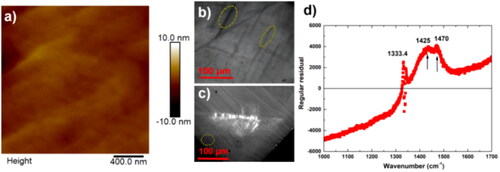
Generally, a sub-nano smooth surface can be obtained after DPF. For example, as shown in , the Ra roughness of 0.56 nm of the D-DFP-60 after polishing for 60 min was realized. However, using SRWBT and CL to examine the crystal quality, the darker color-contrast regions of D-DFP-60 shown in are resulted from the distorted regions and extended defects. Among those defects, the uniform linear distributed color-contrast (the elliptically annotated region in ) is associated with the crystal preferential cleavage, which will be investigated by DSAC-TEM in the following. Using general Raman spectroscopic result, the signal of the non-diamond phase of high-quality SCD usually can be suppressed by the high intensity of the sp3 vibration signal. Therefore, the residual process was introduced to clearly highlight the non-sp3 peaks, as shown in . In addition to the 1333.4 cm−1 of diamond intrinsic peak, the 1425 cm−1 peak is attributed to the amorphous carbon (quasi sp3+sp2), while 1470 cm−1 is associated with the sp2 trans-polyacetylene [Citation25], which are probably transformed from sp3 phase by crystal distortion and friction heat (the temperature would beyond the graphitization point of diamond).
Figure 1. (a) AFM image, (b) SRWBT image, (c) CL images, and (d) processed Raman spectrum showing regular residual of the D-DFP-60.
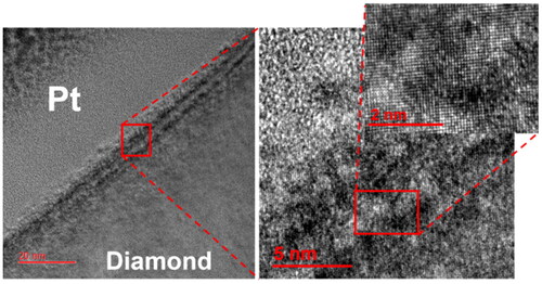
Dislocations which caused by the abrasive induced distortion of the diamond lattice have been observed at the interface between the amorphous layer and the diamond surface. While during DFP without abrasives, amorphous regions are distributed in the near surface ∼20 nm layer after 15 min sliding, as shown in . The interface catalytic action without abrasives between metal and diamond also would give rise to the undamaged lattice near the local amorphous areas, as displayed in the enlarged DSAC-TEM-ABF image and inset in . This would be attributed to the surface graphitization and removal [Citation26]. In this case, the constant mechanical fatigue together with heat accumulation would probably affect the region of near surface areas, especially where have defective lattice structure with higher energy in the SCD. In other words, the discrete amorphous transformation positions probably are associated with as-grown dislocations of SCD or defects resulting from grinding.
Figure 2. The cross-sectional DSAC-TEM image and amplified annular bright field (ABF) image of the subsurface damage of D-DFP-15.
With the continue of DFP, accompanied by energy accumulation and persistent fatigue, the polishing affected region is further extended, as shown in the darker contrast in the DSAC-TEM image in . Magnifying the transition region from the near-surface region and intact diamond matrix, nano-scale lattice crack together with many localized defects, showing in light contrast in the DSAC-TEM-ADF inset, were observed. These crack extension and accompanying localized lattice distortions in the ∼50 nm range would be started from defects and/or amorphous positions in the near-surface region [Citation27]. These detrimental defects would trap and scatter the carriers in diamond devices.
Figure 3. (a) The cross-sectional DSAC-TEM image together with an amplified annular dark field (ADF) inset of the subsurface damage of D-DFP-30, (b) EELS with an inset of enlarged spectrum features of the test point from the top polished surface to the bulk shown in (a).
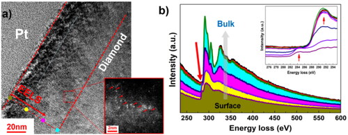
Meanwhile, the fine spectroscopic study of the affected region by EELS is illustrated in to show the types of carbon lattice structures. The test positions were uniformly selected within a depth range extending from the surface to approximately 50 nm (as shown in ). The EELS spectra of diamond crystal have a single loss feature with an onset at about 290 eV, associated with its σ* electronic states. However, compared with the diamond spectra, the EELS spectra of the interface and near surface defective layer have an additional absorption starting at around 285 eV, due to its lower-lying antibonding π* states [Citation28]. The EELS spectrum profile of amorphous carbon is similar to the graphite except for the different intensity. By inspecting typical EELS spectra as presented in , the peak of π* bond disappears gradually, but the peak of σ* bond at around 290 eV turns sharper. This indicates that the dominant phases at positions from the top surface to the bulk in about 50 nm region are graphitic carbons, amorphous carbons, defective diamond, and diamond, gradually.
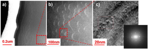
Different to the localized distortion after 15 min and 30 min polishing, longstanding mechanical friction at high sliding speed would also give rise to the crystal cleavage and non-diamond phase, which is much deeper (in micrometers) than the near-surface local defects. As shown in , the apparently light defective stripes in the DSAC-TEM-ADF image in <110> view direction is in the micrometers level. For the cubic diamond structure, when the load stress was applied uniaxially during constant polishing on (100) surface, which has a large angle to the facet, the fracture proceeding along “easy cleavage” planes was expected. This is strongly associated with the difference of lattice crystallographic, inter-planar distance, atomic stacking density, and the symmetry of three crystal planers. Although the easily formed graphite is a benefit to the interface lubrication, its rapid remove rate and high sliding speed inevitably would result in the aggravation of subsurface defects of D-DFP-60 which suffering mechanical shock for a long duration. Further enlarge these damages at the deeper region, as shown in , these cleavages are distributed in a tile-roof-like parallel pattern. As displayed in the inset of , the DSAC-TEM electron diffraction pattern showing an obvious diffraction ring of the cleavage regions of D-DFP-60 in the DSAC-TEM-ABF image indicates the local lattice amorphization.
Figure 4. (a) DSAC-TEM-ADF image and (b) enlarged DSAC-TEM-ADF image of subsurface cleavages as well as the (c) DSAC-TEM-ABF image of defects regions of D-DFP-60 with an inset of electron diffraction pattern.
In fact, during DFP, the phase transformation, length of dislocation lines as well as distribution area of localized shear strain are strongly related to the friction force and the polishing direction transiting from different crystal direction [Citation29]. As the sliding goes on, the repeated sliding of the metal on the diamond surface can cause atom removal, yet with slighter wear and subsurface damage of the diamond substrate. However, if under low pressure and low velocity, the amorphous carbon atoms gradually appear through the interface, affecting surface phase conversion and defect evolution. Based on those evolutionary features, mechanical fatigue and input-energy accumulation associated with constant high-speed mechanical sliding and friction directionality of SCD should be carefully considered and optimized as well in the future works, eg, introducing dynamic control of differentiated polishing processes, graphite assisted and intermittent oxidation enhancement, etc.
4. Conclusion
For further building bases of DFP upgrade and performance consideration of SCD for applications, the SCDs were smoothed by DFP in variable durations. The local amorphization, dislocation extension and subsurface cleavage with the prolonging of sliding duration were observed in the nanoscale. Due to the fatigue and continuous energy input during high-speed DFP, the diamond crystal can be distorted/cracked in varying degrees. Different to the localized distortion with sp2-π* states in the near-surface 10 nm area after 15 min DFP, continuous mechanical friction would result in the dislocation penetration to ∼50 nm. If the sliding lasts to 60 min, preferential cleavage with the non-diamond phase would even be formed in the micrometers range beneath the surface, additionally owing to the (111) crystallographic character of the cubic diamond crystal.
Authors contributions
Sheng Ye: Conceptualization, Methodology, Investigation, Formal Analysis, Device Fabrication, Writing-Original Draft; Yuting Zheng: Data curation, Device Fabrication, Writing-Original draft preparation; Shangman Zhao: Investigation, Device Fabrication; Junjun Wei: Resources, Supervision; Jinlong Liu: Resources, Supervision; Liangxian Chen: Investigation, Device Fabrication; Haitao Ye: Investigation, Device Test; Xiaotong Zhang: Resources, Supervision; Xiaoping Ouyang: Resources, Supervision; Chengming Li: Resources, Supervision.
Disclosure statement
No potential conflict of interest was reported by the authors.
Additional information
Funding
References
- Zheng Y, Ye H, Liu J, et al. Surface morphology evolution of a polycrystalline diamond by inductively coupled plasma reactive ion etching (ICP-RIE). Mater Lett. 2019;253:1–5.
- Crawford KG, Maini I, Macdonald DA, et al. Surface transfer doping of diamond: a review. Prog Surf Sci. 2021;96(1):100613.
- Perez G, MARéCHAL A, Chicot G, et al. Diamond semiconductor performances in power electronics applications. Diamond Relat Mater. 2020;110:108154.
- Tsukioka K. Energy distributions and scattering mechanisms of carriers in diamond. Diamond Relat Mater. 2009;18(5-8):792–795.
- Wade T, Geis MW, Fedynyshyn TH, et al. Effect of surface roughness and H–termination chemistry on diamond’s semiconducting surface conductance. Diamond Relat Mater. 2017;76:79–85.
- Yu X, Li J, Xu H, et al. Influence of diamond matrix morphology on ZnO surface morphology and preferred orientation. Mater Today Commun. 2023;37:107462.
- Long C, Yang LI. Influence of surface roughness on surface acoustic waves. Proceedings of the 2020 15th Symposium on Piezoelectrcity, Acoustic Waves and Device Applications (SPAWDA), p. 16–19. 2021.
- Iriarte GF, RODRíGUEZ JG, Calle F. Synthesis of c-axis oriented AlN thin films on different substrates: a review. Mater Res Bull. 2010;45(9):1039–1045.
- Liang J, Masuya S, Kim S, et al. Stability of diamond/Si bonding interface during device fabrication process. Appl Phys Express. 2019;12(1):016501.
- Liang J, Masuya S, Kasu M, et al. Realization of direct bonding of single crystal diamond and Si substrates. Appl Phys Lett. 2017;110(11):111603.
- Doronin MA, Polyakov SN, Kravchuk KS, et al. Limits of single crystal diamond surface mechanical polishing. Diamond Relat Mater. 2018;87:149–155.
- Wen H, Lu J, Xu S, et al. Mechanical chemical polishing of large-size single-crystal diamond substrates with a sol-gel polishing tool. J Manuf Processes. 2022;80:210–219.
- Zheng Y, Ye H, Thornton R, et al. Subsurface cleavage of diamond after high-speed three-dimensional dynamic friction polishing. Diamond Relat Mater. 2020;101:107600.
- Li Z, Jiang F, Jiang Z, et al. Energy beam-based direct and assisted polishing techniques for diamond: a review. Int J Extreme Manuf. 2024;6(1):012004.
- Yamamura K, Emori K, Sun R, et al. Damage-free highly efficient polishing of single-crystal diamond wafer by plasma-assisted polishing. CIRP Ann. 2018;67(1):353–356.
- Kubota A, Nagae S, Motoyama S. High-precision mechanical polishing method for diamond substrate using micron-sized diamond abrasive grains. Diamond Relat Mater. 2020;101:107644.
- Lu J, Xiao P, Tong R, et al. Precision polishing of single crystal diamond (111) substrates using a Sol-Gel (SG) polishing pad. IEEE Trans. Semicond Manufact. 2019;32(3):341–345.
- Schuelke T, Grotjohn TA. Diamond polishing. Diam Relat Mater. 2013;32:17–26.
- Harris DC. Materials for infrared windows and domes: properties and performance. Bellingham: SPIE Press; 1999.
- Tatsumi N, Harano K, Ito T, et al. Polishing mechanism and surface damage analysis of type IIa single crystal diamond processed by mechanical and chemical polishing methods. Diamond Relat Mater. 2016;63:80–85.
- Haisma J, VAN DER Kruis F J HM, Spierings B A CM, et al. Damage-free tribochemical polishing of diamond at room temperature: a finishing technology. Precis Eng. 1992;14(1):20–27.
- Liu N, Yamada H, Yoshitaka N, et al. Comparison of surface and subsurface damage of mosaic single-crystal diamond substrate processed by mechanical and plasma-assisted polishing. Diamond Relat Mater. 2021;119:108555.
- Volpe P-N, Muret P, Omnes F, et al. Defect analysis and excitons diffusion in undoped homoepitaxial diamond films after polishing and oxygen plasma etching. Diamond Relat Mater. 2009;18(10):1205–1210.
- Chen Y, Zhang LC. Polishing of polycrystalline diamond by the technique of dynamic friction, part 4: establishing the polishing map. Int J Mach Tools Manuf. 2009;49(3-4):309–314.
- Andrea Carlo F, Robertson J. Raman spectroscopy of amorphous, nanostructured, diamond-like carbon, and nanodiamond. Philos Trans A Math Phys Eng Sci. 2004;362(1824):2477–2512.
- Luo H, Ajmal KM, Liu W, et al. Atomic-scale and damage-free polishing of single crystal diamond enhanced by atmospheric pressure inductively coupled plasma. Carbon. 2021;182:175–184.
- Yuan S, Guo X, Zhang S, et al. Influence mechanism of defects on the subsurface damage and structural evolution of diamond in CMP process. Appl Surf Sci. 2021;566:150638.
- Chen Y, Zhang LC, Arsecularatne JA. Polishing of polycrystalline diamond by the technique of dynamic friction. Part 2: material removal mechanism. Int J Mach Tools Manuf. 2007;47(10):1615–1624.
- Yuan S, Guo X, Mao Q, et al. Effects of pressure and velocity on the interface friction behavior of diamond utilizing ReaxFF simulations. Int J Mech Sci. 2021;191:106096.