Abstract
Delete me
To the Editor: Persistent left superior caval vein (SCV) is a rare anatomical variety.Citation[[1]], Citation[[2]] A recently published case report describing the incorrect hemodialysis (HD) catheter position in the internal thoracic vein,Citation[[3]] encouraged us to present the case of HD catheter placement in our patient's persistent left SCV.
T.E., a 31 year old female, suffered from membranoproliferative glomerulonephritis that resulted in end stage renal failure. She was temporarily dialyzed using a 12 Fr double lumen HD catheter (Arrow, USA), placed in superior caval vein through right internal jugular vein. Two attempts to form arteriovenous fistula on her left forearm failed. The skin around the catheter entrance site in the patient's neck showed signs of tunnel infection. In addition, the catheter did not allow adequate blood flow and the patient was referred for catheter replacement.
Replacement by “over guide-wire technique” was not considered, because of the presence of the tunnel infection. Being aware that a few weeks would be needed to create and start to use a permanent vascular access, the attending physician chose the left jugular vein as a second choice approach, instead of the femoral vein.Citation[[4]], Citation[[5]] The catheter from the right jugular vein was withdrawn, and a new catheter was placed into the left jugular vein without any complication during the procedure. Routine control chest X-ray showed the catheter position to be near the pulmonary artery.
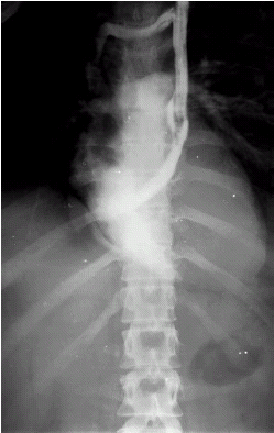
Contrast digital subtraction angiography, showed catheter inside a persistent left SCV, whose confluence was at right atrium, normal right ventricle cavity and pulmonary artery ().
Figure 1. Contrast digital angiography of left superior caval vein.
The catheter was used for maintenance HD during a period of four weeks with blood flow approximately 220 mL/min, until new permanent access (PTFE graft on the left forearm) was created and successfully used for HD. The catheter was subsequently withdrawn without complication.
To the best of our knowledge, this is the first report of a HD catheter placement in persistent left SCV, and its use for HD during a relatively long period of time. This position of HD catheter is not risk free, and serious complication may occur.Citation[[6]], Citation[[7]] This should be borne in mind when considering a second choice approach.Citation[[4]], Citation[[5]] In spite of catheter displacement, we decided to use it, because it was the best choice for our patient.
REFERENCES
- Shumacker H.B., King H., Waldhausen J.A. The Persistent Left Superior Vena Cava. Surgical Implications, with Special Reference to Caval Drainage into the Left Atrium. Ann. Surg. 1967; 165: 797–805
- Agnoleti G., Annecchino F., Preda L., Borghi A. Persistence of the Left Superior Caval Vein: Can it Potentiate Obstructive Lesions of the Left Ventricle? Cardiol. Young 1999; 9: 285–290
- Sotirakopoulos N., Skandalos I., Telemachos T.S., Stambolidou M., Karamachos K., Mauromatidis K. The Incorrect Placement of Hemodialysis Catheters In Veins. The Necessity for Urgent X-ray Evaluation for its Position. Renal Failure 2001; 23: 127–133
- Oliver M.J., Callery S.M., Thorpe K.E., Schwab S.J., Churchill D.N. Risk of Bacteremia from Temporary Hemodialysis Catheters by Site of Insertion and Duration of Use: A Prospective Study. Kidney Int. 2000; 58: 2543–2545
- Schwab S.J., Beathard G. The Hemodialysis Catheter Conundrum: Hate Living with Them, But Can’t Live Without Them. Kidney Int. 1999; 56: 1–17
- Jantsch H., Draxler V., Muhar V., Schlemer M., Waneck R. Pseudodisplacement of the Caval Catheter in Persistent Left Superior Vena Cava. Fortschr Geb Rontgenstr Nuklearmed 1983; 138: 1–44
- Trigano J.A., Torresani J., Pinot J.J. Implantation of a Pacemaker and Superior Vena Cava Anomalies: Value of Perioperative Angiocardiography. Arch. Mal. Coeur. Vaiss. 1982; 75: 817–823