

Abstract
Micromotors and nanomotors are an emerging research field that aims at achieving locomotion on the microscale for a variety of applications such as drug delivery, single cell manipulation, microsensors and lab-on-a-chip devices, just to point out a few. The enthusiastic development of hybrid micromotors harnessing biological power sources for physiologically compatible nano/microdevices has recently brought a lot of attention to the international research community that is looking for a solution for the actuation and locomotion on the microscale. This article describes the potential of sperm-driven micro-bio-robots in the biomedical field such as drug delivery or single cell manipulation. Herein, a specific potential of the sperm-driven micro-bio-robot is described that might have impact on the development of assisted reproductive technologies.
1. Hybrid bio-robots for biomedical applications
Drug-delivery devices need to be powered on the microscale in a physiologically compatible manner. The delivery of therapeutic and diagnostic agents to areas in the body that are difficult to access is the subject of research using micromotors. A very common structure for motion in the natural microscopic world is a flagellum as it can be found in bacteria or spermatozoa. The flagellum is an attractive architecture for the design of drug-delivery machines. Researchers are trying to mimic the motion of such slender filaments, as demonstrated by Zhang et al. Citation[1], Dreyfus et al. Citation[2] or Gao et al. Citation[3], that fabricated artificial magnetic flagella that are propelled by a rotating or oscillating magnetic field. Further, researchers at Cornell University Citation[4] were inspired by the biological motor of a sperm cell and are working on rebuilding its molecular motor with the goal of using it for engineering sperm-powered nanobots. This undertaking includes the design of a metabolic pathway that leads to the conversion of glucose to ATP as fuel using a 10-enzyme glycolysis. There have also been a few approaches using motile cells as actuators of different biohybrid micromotors that are either propelled by motile microorganisms Citation[5] or by contractile cells Citation[6]. All these approaches mainly target the application in the biomedical field, especially drug delivery. The key requirements for application of micromotors for drug delivery are biocompatibility, remote control mechanisms, highly efficient locomotion in the low Reynolds number regime under physiological conditions and cargo pick-up/drop-off mechanisms.
We recently presented a new microcarrier system which moves by the strength of a single flagellum: a bovine sperm cell captured inside a microtube that consists of rolled-up nanomembranes Citation[7], as shown in .
Figure 1. The vision of spermbots: capture and delivery of single spermatozoa to the oocyte by a magnetic microtube that is controlled by an external magnetic field. The inset shows a microscopic image of a bovine spermatozoon entering a 20 µm long microtube. Scale bar 20 µm.
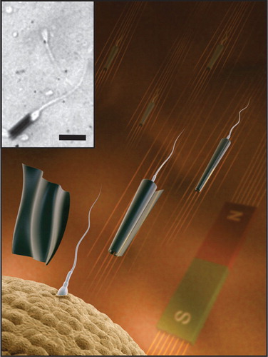
This sperm-driven micro-bio-robot is physiologically compliant and is able to swim in highly viscous media in a low Reynolds numbers regime. The purely mechanical capturing mechanism inside the microtube does not alter the cell compared to other binding mechanisms such as surface functionalization or incorporation of nanoparticles. The integration of a magnetic layer into the microtube enables the magnetic remote control with low external magnetic fields. Further, temperature changes can be used to accelerate and slow down this sperm-driven microtube within the natural temperature tolerance of the bovine spermatozoa. With the cell as a strong power unit and remote control mechanisms, this sperm-driven microtube becomes an interesting device for robotic tasks on the microscale. The spermatozoon serves as biocompatible driving force for the microtube and could remotely be steered through body fluids. If the spermbot is applied for drug delivery, the drug could be loaded on the outer microtube surface. There are various ways to functionalize the surface of microtubes and attach proteins, DNA or other bioactive components as has been shown by many authors previously. The release of the drug could be induced by a slight change in pH or temperature which leads to a change in the drug attachment and unloading from the microtube surface. In addition to drug delivery, the most obvious potential of this system is the development of new assisted reproductive technologies, since we can remotely deliver a single sperm cell to a desired location using the magnetic microtube.
2. Spermbots for assisted in vivo reproduction technology
The success rate of state-of-the-art assisted reproduction techniques such as in vitro fertilization (IVF) and intracytoplasmic sperm injection is still low. These assisted reproduction techniques involve the removal of the oocyte from the body, fertilization in the petri dish, cultivation of embryos and reimplantation of the embryo into the uterus. The vision behind the spermbots for the establishment of alternative assisted reproduction technologies is that the removal of the oocyte from the body and IVF can be bypassed by guiding the selected sperm cells to the oocyte location in the natural environment inside the human body. This is the first approach of keeping the fertilization process completely in its natural surroundings. It is known that the low efficiency of IVFs is due to the artificial stresses that gametes and embryos are exposed to in the in vitro environment and the quality of embryos generated in vitro was shown to be lower than that of their in vivo counterparts Citation[8]. In a usual IVF procedure, gametes and embryos are moved as often as 20 times during the washing and handling steps which induces an immense stress for the cells Citation[8]. The spermbot technology is aiming at higher pregnancy rates since the oocytes and spermatozoa will be kept in their usual microenvironment during fertilization. Up to now, the journey of the sperm to the egg cell and fertilization in vivo is still poorly understood. This might be an additional reason why existing assisted reproductive technologies have a low success rate. The spermbot technology might also help in gaining a better understanding of the journey of the spermatozoa and the obstacles they face on their way. The spermbot consists of a magnetic microtube which acts as an envelope for the cell that can very likely be tracked in vivo, while not interfering with sperm activity. Hence, the spermbot could also serve as explorative device for studying the natural pathways of spermatozoa inside the body. The spermbot might be helpful for studying at which locations in the female reproductive tract spermatozoa are held back, where they encounter obstacles and how long they remain at certain stages. Eventually, this system will help in understanding better what the reasons for different cases of infertility are. It has to be pointed out that the application of spermbots has potential to only treat certain forms of infertility that are originated in poor semen quality, low sperm counts or defects in the chemotactic response of sperm. However, the male infertility accounts for 40% of the cases in which couples remain childless Citation[9].
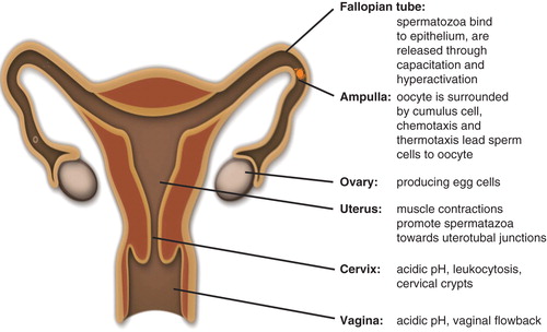
The journey of spermatozoa to the oocyte inside the body is a very complex procedure. illustrates some of the key points of the path of the spermatozoa to the fertilization site in the female reproductive tract. Numerous checkpoints can be found that are believed to impede sperm cells to reach the oocyte, but also represent a selection procedure that ensures that only the most fertile spermatozoa reach the fertilization site Citation[10]. In general, the sperm cells are hostile to the female body and several defense mechanisms exist that keep the vast number of spermatozoa from reaching the egg cell. The cervix has an acidic pH of 5 whereas the semen plasma is about 7. About 30 min after entering the body, < 1% of living sperm cells are remaining in the female reproductive tract due to vaginal flowback, the acidic pH and phagocytosis by leukocytes Citation[11]. The next challenging step is the twisted channels in the cervix with a highly viscous environment through which the cells have to swim in order to reach the uterus. Afterward, the uterotubal junction can only be entered by sperm cells that show a characteristic swimming pattern. Once the fallopian tubes are reached, the conditions are more sperm-friendly. Sperm cells are also known to bind to the mucosal epithelium inside the fallopian tubes and can be stored there Citation[11]. This area is very difficult to study and hence only little is known about the further journey of the sperm cells. However, it is thought that only few sperm cells are left at this stage Citation[10] and that the sperm cells are guided through the ampulla by chemotaxis and thermotaxis to the oocyte location. At this stage, a crucial preparation step for fertilization takes place: capacitation. This is a shedding of proteins from the sperm cell membrane that leads to hyperactivation which results in an increased flagellum beating amplitude and asymmetric beating pattern. This helps the spermatozoa which were stored in the isthmus to release from the epithelial lining of the fallopian tube and promote toward the egg. Only hyperactivated sperm cells are able to pass the outer layers of the egg cell which is covered with cumulus cells. The timing is quite important because once capacitated, a sperm cell only survives a few more hours. It also has to be timed exactly with the ovulation of the egg cell. Finally, the acrosome reaction releases enzymes that are important for the penetration of the zona pellucida, the outer membrane of the oocyte. The journey of spermatozoa to the oocytes belongs to one of the most important processes in nature and is perhaps one of the least well understood. As more and more is discovered about the obstacles that spermatozoa face on their way, important progress can be made in assisted conception technologies that treat increasing infertility.
Figure 2. Schematic representation of female reproductive tract with description of key obstacles of spermatozoa on their journey to the oocyte.
3. Expert opinion: challenges of spermbots
Several key challenges can be defined which need to be tackled in the future in order to promote the impact of the spermbot technology. For a successful fertilization, first, the selection of the most fertile spermatozoa is crucial. Thus, development of sensitive separation and analysis methods of single spermatozoa is subject to many current studies.
The next step will be the highly efficient capture and release of single sperm cells that can be remotely controlled. Optimum cell activity and fertilization capability have to be maintained through the whole process of capture, transport and delivery of the cell. Additionally, in order to avoid the attachment of harmful microbes to the sperm cell, antibacterial agents could be immobilized on the inner surface of the microtube which will protect the single sperm cell from bacterial attacks during the process of delivery. The magnetic microtube must be able to pass the natural obstacles that sperm cells are facing in order to reach the fertilization site. Toxic effects that might be caused by the magnetic material of the microtube have to be ruled out. Further, a challenge will be the controlled timing of initiating the capacitation of the sperm cell to guarantee successful fertilization. In case the acrosome reaction does not take place on its own, the sperm cell needs to be assisted in fusing with the oocyte possibly by functionalizing the microtube with crucial enzymes.
Interestingly, for any in vivo applications of the spermbots, an appropriate tracking technology such as magnetic resonance imaging or ultrasound is necessary for the imaging and guiding of the microtube. Martel et al. demonstrated how magnetic resonance imaging can be used to track and control magnetic microbeads in arteries of live pigs Citation[12] or therapeutic microcarriers inside the tissue Citation[13]. An open question remaining is if the magnetic microtubes have any effect on the human body, even if their materials are proven biocompatible. It is clear that it is best to remove the microtubes from the body after the operation is completed which could be accomplished by applying an external magnetic field. However, current investigations of numerous research groups also target the study of long-term biotransformation of magnetic nanoparticles in the living tissue Citation[14,15]. The uptake of low amounts of magnetic material consisting of biocompatible iron compounds due to the natural iron metabolism might avoid the removal from the body. For example, a human being has a recommended dietary allowance of 18 mg/day of iron. From this point of view, thin nanomembranes from iron oxide might be resorbed by the human body without any harm over a long period of time. This is an interesting approach that avoids the subsequent removal of the microtubes but requires materials that are bioresorbable.
Even though the proposed spermbot technology is still far from being applied as drug delivery or assisted reproduction tool, it offers many interesting aspects for the exploration of remote-controlled processes in biomedical applications. It has high potential to serve in investigations of sperm migration processes in vivo and, eventually, in assisted reproductive technology. Additionally, let us not forget that the sperm flagellum as biological motor has been optimized by nature over millions of years and can be harnessed in many diverse ways as driving force for microdevices.
Declaration of interest
The authors have no relevant affiliations or financial involvement with any organization or entity with a financial interest in or financial conflict with the subject matter or materials discussed in the manuscript. This includes employment, consultancies, honoraria, stock ownership or options, expert testimony, grants or patents received or pending or royalties.
Acknowledgements
Authors thank the Volkswagen Foundation (# 86 362) and the DFG Priority Programme (SPP 1726). V Magdanz thanks Dr Perez Rodriguez for fruitful discussion.
Bibliography
- Zhang L, Abbot JJ, Dong D, et al. Artificial bacterial flagella: fabrication and magnetic control. Appl Phys Lett 2009;94:06410
- Dreyfus R, Baudry J, Roper ML, et al. Microscopic artificial swimmers. Nature 2005;437:862-5
- Gao W, Feng X, Pei A, et al. Bioinspired helical microswimmers based on vascular plants. Nano Lett 2014;14:305-10
- Mukal C, Bergkvist M, Nelson J, Travis AJ. Sequential reactions of surface-tethered glycolytic enzymes. Chem Biol 2009;16:1013-20
- Behkam B, Sitti M. Bacterial flagella-based propulsion and on/off motion control of microscale objects. Appl Phys Lett 2007;90:23902-4
- Williams BJ, Anand SV, Rajagopalan J, Saif MTA. A self-propelled biohybrid swimmer at low Reynolds number. Nature 2014;5:3081
- Magdanz V, Sanchez S, Schmidt OG. Development of a sperm-flagella driven micro-bio-robot. Adv Mater 2013;25(45):6581-8
- Khademhosseini A, Borenstein J, Toner M, Takayama S. Micro and nanoengineering of the cell microenvironment. Artech House, Norwood, USA; 2008
- Quick facts about infertility. American Society for Reproductive Medicine. http://www.reproductivefacts.org/detail.aspx?id=2322 [Last accessed 6 March 2014]
- Scott MA. A glimpse at sperm function in vivo: sperm transport and epithelial interaction in the female reproductive tract. Anim Reprod Sci 2000;60-61:337-48
- Suarez SS, Pacey AA. Sperm transport in the female reproductive tract. Hum Reprod Update 2006;12(1):23-37
- Martel S, Mathieu JB, Felfoul O, et al. Automatic navigation of an untethered device in the artery of a living animal using a conventional clinical magnetic resonance imaging system. Appl Phys Lett 2007;90:114105
- Pouponneau P, Leroux JC, Soulez G, et al. Co-encapsulation of magnetic nanoparticles and doxorubicin into biodegradable microcarriers for deep tissue targeting by vascular MRI navigation. Biomaterials 2011;32:3481-6
- Meijas R, Gutierrez L, Salas G, et al. Long term biotransformation and toxicity of dimercaptosuccinic acid-coated magnetic nanoparticles support their use in biomedical applications. J Control Release 2013;171:225-33
- Levy M, Luciani N, Alloyeau D, et al. Long term in vivo biotransformation of iron oxide nanoparticles. Biomaterials 2011;32:3988-99